Cop
yright
© ABE&M t
odos os dir
eit
os r
eser
vados
.
Lack of association between
polymorphisms in the
UBASH3A
gene and autoimmune thyroid
disease: a case control study
Falta de associação entre polimorismos do gene UBASH3A
e doença tiroidiana autoimune: estudo caso controle
TianTian Cai1, Xuan Wang1, Fatuma-Said Muhali1, RongHua Song1, XiaoHong Shi2, WenJuan Jiang2, Ling Xiao2, DanFeng Li2, JinAn Zhang2
ABSTRACT
Objective: The aim of this study was to investigate UBASH3A gene variation association with
autoimmune thyroid disease and clinical features in a Chinese Han population. Subjects and methods: A total of 667 AITD patients (417 GD and 250 HT) and 301 healthy controls were ge-notyped for two single nucleotide polymorphisms (SNPs) rs11203203, rs3788013 of UBASH3A
gene, utilizing the Matrix Assisted Laser Desorption Ionization-Time of Flight Mass Spectrome-ter (MALDI-TOF-MS) Platform. Results: Between the control group and AITD, GD and HT group, no statistically signiicant difference was observed in the genotypic and allelic frequencies of the two SNPs. There was no signiicant difference in allelic frequencies of the two SNPs be-tween GD with and without ophthalmopathy. There was no signiicant difference in haplotype distributions between the control group and AITD, GD or HT group. Conclusion: Rs11203203 and rs3788013 in UBASH3A gene may not be associated with AITD patients in Chinese Han
population. Arq Bras Endocrinol Metab. 2014;58(6):640-5
Keywords
Autoimmune thyroid disease (AITD); Graves’ disease (GD); Hashimoto’s thyroiditis (HT); ubiquitin associated and SH3 domain containing A (UBASH3A); single nucleotide polymorphism (SNP)
RESUMO
Objetivo: O objetivo deste estudo foi investigar a variação no gene UBASH3A com a doença tiroidiana autoimune e características clínicas na população chinesa Han. Sujeitos e métodos:
Um total de 667 pacientes com DTAI (417 com DG e 250 com TH) e 301 controles saudáveis foi genotipado para dois polimorismos de nucleotídeo simples (SNPs) rs11203203, rs3788013 do gene UBASH3A, usando-se a plataforma MALDI-TOF-MS (Ionização/Dessorção de Matriz Assis-tida por Laser – Tempo de Voo/Espectrômetro de Massa). Resultados: Não foram observadas diferenças signiicativas entre as frequências genotípicas e alélicas dos dois SNPs nos grupos controle e DTAI, DG e TH. Não houve diferenças signiicativas entre as frequências alélicas dos dois SNPs em pacientes com DG com ou sem olftalmopatia. Não houve diferenças signiicati-vas nas distribuições de haplótipos no grupo controle e nos grupos DTAI, DG e TH. Conclusão:
Os SNPs rs11203203 e rs3788013 do gene UBASH3A podem não estar associados a pacientes com DTAI na população chinesa Han. Arq Bras Endocrinol Metab. 2014;58(6):640-5
Descritores
Doença tireoidiana autoimune (DTAI); doença de Graves (DG); tireoidite de Hashimoto (TH); ubiquitina associada e domínio SH3 contendo A (UBASH3A); polimorismos de nucleotídeo simples (SNP)
1 Clinical Research Center,
The First Afiliated Hospital of Medical School, Xi’an Jiaotong University, Xi’an, China
2 Department of Endocrinology,
Jinshan Hospital of Fudan University, Shanghai, China
Correspondence to: JinAn Zhang
Department of Endocrinology, Jinshan Hospital of Fudan University, 1508
201508 – Longhang Road, Shanghai, China
Received on Jan/4/2014 Accepted on Apr/10/2014
Cop
yright
© ABE&M t
odos os dir
eit
os r
eser
vados
.
INTRODUCTION
A
utoimmune thyroid disease (AITD) is one group of organ-speciic autoimmune disease encompas-sing Graves’ disease (GD) and Hashimoto’s thyroiditis (HT), which affects between 2%-4% of women and up to 1% of men (1). Hitherto, the etiology of AITD re-mains largely unknown, but environmental factors and genetic predisposition are believed to be involved in it (2). AITD usually occurs after a series of genetic and environmental factors. It has long been established that major genetic predisposition to this disease is main-ly contributed by variation of several classic immune--related genes, particularly genes of human leukocyte antigen (HLA) (3,4), cytotoxic T-lymphocyte-associa-ted antigen-4 (CTLA-4) (5,6), CD40 (7), and protein tyrosine phosphatase-22 (PTPN22) (8,9). In addition, other thyroid-speciic genes are also reported to be asso-ciated with AITD, such as the genes of thyroid stimula-ting hormone receptor (TSHR) (10,11) and thyroglo-bulin (Tg) (12,13). Currently, genome-wide association (GWA) studies have had a dramatic impact on suscepti-bility locus discovery, leading to more novel regions of the genome emerging as AITD susceptibility loci.The gene UBASH3A encodes one of two family members which belong to the T-cell ubiquitin ligand (TULA) family. UBASH3A is a potent regulator of cellular functions and is expressed predominantly in T-cells, having a suppressing effect on TCR signaling and activation (14). Lack of TULA proteins resulted in hyper-reactivity of T cells (15). Recently, more stu-dies have reported that it is the newly uncovered gene associated with various autoimmune diseases (16-19). In present study, we performed a case-control study of AITD in a Chinese Han population, attempting to ind the potential association of rs11203203 and rs3788013 in the UBASH3A gene with GD and HT.
MATERIALS AND METHODS
Patients and controls
Our study investigated 667 AITD patients, including 417 GD patients and 250 HT patients. All patients were diagnosed according to both clinical and biochemical evidences, and the one with other autoimmune and ge-netic diseases was ruled out. The diagnostic criteria for GD were mainly determined by 1) clinical manifesta-tion and laboratory biochemical proof of hyperthyroi-dism; 2) the presence of diffuse goiter; 3) the presence
of thyroid-associated ophthalmopathy (TAO); 4) the presence of pretibial myxedema; 5) the positive circula-ting TRAb or TPOAb. HT was deined on the basis of enlarged thyroid, and the high level of either TPOAb or TgAb, with or without clinical and biochemical hy-pothyroidism. For the suspicious cases of HT, diagno-ses were conirmed by ine needle aspiration biopsies (FNAC). Presence of AITD family history was deined as the subjects’ irst-degree relatives including parents, children and siblings or second-degree relatives such as grandparents, uncles and aunts who had AITD occur-rence. Another 301 healthy individuals were enrolled as a control group, and the one with thyroid diseases or other autoimmune diseases was excluded from this category. Besides, negative TPOAb necessitates for control individual screening. All the subjects were un-related Chinese Han people and enrolled with infor-med consent from the First Afiliated Hospital of Xi’an Jiaotong University. The research project was approved by the Ethics Committee of the hospital.
Genomic DNA extraction and genotyping
2 ml peripheral venous blood from the subjects was obtained by venipuncture and collected in an EDTA tube. Genomic DNA was extracted by standard proce-dure using RelaxGene Blood DNA System (TIANGEN BIOTECH, BEIJING, China). SNP genotype informa-tion was downloaded from the HapMap CHB popula-tion (corresponding to samples with Beijing of China ancestry). Genotyping rs11203203 and rs3788013 was performed using Matrix Assisted Laser Desorption Io-nization-Time of Flight Mass Spectrometer (MALDI--TOF-MS) Platform from Sequenom (San Diego, CA, USA) in collaboration with Shanghai Benegene Biote-chnologies CO. LTD (Shanghai, China).
Statistical analysis
The clinical data were expressed as M ± SD. The diffe-rence of genotypic and allelic frequencies distributions was analyzed by χ2 test or Fisher’s exact test.
Cop
yright
© ABE&M t
odos os dir
eit
os r
eser
vados
.
RESULTS
Clinical data analysis
Our study investigated 667 AITD patients, including 417 GD patients (29.74% male and 70.26% female, aged 5 - 73, mean age 34.48 ± 13.95) and 250 HT patients (15.60% male and 84.40% female, aged 4 - 77, mean age 31.9 ± 13.10). In GD patients, the avera-ge onset of aavera-ge is 32.31 ± 14.07, 98 individuals had ophthalmopathy and 72 had family history (23.5% and 17.27%, respectively). In HT patients, the avera-ge onset of aavera-ge was 30.29 ± 13.05, 6 individuals had ophthalmopathy and 54 had family history (2.40% and 21.60%, respectively).
Allele and genotyping results
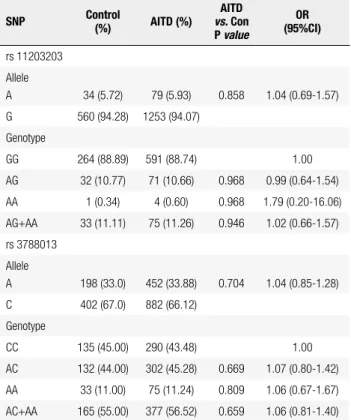
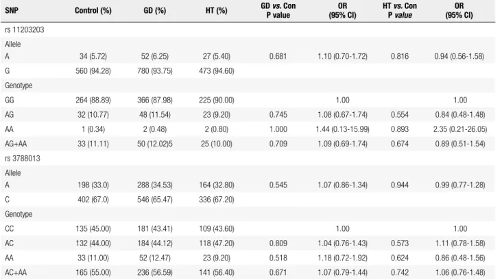
Both of the two SNPs in control group were in HWE (P > 0.05). The distributions of allelic, genotypic fre-quencies and case-control association analysis are shown in table 1 and table 2. For rs11203203, the frequency of minor allele A was higher in AITD and GD group (5.93% and 6.25%, respectively) than that in control group (5.72%), while the frequency in HT group (5.40%) was somehow lower than that of con-trol group. Likewise, for rs3788013, the frequency of minor allele A was also higher in AITD and GD group (33.88% and 34.53%, respectively) than that of con-trol group (33.00%), while frequency in HT (32.80%) was lower than that in control group. However, the differences of these allele frequencies between cases and controls were not found statistically signiicant. The frequency of variant genotype AA of rs11203203 was higher in all the patients (0.60% in AITD, 0.48% in GD, and 0.80% in HT) than that in control group (0.34%). However, when wild-type homozygote GG was used as the reference, no statistically signiicant risk was observed for AITD, GD and HT of variant genotype AA, AG and the combination (AG+AA). The frequency of variant genotype AA of rs3788013 was only higher in AITD and GD group (11.24% in AITD, 12.47% in GD) than that in control group (11.00%). When wild-type homozygote CC was used as the refe-rence, no statistically signiicant risk was observed for AITD, GD and HT of variant genotype AA, AC and the combination (AC+AA).
Given that T-cell induced immunity plays an impor-tant role in the pathogenesis of GO (20), we further investigated the two SNPs for association with the
sub-set of GD patients that had ophthalmopathy. Table 3 shows allele distributions and case-control association analysis in ophthalmopathy/non-ophthalmopathy pa-tients and controls. For rs11203203, the frequencies of allele A were higher in GD patients with and wi-thout ophthalmopathy (7.14% and 5.97%, respectively) than that in control group (5.72%); furthermore, the frequency of allele A was higher in GO than that in GD without ophthalmopathy. For rs3788013, the frequen-cies of allele A were higher in GD patients with and without ophthalmopathy (33.67% and 34.80%, respec-tively) than that in control group (33.00%); however, the frequency of allele A was a little lower in GO than that in GD without ophthalmopathy. None of these differences reached statistical signiicance.
Haplotype analysis
According to the Hapmap CHB data, D’ and r2 for LD
test of the two SNPs were 1.0 and 0.121. Although they were not in one block, we performed haplotype analysis. Four possible haplotypes were detected from the SNPs. As shown in table 4, the frequencies of three halotypes were greater than 0.05, of which GC and GA were the most frequent ones, both in cases and
Table 1. Allele and genotype distributions in AITD patients and controls
SNP Control
(%) AITD (%)
AITD
vs. Con P value
OR (95%CI)
rs 11203203 Allele
A 34 (5.72) 79 (5.93) 0.858 1.04 (0.69-1.57) G 560 (94.28) 1253 (94.07)
Genotype
GG 264 (88.89) 591 (88.74) 1.00 AG 32 (10.77) 71 (10.66) 0.968 0.99 (0.64-1.54) AA 1 (0.34) 4 (0.60) 0.968 1.79 (0.20-16.06) AG+AA 33 (11.11) 75 (11.26) 0.946 1.02 (0.66-1.57) rs 3788013
Allele
A 198 (33.0) 452 (33.88) 0.704 1.04 (0.85-1.28) C 402 (67.0) 882 (66.12)
Genotype
Cop
yright
© ABE&M t
odos os dir
eit
os r
eser
vados
.
Table 2. Allele and genotype distributions in GD, HT patients and controls
SNP Control (%) GD (%) HT (%) GD P valuevs. Con (95% CI)OR HT P vs.value Con (95% CI)OR
rs 11203203 Allele
A 34 (5.72) 52 (6.25) 27 (5.40) 0.681 1.10 (0.70-1.72) 0.816 0.94 (0.56-1.58) G 560 (94.28) 780 (93.75) 473 (94.60)
Genotype
GG 264 (88.89) 366 (87.98) 225 (90.00) 1.00 1.00 AG 32 (10.77) 48 (11.54) 23 (9.20) 0.745 1.08 (0.67-1.74) 0.554 0.84 (0.48-1.48) AA 1 (0.34) 2 (0.48) 2 (0.80) 1.000 1.44 (0.13-15.99) 0.893 2.35 (0.21-26.05) AG+AA 33 (11.11) 50 (12.02)5 25 (10.00) 0.709 1.09 (0.69-1.74) 0.674 0.89 (0.51-1.54) rs 3788013
Allele
A 198 (33.0) 288 (34.53) 164 (32.80) 0.545 1.07 (0.86-1.34) 0.944 0.99 (0.77-1.28) C 402 (67.0) 546 (65.47) 336 (67.20)
Genotype
CC 135 (45.00) 181 (43.41) 109 (43.60) 1.00 1.00 AC 132 (44.00) 184 (44.12) 118 (47.20) 0.809 1.04 (0.76-1.43) 0.573 1.11 (0.78-1.58) AA 33 (11.00) 52 (12.47) 23 (9.20) 0.518 1.18 (0.72-1.92) 0.624 0.86 (0.48-1.56) AC+AA 165 (55.00) 236 (56.59) 141 (56.40) 0.671 1.07 (0.79-1.44) 0.742 1.06 (0.76-1.48)
Table 3. Allele distributions in ophthalmopathy, non-ophthalmopathy patients among GD patients and controls SNP Control (%)
GD patients non-GO vs.
Con P value
OR (95% CI)
GO
vs. Con P value
OR (95%CI)
GO vs. non-GO
P value
OR (95% CI) non-GO (%) GO (%)
rs 11203203
A 34 (5.72) 38 (5.97) 14 (7.14) 0.851 1.05 (0.65-1.69)
0.471 1.27 (0.67-2.41)
0.555 1.21 (0.64-2.28) G 560 (94.28) 598 (94.03) 182 (92.86)
rs 3788013
A 198 (33.00) 222 (34.80) 66 (33.67) 0.505 1.08 (0.86-1.37)
0.862 1.03 (0.73-1.45)
0.772 0.95 (0.68-1.33) C 402 (67.00) 416 (65.20) 130 (66.33)
Table 4. Haplotype analyses in AITD patients and controls Haplotype Control
(Frequency)
AITD (Frequency)
GD (Frequency)
HT (Frequency)
AITD vs.
Con P value
OR (95% CI)
GD
vs. Con P value
OR (95% CI)
HT
vs. Con P value
OR (95% CI)
GC 402 (0.670) 880 (0.664) 544 (0.653) 335 (0.669) 0.672 1.00 0.485 1.00 0.963 1.00 GA 163 (0.272) 375 (0.278) 237 (0.284) 138 (0.277) 0.661 0.602 0.888
AC 0 (0.000) 1 (0.001) 2 (0.002) 2 (0.003) 1.000 0.513 0.207 AA 35 (0.058) 77 (0.057) 51 (0.061) 26 (0.051) 0.960 0.825 0.642
Non-GC 198 (0.330) 453 (0.336) 290 (0.347) 166 (0.331) 0.672
0.96
(0.78-1.17) 0.485
1.08
(0.87-1.35) 0.963
1.00 (0.78-1.30)
controls. All the differences of haplotype distributions were not statistically signiicant. When haplotype GC was used as the reference, the combination of other
Cop
yright
© ABE&M t
odos os dir
eit
os r
eser
vados
.
DISCUSSION
The gene UBASH3A, whose oficial full name is ubi-quitin associated and SH3 domain containing A, is loca-ted on chromosome 21 (21q22.3), spanning 40kb and containing 15 exons. Initially, this gene was isolated and characterized by a study focused on identifying candida-te genes on chromosome 21q22.3 for autosomal reces-sive nonsyndromic deafness (DFNB10) (21). TULA/ STS-2/UBASH3A and TULA-2/STS-1/UBASH3B are both members of the T cell ubiquitin ligand proteins (TULA) family. Both of them are suppressors of TCR signaling and can regulate TCR signaling pathways ne-gatively. The two proteins exhibit a unique architecture comprised of three conserved and functional domain: an N-terminal ubiquitin associated (UBA) domain that can bind mono- and poly-ubiquitin, a central Src homology 3 (SH3) domain which can participate in protein-protein interactions, and a C-terminal PGM (phosphoglycerate mutase) acting as phosphatases or phosphotransferases (14,22). Unlike the other protein,
TULA/STS-2/UBASH3A is only expressed within lym-phocytes and has a low phosphatase activity but a uni-que ability of promoting T-cell apoptosis (23,24). Hen-ce, it may set its suppressing effect on T cell signaling and T cell activation by dephosphorylation of unknown substrates or by apoptosis in T cells. As we all know, the pathologic mechanisms of many autoimmune disorders, including GD and HT, are correlated with immune system abnormality. Under the background of genetic predisposition, environmental factors, such as stress, infection and trauma, may cause the functional and quantitative decrease of suppressor T cells (Ts), which interfere with inhibition of helper T cells (Th) activation within thyroid tissues. Consequently, auto-antigens are presented to Th cells in a recognizable pattern, leading to a pathological immune response and ultimately the activation of B cells which can produce auto-antibodies targeting the thyroid itself. Inadequate mutations in auto-antigens or hyper-response to TCR stimulation could result in the disturbance of self immune toleran-ce. Therefore, we hypothesized that polymorphisms wi-thin the gene UBASH3A could cause changes in the expression and function of the protein. These changes could affect down-regulation of T cell activation and apoptosis, break down a balanced immune response, re-sult in a hyper-reactivity of T cell and inally lead to the occurrence of AITD.
In this study, we investigated the association bet-ween two intronic SNPs (rs11203203 and rs3788013)
of UBASH3A gene and AITD patients in a Chinese Han population. We found that the frequencies of al-lele A in the two SNPs were increased in both AITD and GD group. The AA genotype frequencies of the two SNPs in AITD and GD were also higher than that in control group. Furthermore, we found that the fre-quencies of allele A from rs11203203 were higher in GD patients with and without ophthalmopathy than that in control group; moreover, the frequency of allele A was higher in GO than that in GD without ophthal-mopathy. Nevertheless, it was a pity that none of these above-mentioned differences reached statistical signii-cance. The term haplotype refers to physical arrange-ment of SNP alleles along a chromosome (25). It is demonstrated that analysis based on haplotype would be more powerful and effective to detect an association than those based on individual SNPs. However, our study did not ind any association between the four ha-plotypes and AITD, GD or HT group. To sum up, the-se results have suggested that the allele A of two SNPs may play no role in the pathogenesis of AITD or that its effect may be confounded by other various factors.
It has been generally accepted that a case-control study is eficient enough to detect the association bet-ween candidate genes and diseases. However, this me-thod and design is usually limited to various elements. Inadequate sample size, gender, age, different ethnic group and low power statistical methods are common confounding factors in a case-control study (26-28). Those problems were also shown in our study. Our in-dings provided no signiicant evidence that the gene
UBASH3A was associated with susceptibility to AITD in Chinese Han population. This disaccorded with the analysis of association between 2,477 cases with Gra-ves’ disease and rs3788013 of UBASH3A in white eth-nic group from British cohort (29). This dichotomy could be attributed to ethnic diversity, relative small sample size and other different elements. Therefore, we are motivated to investigate more loci in this gene with a larger sample size and in different nations among Chinese people in order to capture the genetic contri-butions by UBASH3A in the pathogenesis of AITD.
Acknowledgement: this work was supported by grants from the National Natural Science Foundation of China (81270871, 81070627).
Cop
yright
© ABE&M t
odos os dir
eit
os r
eser
vados
.
REFERENCES
1. Vaidya B, Kendall-Taylor P, Pearce SH. The genetics of autoimmu-ne thyroid disease. JCEM. 2002;87:5385-97.
2. Tomer Y, Davies TF. Searching for the autoimmune thyroid disease susceptibility genes: from gene mapping to gene function. Endo-cr Rev. 2003;24:694-717.
3. Simmonds MJ, Howson JM, Heward JM, Carr-Smith J, Franklyn JA, Todd JA, et al. A novel and major association of HLA-C in Gra-ves’ disease that eclipses the classical HLA-DRB1 effect. Hum Mol Genet. 2007;16:2149-53.
4. Simmonds MJ, Howson JM, Heward JM, Cordell HJ, Foxall H, Carr-Smith J, et al. Regression mapping of association between the human leukocyte antigen region and Graves disease. Am J Hum Genet. 2005;76:157-63.
5. Kavvoura FK, Akamizu T, Awata T, Ban Y, Chistiakov DA, Frydecka I, et al. Cytotoxic T-lymphocyte associated antigen 4 gene polymor-phisms and autoimmune thyroid disease: a meta-analysis. J Clin Endocrinol Metab. 2007;92:3162-70.
6. Yang J, Qin Q, Yan N, Zhu YF, Li C, Yang XJ, et al. CD40 C/T(-1) and CTLA-4 A/G(49) SNPs are associated with autoimmune thyroid diseases in the Chinese population. Endocrine. 2012;41:111-5. 7. Mukai T, Hiromatsu Y, Fukutani T, Ichimura M, Kaku H, Miyake I, et
al. A C/T polymorphism in the 5’ untranslated region of the CD40 gene is associated with later onset of Graves’ disease in Japane-se. Endocr J. 2005;52:471-7.
8. Velaga MR, Wilson V, Jennings CE, Owen CJ, Herington S, Donal-dson PT, et al. The codon 620 tryptophan allele of the lymphoid tyrosine phosphatase (LYP) gene is a major determinant of Gra-ves’ disease. J Clin Endocrinol Metab. 2004;89:5862-5.
9. Heward JM, Brand OJ, Barrett JC, Carr-Smith JD, Franklyn JA, Gough SC. Association of PTPN22 haplotypes with Graves’ dise-ase. J Clin Endocrinol Metab. 2007;92:685-90.
10. Brand OJ, Barrett JC, Simmonds MJ, Newby PR, McCabe CJ, Bruce CK, et al. Association of the thyroid stimulating hormone receptor gene (TSHR) with Graves’ disease. Hum Mol Genet. 2009;18:1704-13.
11. Liu L, Wu HQ, Wang Q, Zhu YF, Zhang W, Guan LJ, et al. Associa-tion between thyroid stimulating hormone receptor gene intron polymorphisms and autoimmune thyroid disease in a Chinese Han population. Endocr J. 2012;59:717-23.
12. Tomer Y, Greenberg DA, Concepcion E, Ban Y, Davies TF. Thyro-globulin is a thyroid speciic gene for the familial autoimmune thyroid diseases. J Clin Endocrinol Metab. 2002;87:404-7. 13. Ban Y, Tozaki T, Taniyama M, Skrabanek L, Nakano Y, Ban Y, et al.
Multiple SNPs in intron 41 of thyroglobulin gene are associated with autoimmune thyroid disease in the Japanese Population. PLoS One. 2012;7:e37501.
14. Tsygankov AY. Multidomain STS/TULA proteins are novel cellular regulators. IUBMB Life. 2008;60:224-31.
15. Tsygankov AY. TULA-family proteins: an odd couple. Cell Mol Life Sci. 2009;66:2949-52.
16. Concannon P, Onengut-Gumuscu S, Todd JA, Smyth DJ, Pociot F, Bergholdt R, et al. A human type 1 diabetes susceptibility locus maps to chromosome 21q22.3. Diabetes. 2008;57:2858-61. 17. Grant SF, Qu HQ, Bradield JP, Marchand L, Kim CE, Glessner JT,
et al. Follow-up analysis of genome-wide association data identi-ies novel loci for type 1 diabetes. Diabetes. 2009;58:290-5. 18. Jin Y, Birlea SA, Fain PR, Gowan K, Riccardi SL, Holland PJ et al.
Variant of TYR and autoimmunity susceptibility loci in generalized vitiligo. N Engl J Med. 2010;362:1686-97.
19. Zhernakova A, Stahl EA, Trynka G, Raychaudhuri S, Festen EA, Franke L, et al. Meta-analysis of genome-wide association studies in celiac disease and rheumatoid arthritis identiies fourteen non--HLA shared Loci. PLoS Genet. 2011;7:e1002004.
20. Bahn RS. Clinical review 157: pathophysiology of Graves’ ophthalmopathy: the cycle of disease. J Clin Endocrinol Metab. 2003;88:1939-46.
21. Wattenhofer M, Shibuya K, Kudoh J, Lyle R, Michaud J, Rossier C, et al. Isolation and characterization of the UBASH3A gene on 21q22.3 encoding a potential nuclear protein with a novel combi-nation of domains. Hum Genet. 2001;108:140-7.
22. San Luis B, Sondgeroth B, Nassar N, Carpino N. Sts-2 is a phosphatase that negatively regulates zeta-associated protein (ZAP)-70 and T cell receptor signaling pathways. J Biol Chem. 2011;286:15943-54.
23. Carpino N, Chen Y, Nassar N, Oh HW. The Sts proteins target tyro-sine phosphorylated, ubiquitinated proteins within TCR signaling pathways. Mol Immunol. 2009;46:3224-31.
24. Collingwood TS, Smirnova EV, Bogush M, Carpino N, Annan RS, Tsygankov AY. T-cell ubiquitin ligand affects cell death through a functional interaction with apoptosis-inducing factor, a key factor of caspase-independent apoptosis. J Biol Chem. 2007;282:30920-8. 25. Olivier M. A haplotype map of the human genome. Physiol
Ge-nomics. 2003;13:3-9.
26. Mao R, Fan Y, Zuo L, Geng D, Meng F, Zhu J, et al. Association stu-dy between methylenetetrahydrofolate reductase gene polymor-phisms and Graves’ disease. Cell Biochem Funct. 2010;28:585-90. 27. Schaid DJ. Transmission disequilibrium, family controls, and
gre-at Expectgre-ations. Am J Hum Genet. 1998;63:935-41.
28. Collins A. Approaches to the identiication of susceptibility genes. Parasite Immunol. 2009;31:225-33.