Nodular glomerulosclerosis in a non-diabetic hypertensive,
dyslipidemic, smoker patient: a case report
Glomeruloesclerose nodular em paciente fumante não diabético,
hipertenso e dislipidêmico: relato de caso
Authors
Liliane Silvano Araújo 1
Alessandro Alves Queiroz 2
Maria Luíza Reis Monteiro 1
Crislaine Aparecida Silva 1
Lívia Helena de Morais Pereira 1
Mariana Molinar Mauad Cintra 1
Claudia Renata Bibiano Borges 1
Juliana Reis Machado 3
Marlene Antônia dos Reis 1
1 Universidade Federal do Triângulo Mineiro. 2 Hospital Geral Clériston Andrade.
3 Universidade Federal de Goiás.
Submitted on: 12/11/2015. Approved on: 01/27/2016.
Correspondence to: Lívia Helena de Morais Pereira. Universidade Federal do Triângulo Mineiro.
Praça Manoel Terra, nº 330, Centro, Uberaba, MG, Brazil. CEP: 38015-050
E-mail: [email protected] O trabalho teve o suporte financeiro da Universidade Federal do Triângulo Mineiro (UFTM), Empresas Brasileiras de Serviços Hospitalares (EBSERH), Fundação de Amparo à Pesquisa do estado de Minas Gerais (FAPEMIG), Conselho Nacional de Desenvolvimento Científico e Tecnológico (CNPq) e Fundação de Ensino e Pesquisa de Uberaba (FUNEPU).
I
NTRODUCTIONIdiopathic nodular glomerulosclero-sis (ING) is a rare condition associated
DOI: 10.5935/0101-2800.20160076
Introduction: This is a case report of a patient with idiopathic nodular glomeru-losclerosis whose pathogenesis and mor-phology are similar to diabetic nephrop-athy. Case presentation: A 64-year-old Brazilian man, leukoderma, dyslipidemic, obese with chronic obstructive pulmo-nary disease secondary to tobacco smok-ing, known to be hypertensive for five years and he had no history of diabetes. He was admitted with sudden anasarca, rapid loss of renal function and needed to start hemodialysis immediately. Renal biopsy was performed, and the sections were examined by light microscopy, im-munofluorescence and electron micros-copy. Morphological and ultrastructural findings showed that the profile of the disease studied herein strongly resembles diabetic nephropathy. However, the ab-sence of diabetes mellitus, the presence of arteriolar hyalinosis in renal arterioles, tobacco smoking, and other clinical fac-tors observed can play a significant role in nodular formation. Conclusion: The clinical features of the patient, and most importantly, the fact that he is a smoker, favor the diagnosis of "nodular glomeru-losclerosis associated with smoking", a nomenclature proposed by some authors as an alternative to the term idiopathic nodular glomerulosclerosis. This clinical case report highlights idiopathic nodular glomerulosclerosis as a rare disease of lit-tle known etiopathogenesis; thus, further studies are necessary in order to elucidate the causes of this disease.
A
BSTRACTKeywords: diabetic nephropathies; hyper-tension; smoking.
Introdução: Este é um relato de caso de um paciente com glomeruloesclerose no-dular idiopática, cuja patogênese e morfo-logia são semelhantes à nefropatia diabé-tica. Apresentação do caso: Homem, 64 anos de idade, leucodermo, com dislipi-demia, obesidade, doença pulmonar obs-trutiva crônica secundária ao tabagismo, hipertenso há cinco anos e sem história de
diabetes mellitus. Ele foi internado com anasarca súbita, perda rápida da função renal com necessidade de hemodiálise imediata. A biópsia renal foi realizada, e as seções foram examinadas por copia luz, imunofluorescência e micros-copia eletrônica. Achados morfológicos e ultraestruturais mostraram que o perfil da doença estudado fortemente se assemelha à nefropatia diabética. No entanto, a au-sência de diabetes mellitus, a presença de hialinose arteriolar em arteríolas renais, o fumo do tabaco, e outros fatores clíni-cos observados podem desempenhar um papel significativo na formação nodular.
Conclusão: As características clínicas do paciente e, o mais importante, o fato de que ele é fumante favorecem o diagnóstico de "glomeruloesclerose nodular associada ao tabagismo", uma nomenclatura pro-posta por alguns autores como uma alter-nativa para o termo glomeruloesclerose nodular idiopática. Este relato de caso clínico realça glomeruloesclerose nodular idiopática como uma doença rara, de etio-patogenia pouco conhecida. Desse modo, mais estudos são necessários para elucidar as causas desta doença.
R
ESUMOPalavras-chave: hábito de fumar; hiper-tensão; nefropatias diabéticas.
with smoking, hypertension,1-4 and,
Caucasian elderly male smokers with normal blood glucose levels, hypertension, dyslipidemia, peripheral vascular complications, and significant levels of azo-temia, renal insufficiency, and proteinuria.3 The
his-tological pattern of ING includes glomerular injury characterized by nodular expansion of the mesangial matrix (MM), arteriolar hyalinosis (AH), absence of immune complex deposition and, in some cases, for-mation of microaneurysms.4,9,10
This condition is diagnosed by the exclusion of other causes of nodular sclerosis, previously characterized as diabetic nephropathy (DN) class III.11 It repeats the same morphology pattern seen in
renal biopsies specimens of membranoproliferative glomerulonephritis, dysproteinemia, amyloidosis, light-chain deposition disease, renal diseases with organized deposits, and in contexts of hypoxia or chronic ischemia, as in cases of Takayasu arteritis with renal artery stenosis, among others.3,10
Smoking has been strongly associated with the development of ING, renal vascular disease, and chronic hypertension.12 Differential diagnosis from
glomerulonephritis is facilitated by renal biopsy with light (LM) or scanning electron microscopy (SEM), immunofluorescence (IF) and Congo Red staining. Given the rarity of this condition, this paper aimed to report the case of a patient with clinical and morphological findings consistent with ING.
C
ASE REPORTThe patient was a 64-year old Caucasian man, diagnosed with hypertension for five years, dyslipidemia, obesity, and chronic obstructive pulmonary disease (COPD) secondary to heavy smoking. He had no history of diabetes and no records of renal disease in his family.
At admission, he presented with exacerbation of COPD, anasarca and hypertension. He suffered from progressive loss of renal function requiring hemodialysis. The patient was started on prednisone 60 mg/day, with no improvements (creatinine clearance < 10 ml/min). The workup performed at admission included the following: creatinine = 2.1 mg/dL; blood urea nitrogen = 189 mg/dL; blood glucose = 83 mg/dL; total cholesterol = 363 mg/dL; LDL = 162 mg/dL; triglycerides = 99 mg/dL; serum albumin = 2.6 g/dL; 24-hour proteinuria = 1.8 g; urine I = 4+ proteinuria; total complement activity,
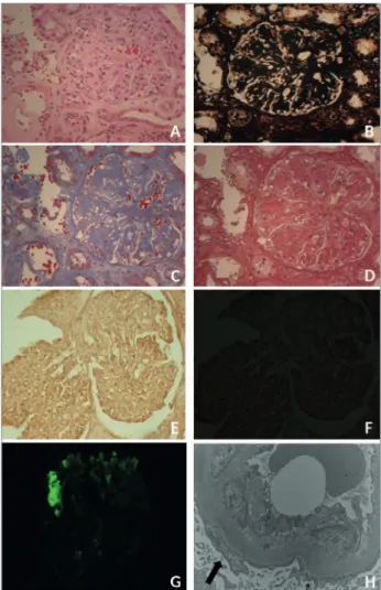
A renal biopsy was performed. The specimen was processed and treated with the following staining protocols: Hematoxylin-Eosin (HE) (Plate 1A); periodic acid silver methenamine stain (PAMS) (Plate 1B); Masson’s trichrome (MT) (Plate 1C); picro-sirius red (PSR) (Plate 1D); and Congo red (Plates 1E and 1F) (Figure 1).
Figure 1. Histology changes in Idiopathic Nodular Glomerulosclerosis. Plate 1. Renal biopsy histology specimens diagnosed with Idiopathic Nodular Glomerulosclerosis (ING). LM showing diffuse mesangial expansion in (A) HE; (B) PAMS; (C) MT; (D) PRS; (E) Congo Red without potassium permanganate, negative for amyloidosis, (F) Congo Red under non-birefringent polarized light. Immunofluorescence, positive for anti-IgM (G). SEM showing preserved areas (small arrow) and areas (large arrow) of foot process effacement (H) (12000x).
LM revealed three utterly sclerotic glomeruli; three had segmental sclerosis and MM expansion, collapsed adjacent capillary loops, and adhesions to Bowman’s capsule; two had mesangial expansion with nodular intercapillary glomerulosclerosis and open capillaries at the periphery; and in one an aneurysm appeared to form.
and interstitial compartments presented moderate interstitial fibrosis accompanied by atrophic tubules, foci of mild mononuclear inflammatory infiltrate, and some tubules with mild tubulitis and dilatation. A minor area with calcification and some hyaline casts were also seen.
The vascular compartment contained an arteriole with marked hyaline deposits on the wall and an interlobular artery with significant intimal fibroelastic thickening. There were no arcuate arteries to be found. The negative result in the Congo red staining was confirmed negative birefringence in polarized light (Plate 1F) (Figure 1).
IF yielded the following results: 1) anti-IgA serum was negative in the glomeruli and positive in the casts; 2) anti-IgG serum was negative; 3) anti-IgM serum (Plate 1G) (Figure 1): irregular segmental entrapment in three glomeruli, in sclerotic glomeruli, and in the vascular wall; 4) anti-C1q serum: mild irregular segmental entrapment in a glomerulus and in sclerotic glomeruli, the Bowman’s capsule, and the vascular wall; 5) anti-C3 serum: moderate irregular segmental entrapment in a glomerulus, in sclerotic glomeruli, in the vascular wall, and in the peritubular capillary wall; 6) mild segmental irregular positive serum test for anti-Kappa in a glomerulus and positive in the casts; 7) mild segmental irregular positive serum test for anti-Lambda in a glomerulus and in the casts; 8) anti-fibrinogen serum was negative.
SEM (Plate 1H) (Figure 1) did not reveal subendothelial, subepithelial, or intramembranous structured fibrillar deposits in the mesangium or in the capillary loops. A capillary loop had non-specific subendothelial electron-dense material. GBM thickness morphometric analysis measured the distance between the cytoplasmic membrane in the endothelium to the cytoplasmic membrane in the foot process/podocyte forty times and yielded mean and median distances of 409.06 ± 86.41nm and 375.63 (295.12 - 650.50) nm, respectively. Although some foot processes were preserved, alterations were seen in podocyte cytoplasm, such as non-formation, widening, or narrowing of foot processes.
D
ISCUSSIONThe clinical signs identified in this patient were consistent with nephrotic syndrome, characterized
by important comorbidities, rapid loss of renal function, and need for dialysis. These signs may be found in most of the glomerular diseases considered in the differential diagnosis for ING; however, LM findings showed a pattern similar to DN, in which characteristically argyrophilic collagenous sclerotic nodules stained by MT and PSR are seen. Since there were no clinical or workup evidences of DM, the patients was diagnosed with ING.4,8
IF showed no typical deposits; only non-specific C3 deposits in vessels and sclerotic sites and poor linear pattern IgG deposits in glomeruli were seen. SEM revealed evidences of GBM thickening, significant MM expansion, and areas of podocyte and foot process effacement; no organized or characteristic deposits were seen. There were great similarities with DN, including findings such as fibrin caps and capsular drops.13
Clinical and pathology testing performed on 15 patients with ING revealed combined AH in the afferent and efferent arterioles, but no cases of capsular drop lesion. Although the mechanism of arteriolosclerosis formation in these arterioles is uncertain, AH is believed to play a significant role in the progression to nodular formation in ING.2
Another study described changes similar to ours, showing ten patients with a long history of smoking, hypertension, and proteinuria. Six individuals were diagnosed with diffuse mesangial sclerosis, four with nodular glomerulosclerosis, and eight with AH; GBM thickening and foot process effacement were also detected.12
The most accepted explanation for the patho-physiology of ING development involves smoking and mechanisms inducing oxidative stress, forma-tion of advanced glycaforma-tion end-products (AGEs), angiogenesis, and alteration of intrarenal hemo-dynamics.3,12 Oxidative stress may be caused by
free radicals present in tobacco smoke. The ensu-ing lesion causes the MM to expand via activation of tumor growth factor β (TGFβ) and insulin-like growth factor.3
protein kinases, JAK/STAT and SMAD pathway, to promote mesangial cell proliferation and the synthesis of fibrogenic cytokines, such as PDGF and TGFβ. Reactive glycation products are present in tobacco aqueous extracts and smoke, and may react rapidly with proteins to form AGEs.3,5
Nicotine appears to activate angiogenesis by binding directly to endothelial cell acetylcholine receptors. Activation leads to vasodilation associated with the deregulation in the production of type IV collagen, thus promoting GBM thickening and inducing albuminuria.3
Studies have shown that smoking increases urinary albumin excretion in the general population, even in non-diabetic and normotensive patients.5,14
However, most renal function studies looking into in long-term smokers have reported a high prevalence of hypertension and DM.15
A study described the case of a 70-year-old woman with a long history of hypertension and heavy smoking (60 pack-years), a habit she had ceased for 20 years then. She had no history of DM but had severe COPD and chronic kidney disease. She was quite similar to the patient in our study, in that her clinical signs were suggestive of “nodular glomerulosclerosis associated with smoking,” a designation proposed by the authors to replace the term ING.3
Concomitant smoking and hypertension, in addition to genetic and environmental susceptibility, may increase the risk of developing chronic kidney disease.12 Hypoxia induced in patients with COPD
also appears to contribute to ING. Decreased levels of oxygen may activate the sympathetic nervous system and the renin angiotensin system, which induce vascular remodeling, accompanied by tissue hypoxia, endothelial lesion, and decreased glomerular filtration rates. This same mechanism of hypoxia induced by COPD may explain the intrarenal hemodynamic alterations caused by smoking.3
ING is a rare condition, and little is known about its pathogenesis. However, new case reports have helped to understand the disease and to sketch a profile for patients suffering from the condition. Smoking has been established as the cause of ING, a fact that has called for changes in the designation of the disease. However, other pathways to the development of the
disease have not been entirely ruled out, as many mechanisms appear to collaborate with the formation of nodular glomerulosclerosis. Further research is needed in order to more specifically assess the factors causing ING.
R
EFERENCES1. Halmai R, Degrell P, Szijártó IA, Mátyás V, Molnár GA, Kovács T, et al. Smoking as the potential link between Kim-melstiel-Wilson lesion and non-diabetic nodular glomerulo-sclerosis in male patients - a single center retrospective. Clin Nephrol 2013;80:23-8. DOI: http://dx.doi.org/10.5414/ CN107812
2. Li W, Verani RR. Idiopathic nodular glomerulosclero-sis: a clinicopathologic study of 15 cases. Hum Pathol 2008;39:1771-6. DOI:http://dx.doi.org/10.1016/j.hump-ath.2008.05.004
3. Nasr SH, D’Agati VD. Nodular glomerulosclerosis in the nondiabetic smoker. J Am Soc Nephrol 2007;18:2032-6. DOI:http://dx.doi.org/10.1681/ASN.2006121328
4. Markowitz GS, Lin J, Valeri AM, Avila C, Nasr SH, D’Agati VD. Idiopathic nodular glomerulosclerosis is a distinct clinicopathologic entity linked to hypertension and smok-ing. Hum Pathol 2002;33:826-35. DOI: http://dx.doi. org/10.1053/hupa.2002.126189
5. Lópes-Revuelta K, Abreu AA, Gerrero-Márquez C, Stanescu RI, Marín MI, Fernández EP. Diabetic Nephropathy with-out Diabetes. J Clin Med 2015;4:1403-27. DOI: http:// dx.doi.org/10.3390/jcm4071403
6. Predosa AF, Gomes dos Santos WA, Filho MA, Farias FT, Modesto dos Santos V. Nodular glomerulosclerosis in a non-diabetic hypertensive smoker with dyslipidemia. An Sist Sanit Navar 2011;34:301-8.
7. Souraty P, Nast CC, Mehrotra R, Barba L, Martina J, Adler SG. Nodular glomerulosclerosis in a patient with meta-bolic syndrome without diabetes. Nat Clin Pract Nephrol 2008;4:639-42. DOI: http://dx.doi.org/10.1038/ncp-neph0946
8. Müller-Höcker J, Weiss M, Thoenes GH, Grund A, Nerlich A. A case of idiopathic nodular glomerulosclerosis mimick-ing diabetic glomerulosclerosis (Kimmelstiel-Wilson type). Pathol Res Pract 2002;198:375-9. PMID: 12092775 DOI: http://dx.doi.org/10.1078/0344-0338-00269
9. Alpers CE, Biava CG. Idiopathic lobular glomerulonephritis (nodular mesangial sclerosis): a distinct diagnostic entity. Clin Nephrol 1989;32:68-74. PMID: 2766585
10. Herzenberg AM, Holden JK, Singh S, Magil AB. Idiopathic nodular glomerulosclerosis. Am J Kidney Dis 1999;34:560-4. PMID: 10469869DOI: http://dx.doi.org/10.1016/S0272-6386(99)70086-7
11. Tervaert TW, Mooyaart AL, Amann K, Cohen AH, Cook HT, Drachenberg CB, et al.; Renal Pathology Society. Pathologic classification of diabetic nephropathy. J Am Soc Nephrol 2010;21:556-63. DOI: http://dx.doi.org/10.1681/ ASN.2010010010
12. Salvatore SP, Troxell ML, Hecox D, Sperling KR, Seshan SV. Smoking-related glomerulopathy: expanding the morpho-logic spectrum. Am J Nephrol 2015;41:66-72. DOI: http:// dx.doi.org/10.1159/000371727
14. Hillege HL, Janssen WM, Bak AA, Diercks GF, Grobbee DE, Crijns HJ, et al.; Prevend Study Group. Microalbuminuria is common, also in a nondiabetic, nonhypertensive population, and an independent indicator of cardiovascular risk factors and cardiovascular morbidity. J Intern Med 2001;249:519-26. DOI: http://dx.doi.org/10.1046/j.1365-2796.2001.00833.x