PESQUIS
A
ORIGINAL
Correspondence to: Raquel Britto – Avenida Antonio Carlos, 6627 – CEP: 31370-901 – Belo Horizonte (MG), Brasil – E-mail: [email protected]
Presentation: Sept. 2012 – Accepted for publication: Mar. 2013 – Financing source: FAPEMIG – Proccess PPM-072-09 and CNPq 307597/2011-3 – Conflict of interest: nothing to declare – Approval at the Ethics Committee n. 0515.0.203.000-10.
ABSTRACT | The myocardial infarction (MI) alters left ven-tricle diastolic function (LVDF) in different grades, which may reflect on functional capacity (FC). This study aimed to assess, in patients with recent MI, the relation between LVDF and FC evaluated by the distance covered during the six minute walking test (6MWT). Fifty-six uncomplicat-ed MI inpatients were selectuncomplicat-ed after discharge from the coronary care unit and submitted to tests. Statistic analy-ses were carried out considering all patients for correla-tions and groups according to the classification of LVDF to comparison. It was found correlation between lateral wave a’ (later diastole) and 6MWD (r=-0.320; p=0.023) and no difference between FC and LVDF between groups. Blood pressure and heart rate had physiologic responses. The correlation indicates that the impairment of early diastole expands the role of atrial contraction in CF, reinforcing the need for evaluation of these patients still in the hospital. The physiological responses related to the six minute walk-ing test reinforce the feasibility of its use after recent MI. Keywords | exercise test; echocardiography; myocardial infarction; diastole.
RESUMO | O infarto do miocárdio (IM) altera a função dias-tólica (FD) do ventrículo esquerdo (VE) em diferentes graus, o que pode refletir na capacidade funcional (CF). O objetivo deste estudo foi avaliar, após IM recente, a relação entre a FD do VE por meio de ecocardiografia Doppler e a CF estimada por meio da distância percorrida no teste de caminhada de
Relationship between functional capacity and
diastolic function in early myocardial infarction
Relação entre capacidade funcional e função diastólica no infarto recente
Relación entre capacidad funcional y función diastólica en el infarto reciente
Lívia Santos Diniz
1, Danielle Aparecida Pereira Gomes
2, Victor Ribeiro Neves
3, Marconi Gomes da Silva
4,
Maria do Carmo Pereira Nunes
4, Raquel Rodrigues Britto
2Study conducted at Hospital das Clínicas of Universidade Federal de Minas Gerais (UFMG) – Belo Horizonte (MG), Brazil.
1Resident at the Multiprofessional Residency Program of Hospital das Clínicas at UFMG – Belo Horizonte (MG), Brazil.
2Professor at the Physical Therapy Department and at the Postgraduate program of Rehabilitation Sciences at UFMG – Belo Horizonte
(MG), Brazil.
3Post doctorate at the Telehealth Program of Hospital das Clínicas of UFMG – Belo Horizonte (MG), Brazil. 4Cardiologists and Echocardiographists at Hospital das Clínicas of UFMG – Belo Horizonte (MG), Brazil.
seis minutos (DP6). Cinquenta e seis pacientes com IM não complicado foram selecionados após a alta da unidade co-ronariana e submetidos aos testes. Foi realizada análise de correlação considerando todos os pacientes e de compara-ção entre grupos definidos de acordo com a classificacompara-ção da FD do VE. Foi observada correlação entre a onda a’ lateral (referente à diástole tardia) e a DP6 (r=-0,320; p=0,023) e não houve diferença entre a CF dos grupos classificados confor-me a FD do VE. As respostas de pressão arterial e frequência cardíaca ao teste foram fisiológicas. A correlação encontrada indica que o comprometimento da diástole precoce amplia o papel da contração atrial na CF, reforçando a necessidade de avaliação desses pacientes ainda no hospital. A resposta fisiológica ao TC6 reforça a viabilidade de sua utilização após IM recente.
Descritores | teste de esforço; ecocardiografia; infarto do miocárdio; diástole.
RESUMEN | La relación entre la capacidad funcional (CF)
INTRODUCTION
Myocardial infarction (MI) is deined by the total
inter-ruption of the coronary low with consequent ischemia and
myocardial necrosis
1. In MI, the diastolic function (DF) of
the left ventricle (LV) is rapidly altered
2,3, since part of the
diastole depends on energy
4-6. DF of the LV is associated
with functional capacity (FC) in diferent populations
7-10,
but it is little investigated in acute conditions
11.
he six-minute walking test (6MWT) is a
submaxi-mal, reliable, practical and low cost test
12-15to assess the
FC of healthy subjects, as well as cardiac
16and lung
dis-ease patients
17. Its use in MI before hospital discharge is
recent
18and little discussed
19. he evaluation of FC in the
hospital assists risk stratiication and medical
therapeu-tics and, especially, the proper prescription of exercise
15-20.
It is likely that patients with higher degree of
dia-stolic dysfunction (reduced DF of the LV) present
re-duced FC, proportional to the severity. hus, the main
objective of this study was to assess the relation between
FC and DF of the LV in patients after recent MI, and
also to identify the behavior of physiological variables
during the application of the 6MWT.
MATERIALS AND METHODS
Cross-sectional study that assessed volunteers
diag-nosed with MI with or without the elevation of the
ST segment (MI CSST and MI SSST),
uncomplicat-ed, after discharge from the coronary unit and
medi-cal. MI was deined by: ischemic symptoms, elevated
troponin and evaluation of electrocardiogram (ECG).
MI CSST was characterized by the presence of SST
in at least two consecutive derivations in ECG and
MI SSST due to the absence of changes in the ECG
or the presence of changes indicating ischemia,
dif-ferent from SST
21. Patients higher than I in the Killip
classiication
22were excluded (clinical signs of
ven-tricular dysfunction), as well as those with persistent
arrhythmias, myocardial ischemia at the ECG,
in-stability of pressure levels, moderate to severe mitral
valve insuiciency
23,24or diiculties for walking. he
patients were divided into groups according to the DF
classiication of the LV. he study was approved by the
Research Ethics Committee of the institution (Report
n. 0515.0.203.000-10), and all the volunteers signed
the informed consent form.
•
6MWT. FC was assessed by means of distance
wal-ked in the 6MWT (6DW), according to the
gui-delines by the American horacic Society (ATS)
12.
Heart rate (HR), blood pressure, saturation of
peri-pheral oxygen (SpO
2), and perceived exertion were
assessed before and after the test (at the end and
after ive minutes), by means of the cardio frequency
meter (Polar®,
FS2c, Finland), auscultatory method,
wrist oximeter (Mindray®, PM50, China) and the
modiied Borg Scale
23, respectively. HR and SpO
2
were monitored also during the performance of the
test. 6MWT was performed by the same examiner
twice (due to the learning efect
12,13) on the same day,
with rest interval
12,13. he longest distance walked
was considered. Signs and symptoms of exertional
intolerance
12and elevated HR, higher than 85% in
relation to the estimated HR for the age, were used
as criteria to interrupt the test
15.
•
ECHO. he diastolic function (DF) of the left ventricle
(LV) can be assessed by diferent parameters, such as the
E/A ratio and the E/e’ ratio
25,26. he E and A waves refer
to the blood low in the mitral valve during early and
late diastole, respectively. he e’ wave and the a’ wave
re-present, respectively, the myocardial displacement in the
mitral ring during early and late diastole. he E/e’ ratio
has been the most used parameter to assess DF and LV,
since it is less inluenced by other physiological
varia-bles
25,26. Examinations were performed by experienced
echocardiographists who did not have access to clinical
data. he echocardiography system Philips IE 33 (USA,
2010) was used, with a multifrequency transducer of 2 to
4 mHz to obtain bidimensional images of the M mode
sometidos a los tests. Fue realizado el análisis estadístico consi-derando todos los pacientes y por grupos, de acuerdo a la clasi-ficación de la FD del VI. Fue observada una correlación entre la onda a’ lateral (referente a la diástole tardía) y la DTM6 (r=-0,320; p=0,023). Sin embargo, no hubo asociación entre la CF y la FD del VI en el análisis por grupos. La correlación entre la DTM6 y la onda a´ lateral indica asociación entre la diástole tardía y la
CF en estos pacientes, sugiriendo una mayor contribución de la contracción auricular para la promoción del llenado del VI en esta población. Estos datos proporcionan una asignación adicio-nal para la utilización del TM6 en la evaluación de la CF después de un IAM reciente.
and with pulsatile Doppler. Mitral inlow velocities were
registered with the pulsatile Doppler in the apical four
chamber cut, with a 5 mm sample volume of at the tip
of the mitral lealets. he velocities of early (E) and late
(A) diastole were measured, and the E/A ratio was also
calculated. A 2 mm sample volume was placed at the
junction of the LV wall and the mitral ring, in the septal
and lateral regions, for the tissue Doppler register,
deri-ving the velocities during systole (s’), early diastole (e’)
and late diastole (a’).
he left ventricular ejection fraction (LVEF) was
calculated by the Simpson method
24. he
compro-mised systolic function was considered as LVEF
<50%
27. he DF of the LV was classiied, according to
the E/A ratio, as normal, abnormal diastolic relaxation
(ADR), pseudonormal relaxation (PR) or restrictive
(RE)
26. he E/e’ ratio was calculated to estimate LV
illing pressures
26. he E/e’ ratio <8 is associated with
normal LV illing pressures, and the E/e’ ratio >13 is
associated with high LV illing pressures in patients
with normal LVEF, when the mean of septal and
lat-eral e’ was used for calculation
26.
Statistical analysis
An assessment of normality of data distribution by
the was conducted by the Kolmogorov-Smirnov test,
ANOVA one-way and the least signiicant diference
test or the
χ
2test, to compare between groups. Pearson
or Spearman correlation was used to assess the
rela-tion between the 6MWT and the echocardiographic
parameters. Data are presented as absolute frequency,
percentage and mean
±
deviation, considering
α
=5% as
being signiicant.
RESULTS
Seventy-two patients were selected, and some of them
were excluded: 3 due to compromised deambulation, 8
for not being submitted to ECHO during hospital stay,
4 for using another device, and 1 due to severe mitral
dysfunction. herefore, 56 subjects were included and
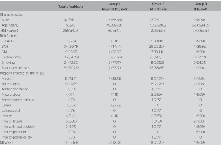
their characteristics are presented in Table 1. Most
pa-tients (71.42%) underwent revascularization by means
Table 1. Characteristics of subjects
Values expressed as absolute frequency and percentage (%) or mean±standard deviation (age and BMI). No significant diferences were observed between the grouops by the ANOVA or the χ2 test.
DF: diastolic function; ADR: abnormal diastolic relaxation; PR: pseudonormal relaxation; BMI: body mass index; FH ACD: family history of atherosclerosis coronary disease; SAH: systemic arterial hypertension; DM: diabetes mellitus; MI EST: myocardial infarction with elevated ST segment; MI WEST: myocardial infarction without elevated ST segment; RV: right ventricle
Total of subjects Group 1 Group 2 Group 3
(normal DF) n=9 (ADR) n=36 (PR) n=11
Characteristics
Male 42 (75) 6 (66.66) 27 (75) 9 (81.81) Age (years) 56±10 49.88±7.93 57.50±8.62 57.00±14.39 BMI (kg/m2) 26.91±4.52 26.12±4.15 27.63±5.13 27.00±3.54
Risk factors
FH ACD 7 (12.5) 1 (11.11) 5 (13.88) 1 (9.09) SAH 34 (60.71) 4 (44.44) 26 (72.22) 4 (36.36) DM 10 (17.85) 2 (22.22) 7 (19.44) 1 (9.09) Dyslipidemia 36 (64.28) 6 (66.66) 22 (61.11) 8 (72.72) Smoking 24 (42.85) 7 (77.77) 11 (30.55) 6 (54.54) Sedentary lifestyle 50 (89.28) 7 (77.77) 32 (88.88) 11 (100) Regions afected by the MI EST
Anterior 13 (23.21) 3 (33.33) 8 (22.22) 2 (18.18) Anteroseptal 10 (17.85) 0 8 (22.22) 2 (18.18) Ânterior-posterior 1 (1.78) 0 1 (2.77) 0 Anterolateral 4 (7.14) 1 (11.11) 2 (5.55) 1 (9.09) Ânterior-lateral-inferior 1 (1.78) 0 1 (2.77) 0
Lateral 2 (3.57) 2 (22.22) 0 0
of thrombolysis (30.35%), angioplasty (64.28%) or both,
before the performance of the 6MWT, and 53 (94.64%)
were on beta blockers.
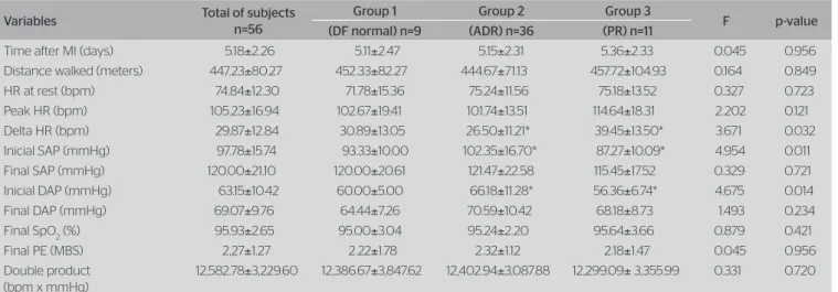
The echocardiographic analysis (Table 2) showed
that 18 patients (32.14%) had systolic dysfunction,
and 47 (83.93%), diastolic dysfunction. Only 8
(14.28%) had high LV filling pressures; all of these
presented with diastolic dysfunction, being 3 with
PR and 5 with ADR. According to the classification
of the DF of the LV, the following proportion was
observed: normal FD: 16.07% (9 patients), ADR:
64.29% (36 patients), PR: 19.64% (11 patients) and
RE: 0% (no patient). Therefore, three groups were
considered for comparison (1 to 3). There was no
statistical difference between the base
characteris-tics of the groups (Table 1). However, as expected,
differences were observed in echocardiographic
variables (Table 2).
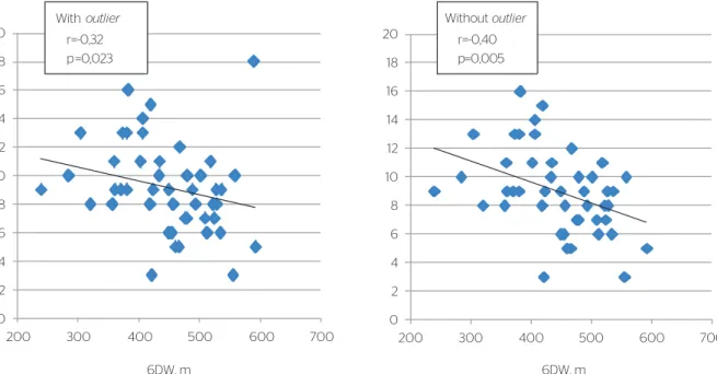
here was correlation (Figure 1) betwen the
lat-eral a’ wave and the 6MWT. here was no diference
in the 6MWT in the analysis per group (Table 3). In
the 6MWT, only one patient surpassed the deined HR
limit. he others presented physiological behavior of
the assessed variables (Table 3).
DISCUSSION
his study indicates that: 1) the correlation between the
6MWT and the lateral a’ wave (late diastole), even if
with low magnitude, suggests a greater contribution of
the atrial contraction for the LV illing; 2) there is no
diference in the 6MWT in groups with diferent DF
of the LV; and 3) no adverse physiological response to
the 6MWT was found in the assessed patients.
Table 2. Echocardiographic characteristics
Parameters Total of subjects Group 1 Group 2 Group 3 F p-value
(DF normal) n=9 (ADR) n=36 (PR) n=11
Ejection fraction (%) 53.36±10.65 59.25±7.74 54.63±10.12 47.91±11.19 2.799 0.070 Velocity mitral E wave (cm/s) 71.10±18.48 84.12±10.11 60.46±13.72* 90.63±15.91 26.965 <0.001 Velocity mitral A wave (cm/s) 70.59±19.02 55.12±7.58 80.23±15.89* 56.45±20.51# 12.553 <0.001
E/A ratio 1.12±0.62 1.54±0.30 0.77±0.18* 1.84±0.92# 27.457 <0.001
E/e’ ratio 9.67±4.11 7.75±1.48 9.13±3.41 12.90±6.30*# 5.370 0.008
Velocities of the mitral ring
Velocity e’ septal region (cm/s) 6.28±2.43 9.75±3.15 5.30±1.57* 6.09±1.64* 20.481 <0.001 Velocity e’ lateral region (cm/s) 8.25±3.51 11.87±3.97 7.23±2.81* 8.45±3.75* 7.149 0.002 Velocity a’ septal region (cm/s) 8.35±2.22 8.37±1.68 8.83±2.10 6.90±2.70# 3.491 0.038
Velocity a’ lateral region (cm/s) 9.19±3.04 8.37±3.37 10.00±2.25 7.81±4.30 2.328 0.108 Velocity s’ septal region (cm/s) 6.18±1.95 7.12±2.53 5.96±1.75 6.18±2.22 1.641 0.204 Velocity s’ lateral region (cm/s) 7.01±2.67 7.87±2.35 6.60±2.34 7.00±3.13 0.496 0.612
Values expressed as mean±standard deviation. DF: diastolic function; ADR: abnormal diastolic relaxation; PR: pseudonormal relaxation *p<0.05: comparison of Groups 2 and 3 in relation to Group 1 with normal DF
#p<0.05: comparison between Groups 2 and 3, ANOVA and least significant diference
Table 3. Data from the 6MWT
Values expressed as mean±standard deviation. DF: diastolic function; ADR: abnormal diastolic relaxation; PR: pseudonormal relaxation; MI: myocardial infarction; HR: heart rate; bpm: beats per minute; SAP: systolic arterial pressure; DAP: diastolic arterial pressure; SpO2: peripheral oxygen saturation; PE: perceived exertion; MBS: modified Borg Scale; *p<0.05. ANOVA and least significant diference.
Variables Total of subjects
n=56
Group 1 Group 2 Group 3
F p-value
(DF normal) n=9 (ADR) n=36 (PR) n=11
Time after MI (days) 5.18±2.26 5.11±2.47 5.15±2.31 5.36±2.33 0.045 0.956 Distance walked (meters) 447.23±80.27 452.33±82.27 444.67±71.13 457.72±104.93 0.164 0.849 HR at rest (bpm) 74.84±12.30 71.78±15.36 75.24±11.56 75.18±13.52 0.327 0.723 Peak HR (bpm) 105.23±16.94 102.67±19.41 101.74±13.51 114.64±18.31 2.202 0.121 Delta HR (bpm) 29.87±12.84 30.89±13.05 26.50±11.21* 39.45±13.50* 3.671 0.032 Inicial SAP (mmHg) 97.78±15.74 93.33±10.00 102.35±16.70* 87.27±10.09* 4.954 0.011 Final SAP (mmHg) 120.00±21.10 120.00±20.61 121.47±22.58 115.45±17.52 0.329 0.721 Inicial DAP (mmHg) 63.15±10.42 60.00±5.00 66.18±11.28* 56.36±6.74* 4.675 0.014 Final DAP (mmHg) 69.07±9.76 64.44±7.26 70.59±10.42 68.18±8.73 1.493 0.234 Final SpO2 (%) 95.93±2.65 95.00±3.04 95.24±2.20 95.64±3.66 0.879 0.421 Final PE (MBS) 2,27±1.27 2.22±1.78 2.32±1.12 2.18±1.47 0.045 0.956 Double product
(bpm x mmHg)
he diastole is divided into isovolumetric relaxation,
early diastole, diastasis and late diastole (atrial
contrac-tion)
26. In the MI, the early diastolic relaxation is the irst
stage to be altered, since it is a process that depends on
energy
17,4-6, thus generating more dependency on atrial
contraction to promote ventricular illing
28. his can be
observed in ECHO by the increased velocity of the A
wave of the mitral low. Such change consists of the irst
stage of diastolic dysfunction: the ADR, which is
com-mon in the early stage of most heart conditions
25,29.
he lateral a’ wave presented higher values
(9.19
±
3.04) than the mean of the national population
(7.3
±
1.5)
30, and was inversely correlated with FC. In
patients with with ADR and normal LV illing
pres-sures, the a’ wave is related to the systolic function of
the left atrium
31. According to Nagueh et al (2001),
the compromised early diastolic relaxation increases
the left atrial preload, leading to the increased muscle
contraction, by the frank-Starling mechanism, which
results in the increased a’ wave velocity.
he found correlation can be related to the regions
that are most compromised by the MI. Since the
an-terior and septal regions presented with compromised
mobility for most subjects, the other regions can
pres-ent with hyperkinesias as an attempt to attenuate the
compromise of the ventricular function
32. his
com-pensation mechanism may have increased the
veloc-ity of the lateral a’ wave. herefore, this study, despite
the low correlation magnitude, draws attention to the
need for the early assessment of FC, especially among
subjects who present with these echocardiographic
characteristics, and also to the need for additional
stud-ies. Echocardiography is a routine procedure for these
patients, being little explored from the functional
ca-pacity point of view.
According to ATS
12, the recent MI (up to 30 days)
is a contraindication to perform the 6MWT;
how-ever, there is no evidence to prove such restriction
19.
On the contrary, the test is widely used in conditions
of reduced FC, such as heart failure
16. Besides, the
6MWT has been used with patients with recent MI,
without intercurrences, in other studies
18,19. he early
assessment of FC guides the prescription of physical
exercise in cardiac rehabilitation
15,20, thus contributing
for the return to activities
33and the improvement in
quality of life
34.
Concerning the absence of 6MWT diferences
be-tween groups with diferent DF of the LV, the type II
error cannot be put aside, since the sample size was not
calculated due to the absence of a clinically important
reference in previous studies. Besides, the uneven
num-ber of patients per group and the lack of details on the
used medication point to the need for new studies with
the same focus.
In conclusion, even though no diferences were
observed between the groups with diferent degrees
of DF, the found correlation indicates that the
com-promised early diastole increases the role of atrial
contraction in the FC, reinforcing the need to
as-sess these patients in the hospital. he physiological
response to the 6MWT reinforces the viability of its
use after recent MI.
Figure 1. Graphic representation of the Spearman correlation between the distance walked in 6 minutes (6DW) and a’ lateral velocity with (left) and without outlier (right)
0 2 4 6 8 10 12 14 16 18 20
200 300 400 500 600 700
outlier
r=-0,32 p=0,023
0 2 4 6 8 10 12 14 16 18 20
200 300 400 500 600 700
outlier
p=0,005 r=-0,40
6DW, m 6DW, m
ACKNOWLEDGEMENT
To Dr. Luiz Ricardo de Ataíde Castro, for the
colabora-tion during the seleccolabora-tion of patients.
REFERENCES
1. Davies M, J. The pathophysiology of acute coronary syndromes. Heart. 2000;83(3):361-6.
2. Poulsen SH. Clinical aspects of left ventricular diastolic function assessed by Doppler echocardiography following acute myocardial infarction. Dan Med Bull. 2001;48(4):199-210.
3. Poulsen SH, Jensen SE, Egstrup K. Longitudinal changes and prognostic implications of left ventricular diastolic function in first acute myocardial infarction. Am Heart J. 1999;137(5):910-8. 4. Fabbiocchi F, Galli C, Sganzerla P, Montorsi P, Loaldi A, de Cesare
N, et al. Changes in right ventricular filling dynamics during left anterior descending, left circumflex and right coronary artery balloon occlusion. Eur Heart J. 1997;18(9):1432-7.
5. de Bruyne B, Lerch R, Meier B, Schlaepfer H, Gabathuler J, Rutishauser W. Doppler assessment of left ventricular diastolic filling during brief coronary occlusion. Am Heart J. 1989;117(3):629-35. 6. Bowman LK, Cleman MW, Cabin HS, Zaret BL, Jaffe CC. Dynamics
of early and late left ventricular filling determined by Doppler two-dimensional echocardiography during percutaneous transluminal coronary angioplasty. Am J Cardiol. 1988;61(8):541-5.
7. Otto ME, Pereira MM, Beck AL, Milani M. [Correlation between diastolic function and maximal exercise capacity on exercise test]. Arq Bras Cardiol. 2011;96(2):107-13. English, Portuguese, Spanish. 8. Okura H, Inoue H, Tomon M, Nishiyama S, Yoshikawa T, Yoshida
K, et al. Impact of Doppler-derived left ventricular diastolic performance on exercise capacity in normal individuals. Am Heart J. 2000;139(4):716-22.
9. Bajraktari G, Elezi S, Berisha V, Lindqvist P, Rexhepaj N, Henein MY. Left ventricular asynchrony and raised filling pressure predict limited exercise performance assessed by 6 minute walk test. Int J Cardiol. 2011;146(3):385-9.
10. Miyashita T, Okano Y, Takaki H, Satoh T, Kobayashi Y, Goto Y. Relation between exercise capacity and left ventricular systolic versus diastolic function during exercise in patients after myocardial infarction. Coron Artery Dis. 2001;12(3):217-25.
11. Urek R, Cubrilo-Turek M, Crncević-Urek M. The relationship between left ventricular filling shortly after an uncomplicated myocardial infarction and subsequent exercise capacity. Coll Antropol. 2001;25(1):279-87.
12. ATS Committee on Proficiency Standards for Clinical Pulmonary Function Laboratories. ATS statement: guidelines for the six-minute walk test. Am J Respir Crit Care Med. 2002;166(1):111-7. 13. Solway S, Brooks D, Lacasse Y, Thomas S. A qualitative systematic
overview of the measurement properties of functional walk tests used in the cardiorespiratory domain. Chest. 2001;119(1):256-70. 14. Noonan V, Dean E. Submaximal exercise testing: clinical application
and interpretation. Phys Ther. 2000;80(8):782-807.
15. Arena R, Myers J, Williams MA, Gulati M, Kligfield P, Balady GJ, et al. Assessment of functional capacity in clinical and research settings:
a scientific statement from the American Heart Association Committee on Exercise, Rehabilitation, and Prevention of the Council on Clinical Cardiology and the Council on Cardiovascular Nursing. Circulation. 2007;116(3):329-43.
16. Pollentier B, Irons SL, Benedetto CM, Dibenedetto AM, Loton D, Seyler RD, et al. Examination of the six minute walk test to determine functional capacity in people with chronic heart failure: a systematic review. Cardiopulm Phys Ther J. 2010;21(1):13-21. 17. Cote CG, Pinto-Plata V, Kasprzyk K, Dordelly LJ, Celli BR. The 6-min
walk distance, peak oxygen uptake, and mortality in COPD. Chest. 2007;132(6):1778-85.
18. Nogueira PA, Leal AC, Pulz C, Nogueira ID, Filho JA. Clinical reliability of the 6 minute corridor walk test performed within a week of a myocardial infarction. Int Heart J. 2006;47(4):533-40. 19. Sancho AG, Bacelar SC, Cader SA, Caldeira JB, Pereira CCL, Lima
Júnior NA Significance of in-Hospital Evaluation of Functional Capacity in Acute Coronary Syndrome. Rev Bras Cardiol. 2011;24(5):282-90.
20. Antman EM, Hand M, Armstrong PW, Bates ER, Green LA, Halasyamani LK, et al. 2007 focused update of the ACC/ AHA 2004 guidelines for the management of patients with ST-elevation myocardial infarction: a report of the American College of Cardiology/American Heart Association Task Force on Practice Guidelines. Circulation. 2008;117(2):296-329.
21. Thygesen K, Alpert JS, Jaffe AS, Simoons ML, Chaitman BR, White HD, et al. Third universal definition of myocardial infarction. J Am Coll Cardiol. 2012;60(16):1581-98.
22. Killip T 3rd, Kimball JT. Treatment of myocardial infarction in a coronary care unit. A two year experience with 250 patients. Am J Cardiol. 1967;20(4):457-64.
23. Borg GA. Psychophysical bases of perceived exertion. Med Sci Sports Exerc. 1982;14(5):377-81.
24. Lang RM , Bierig M, Devereux RB, Flachskampf FA, Foster E, Pellikka PA, et al. Recommendations for chamber quantification: a report from the American Society of Echocardiography’s Guidelines and Standards Committee and the Chamber Quantification Writing Group, developed in conjunction with the European Association of Echocardiography, a branch of the European Society of Cardiology. J Am Soc Echocardiogr. 2005;18(12):1440-63.
25. Mottram PM, Marwick TH. Assessment of diastolic function: what the general cardiologist needs to know. Heart. 2005;91(5):681-95. 26. Nagueh SF, Appleton CP, Gillebert TC, Marino PN, Oh JK, Smiseth
OA, et al. Recommendations for the evaluation of left ventricular diastolic function by echocardiography. J Am Soc Echocardiogr. 2009;22(2):107-33.
27. Dickstein K, Cohen-Solal A, Filippatos G, McMurray JJ, Ponikowski P, Poole-Wilson PA, et al. ESC guidelines for the diagnosis and treatment of acute and chronic heart failure 2008. Eur J Heart Fail. 2008;10(10):933-89.
28. Mesquita ET, Socrates J, Rassi S, Villacorta H, Mady C. [Heart failure with preserved systolic function]. Arq Bras Cardiol. 2004;82(5):494-500.
29. Poulsen SH, Jensen SE, Gøtzsche O, Egstrup K. Evaluation and prognostic significance of left ventricular diastolic function assessed by Doppler echocardiography in the early phase of a first acute myocardial infarction. Eur Heart J. 1997;18(12):1882-9. 30. Pedone MD, Castro I, Hatem D, Haertel JC, Feier F, Pandolfo F.
31. Nagueh SF, Sun H, Kopelen HA, Middleton KJ, Khoury DS. Hemodynamic determinants of the mitral annulus diastolic velocities by tissue Doppler. J Am Coll Cardiol. 2001;37(1):278-85.
32. Gurudevan SV, Mahmud E, Blanchard DG, Strachan GM, Dittrich T, Mathews C, et al. Usefulness of compensatory hyperkinesis in the noninfarcted left ventricular wall in separating single from multivessel
coronary artery disease in patients with initial ST-elevation acute myocardial infarction. Am J Cardiol. 2004;93(2):201-3.
33. Lavie CJ, Milani RV. Benefits of cardiac rehabilitation and exercise training. Chest. 2000;117(1):5-7.