BR IEF COMMUNIC ATION
368 J Vasc Bras. 2015 Oct.-Dec.; 14(4):368-371 http://dx.doi.org/10.1590/1677-5449.004515
Do the femoral veins of female Wistar rats have valves?
Existem válvulas na veia femoral em ratas Wistar?
Renan Kleber Costa Teixeira1
*
, Vitor Nagai Yamaki1, André Lopes Valente1, Denilson José Silva Feitosa Júnior1, Mauricio Fortuna Pinheiro1, José Maciel Caldas dos Reis1, Edvaldo Lima Silveira1, Rui Sergio Monteiro de Barros1
Abstract
he femoral veins of 30 female rats of the Wistar lineage were studied using histological methods with the objective of determining whether they have valves. Histological analysis did not detect any endothelial projections or valve recesses that would suggest the presence of venous valves in this species of animal.
Keywords: rats; femoral vein; valves.
Resumo
As veias femorais de 30 ratas da linhagem Wistar foram estudadas por método histológico com objetivo de investigar a presença de válvulas. Na análise histológica não foram identiicadas projeções do endotélio ou recessos valvares que poderiam sugerir a presença de válvulas venosas nessa espécie de animal.
Palavras-chave: ratos; veia femoral; válvulas.
1 Universidade do Estado do Pará – UEPA, Belém, PA, Brazil.
Financial support: None.
Conlicts of interest: No conlicts of interest declared concerning the publication of this article. Submitted: July 09, 2015. Accepted: September 25, 2015.
369 J Vasc Bras. 2015 Oct.-Dec.; 14(4):368-371 Renan Kleber Costa Teixeira, Vitor Nagai Yamaki et al.
INTRODUCTION
Hieronymous Fabricius Ab Acquapendente discovered the existence of venous valves in 16031,2
and since then these important structures have been studied in the context of blood circulation, in particular with relation to their role in the physiology of venous
return, impeding relux of blood,1,3 with particular
importance in the large caliber veins of the lower limbs.1,2,4 From a histological point of view, valves
are paired invaginations of the tunica intima of the veins, creating saliencies into the interior of the vessel.5
Venous grafts are widely used in surgical practice
to reestablish blood low in many different specialties
and care must taken to ensure that the graft is implanted in the correct direction, so that the venous valves do
not impede blood low.5,6 Nowadays, veins are also
grafted to reestablish the continuity of peripheral nerves, in cases of nerve tissue loss, as though they were conduits within which nerve regeneration can proceed.7,8 They also play a role in the pathophysiology of diseases such as chronic venous insuficiency3,5
and, because of this, experimental animal models employing the femoral vein, the saphenous vein and others are widely employed.7,9
Even in small animals such as rats, primarily of the Wistar lineage, the morphofunctional role played by venous valves in the venous drainage system is recognized. However, it can be observed that reports in the literature on the presence10-12 or absence of
valves7,13,14 or providing anatomic details of the valves
in the deep and peripheral system veins of female rats are rare and of debatable merit. In smaller quadruped animals, in which the pelvic veins of the limbs are of microscopic diameters, absence of vein valves may be a common occurrence.15 This study was therefore
conducted with the objective of identifying the presence or absence of venous valves in the femoral veins of Wistar female rats.
METHODS
This study was approved by the Animal Research Ethics Committee at the Universidade do Estado do Pará (UEPA), Belém, PA, Brazil. Thirty female rats
(Rattus norvegicus) of the Wistar lineage weighing
200-240 grams were acquired from the animal house at the university’s Experimental Surgery Laboratory. They were housed in temperature humidity controlled conditions and provided with food and water ad libitum throughout the study.
The animals were anesthetized with an intraperitoneal injection of ketamine (70 mg/kg) and xylazine (10 mg/kg). Once adequate anesthesia had been
conirmed, the right inguinal region was dissected and the femoral vein was identiied. The vein was
carefully dissected and isolated cranially and caudally before being sectioned between single nylon 10-0
monoilament ligatures, maintaining the blood content
inside. At this poin, analysis of the presence/absence of valves in the femoral vein was conducted with a surgical microscope.
The femoral vein was immediately ixed in 10% buffered formaldehyde, set in parafin, cleaved with
4 µm longitudinal slices and stained with Hematoxylin and Eosin. A histological analysis with an optical microscope was then conducted to attempt to identify valves along the length of the femoral vein.
The animals were maintained alive for use in microsurgery training by interns at the UEPA Experimental Surgery Laboratory. They were then euthanized with an overdose of intraperitoneal xylazine.
RESULTS
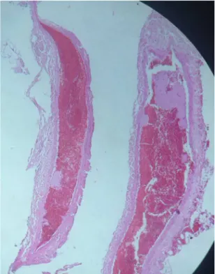
The femoral veins of all of the animals studied were patent and contained large quantities of red blood cells. No endothelial projections or valve recesses
compatible with venous valves were identiied under
the surgical microscope or the optical microscope (Figure 1).
Figure 1. Photomicrograph showing a segment of femoral vein
370 J Vasc Bras. 2015 Oct.-Dec.; 14(4):368-371 Do the femoral veins of female rats have valves?
DISCUSSION
Anatomic knowledge is extremely important for reproducing clinical situations in animals for validation of experimental models of diseases and surgical conditions. In humans, the anatomical and physiological notion of venous valves is vital,3,5
primarily in cardiac and vascular procedures, given
that the low direction of veins should be carefully
respected when fragments of veins are connected into the circulation.1,4,8
The fact that the femoral veins of female Wistar rats do not contain valves could compromise many microsurgery research streams, since experimental models do not reproduce the situation found human beings: the constant presence of valves. Notwithstanding, this should not necessarily affect training in the microsurgical skills of anastomosis and interposition of vein grafts and neurotubes.
A review of the literature did not locate any studies that had analyzed whether venous valves exist in the femoral vein, but several studies suggested that they do exist in Wistar rats, by analogy with the human system.7-10 It is speculated that the absence of valves
is because of the small dimensions of the animal, since Caggiati et al.15 state that vessels smaller than
2 mm do not have valves, but it is also possible that there are microvalves that could not be detected using histological methods, and that other methods such as electron microscopy are necessary.
The rat femoral vein is the most commonly used venous graft in microsurgery,7-9 because of its caliber
and ease of access and because when removed it does not compromise the limb, since there is a rich system of collateral drainage from the foot. Studies that are designed to analyze the effects of venous valves should therefore seek other models, because this one does not reproduce human anatomy and physiology. Further studies are needed to determine whether other veins in these animals’ bodies have valves, including studies employing electron microscopy.
In summary, on the basis of the methodology used in this study, the authors conclude that no valves were found in segments of femoral veins from female Wistar rats.
REFERENCES
1. Bestetti RB, Restini CBA, Couto LB. Evolução do conhecimento anatomofisiológico do sistema cardiovascular: dos egípcios a Harvey. Arq Bras Cardiol. 2014;103(6):538-45. PMid:25590934.
2. Medeiros CAF. Cirurgia de varizes: história e evolução. J Vasc Bras. 2006;5(4):295-302. http://dx.doi.org/10.1590/S1677-54492006000400009.
3. Piazza G. Varicose veins. Circulation. 2014;130(7):582-7. http:// dx.doi.org/10.1161/CIRCULATIONAHA.113.008331. PMid:25114187. 4. Santos CAS, Figueiredo LFP, Gusmão LCB, Pitta GBP, Miranda F Jr. Válvulas da veia braquial comum: estudo anatômico. J Vasc Bras. 2007;6(1):35-41. http://dx.doi.org/10.1590/S1677-54492007000100006. 5. Oklu R, Habito R, Mayr M, et al. Pathogenesis of varicose veins.
J Vasc Interv Radiol. 2012;23(1):33-9, quiz 40. http://dx.doi. org/10.1016/j.jvir.2011.09.010. PMid:22030459.
6. Bazigou E, Makinen T. Flow control in our vessels: vascular valves make sure there is no way back. Cell Mol Life Sci. 2013;70(6):1055-66. http://dx.doi.org/10.1007/s00018-012-1110-6. PMid:22922986. 7. Santos EB, Fernandes M, Santos JBG, Leite VM, Valente SG, Faloppa F. Estudo da regeneração de nervos tibiais de ratos Wistar em sutura primária com “gap” e sem “gap”, cobertos por segmentos de veia. Acta Ortop Bras. 2012;20(3):165-9. http://dx.doi.org/10.1590/ S1413-78522012000300006. PMid:24453597.
8. Strauch RJ, Strauch B. Nerve conduits: an update on tubular nerve repair and reconstruction. J Hand Surg Am. 2013;38(6):1252-5, quiz 1255. http://dx.doi.org/10.1016/j.jhsa.2013.02.034. PMid:23602436.
9. Thomas AC. Animal models for studying vein graft failure and therapeutic interventions. Curr Opin Pharmacol. 2012;12(2):121-6. http://dx.doi.org/10.1016/j.coph.2012.01.002. PMid:22281067.
10. Fernandes M, Valente SG, Amado D, et al. Estudo comparativo entre enxerto autógeno e enxerto muscular coberto com tubo de veia autógeno em nervos tibiais de ratos wistar, utilizando o fluoro-gold® como marcador neuronal. Acta Ortop Bras. 2007;15(2):97-100. http://dx.doi.org/10.1590/S1413-78522007000200008.
11. Takase S, Pascarella L, Bergan JJ, Schmid-Schönbein GW. Hypertension-induced venous valve remodeling. J Vasc Surg. 2004;39(6):1329-34. http://dx.doi.org/10.1016/j.jvs.2004.02.044. PMid:15192576.
12. Pascarella L, Schmid-Schönbein GW, Bergan J. An animal model of venous hypertension: the role of inflammation in venous valve failure. J Vasc Surg. 2005;41(2):303-11. http://dx.doi.org/10.1016/j. jvs.2004.10.038. PMid:15768014.
13. Petrov M, Malik A, Mead A, Bridges CR, Stedman HH. Gene transfer to muscle from the isolated regional circulation. Methods Mol Biol. 2011;709:277-86. http://dx.doi.org/10.1007/978-1-61737-982-6_18. PMid:21194035.
14. Jeon WJ, Kang JW, Park JH, et al. Clinical application of inside-out vein grafts for the treatment of sensory nerve segmental defect. Microsurgery. 2011;31(4):268-73, discussion 274-5. http://dx.doi. org/10.1002/micr.20850. PMid:21557305.
371 J Vasc Bras. 2015 Oct.-Dec.; 14(4):368-371 Renan Kleber Costa Teixeira, Vitor Nagai Yamaki et al.
*
Correspondence
Renan Kleber Costa Teixeira Rua dos Mundurucus, 2256/1401 CEP 66035-360 – Belém (PA), Brazil E-mail: [email protected]
Author information
RKCT - Physician, MSc candidate at the Graduate Program in Experimental Surgery and Research, Universidade do Estado do Pará (UEPA). VNY - Medical student (6th year) at Universidade do Estado do Pará (UEPA). ALV and DJSFJ - Medical student (4th year) at Universidade do Estado do Pará (UEPA). MFP - Cardiovascular surgeon, MSc candidate at the Graduate Program in Experimental Surgery and Research, Universidade do Estado do Pará (UEPA). JMCR - Vascular surgeon, MSc candidate at the Graduate Program in Experimental Surgery and Research, Universidade do Estado do Pará (UEPA). ELS - Pathologist, PhD candidate at the Graduate Program in Infectious and Parasitic Diseases, Associate professor IV at Universidade do Estado do Pará (UEPA). RSMB - Orthopedist and hand surgeon, PhD in Orthopedics, Adjunct professor III at Universidade do Estado do Pará (UEPA).
Author contributions
Conception and design: RKCT, RSMB. Analysis and interpretation: ALV, MFP. Data collection: RKCT, ELS. Writing the article: RKCT, VNY, ALV, DJSFJ, JMCR, ELS. Critical revision of the article: MFP, JMCR. Final approval of the article*: RKCT, VNY, ALV, DJSFJ, MFP, JMCR, ELS, RSMB. Statistical analysis: VNY. Overall responsibility: RKCT.