Evaluation of great saphenous vein occlusion rate and clinical outcome
in patients undergoing laser thermal ablation with a 1470-nm bare
fiber laser with low linear endovenous energy density
Avaliação da taxa de obliteração da veia safena magna e da evolução clínica de
pacientes submetidos a termoablação com laser 1470 nm, fibra linear e baixa
densidade de energia endovenosa linear
Walter Junior Boim Araujo1
*
, Jorge Ruino Ribas Timi1, Carlos Seme Nejm Júnior1, Filipe Carlos Caron1
Abstract
Background: Water-speciic 1470-nm lasers enable vein ablation at lower energy densities and with fewer side efects because they target interstitial water in the vessel wall. Objectives: To determine great saphenous vein (GSV) occlusion rate after thermal ablation with 1470-nm laser using 7W power and to evaluate clinical outcomes and complications. Method: Nineteen patients (31 GSVs) underwent thermal ablation. Follow-up duplex scanning, clinical evaluation using the Venous Clinical Severity Score (VCSS), and evaluation of procedure-related complications were performed at 3-5 days after the procedure and at 30 and 180 days. Results: Mean patient age was 46 years and 17 of the patients were female (89.47%). Of 31 limbs treated, 2 limbs were clinical class C2, 19 were C3, 9 were C4, and 1 limb was C5 according to the Clinical-Etiology-Anatomy-Pathophysiology (CEAP) classiication. Mean linear endovenous energy density was 33.53 J/cm. he GSV occlusion rate was 93.5% immediately after treatment, 100% at 3-5 days and 100% at 30 days after treatment and 87.1% 180 days after treatment. here was a signiicant reduction in VCSS at all time points. Conclusions: he data from this study support the possibility that the incidence of complications can be reduced without signiicantly afecting the clinical outcomes, by using lower energy density. However, this appears to be at the cost of reduced eicacy in terms of GSV occlusion rates.
Keywords: ablation techniques; laser therapy; varicose veins.
Resumo
Contexto: O laser de diodo 1470 nm, com comprimento de onda especíico para água, tendo como alvo a água intersticial da parede venosa, poderia causar ablação venosa a densidades de energia menores e com menos efeitos colaterais. Objetivos: Determinar a taxa de obliteração da veia safena magna (VSM) após termoablação com laser 1470 nm utilizando 7 W de potência e avaliar a evolução clínica e as complicações. Métodos: Dezenove pacientes (31 VSMs) foram submetidos a termoablação e reexaminados através de ecodoppler, avaliação clínica utilizando o Venous Clinical Severity Score (VCSS) e avaliação das complicações do procedimento entre 3 e 5 dias e aos 30 e 180 dias de pós-operatório. Resultados: A média de idade dos pacientes foi de 46 anos; 17 eram mulheres (89,47%). De acordo com a classiicação de Clinical-Etiology-Anatomy-Physiopathology (CEAP), 2 dos 31 membros tratados eram C2, 19 eram C3, 9 eram C4 e 1 membro era C5. A densidade de energia linear endovenosa média foi de 33,53 J/cm. A taxa de obliteração da VSM foi de 93,5% no pós-operatório imediato, de 100% entre 3 e 5 dias e aos 30 dias, e de 87,1% aos 180 dias. Houve uma redução signiicativa dos valores de VCSS em todos os momentos de avaliação. Conclusões: Os dados deste estudo apoiam a possibilidade de que, utilizando baixa densidade de energia, podemos reduzir a incidência de complicações sem afetar signiicativamente o resultado clínico. No entanto, isso parece ocorrer às custas da diminuição da eicácia em termos de taxa de obliteração da VSM.
Palavras-chave: técnicas de ablação; terapia a laser; varizes.
1 Universidade Federal do Paraná – UFPR, Department of Surgery, Curitiba, PR, Brazil.
Financial support: None.
Conlicts of interest: No conlicts of interest declared concerning the publication of this article. Submitted: June 05, 2015. Accepted: June 26, 2015.
INTRODUCTION
Chronic venous insuficiency (CVI) caused by varicose veins is a common medical condition and prevalence rates in adults can be as high as 28% to 35%.1 The impact of CVI on patients’ quality of life is
comparable to that of other common chronic diseases, such as arthritis, diabetes, and cardiovascular disease.2
Conventional treatment of great saphenous vein (GSV) insuficiency includes high ligation at the saphenofemoral junction (SFJ) combined with stripping of the GSV. However, the morbidity and patient dissatisfaction associated with this treatment have prompted development of alternative techniques.3
The irst study investigating endovenous laser ablation (EVLA) was published by Charles Boné4 in
1999 and an 810-nm diode laser was used.However, it was only in 2001, when Navarro et al.5 published the
irst major study on the use of endovenous laser for saphenous vein ablation, that this technique attracted the attention of phlebologists.Many studies have been published subsequently and, since then, endovenous laser treatment, which aims to irreversibly destroy the vein with relux, has become a minimally invasive alternative to surgery.
The 1470-nm diode laser operates at a wavelength that is relatively new to thermal ablation therapy and has been in use since 2006. The irst successful results were published by Pannier et al.6 In comparison with
the 980-nm wavelength, the 1470-nm wavelength is 40 times more preferentially absorbed by water. In theory, targeting the interstitial water in the vessel wall should achieve vein ablation at lower energy densities and with fewer side effects.7 Given the recent
commercial release of the 1470-nm diode laser, there are currently no well-deined protocols for treatment with this laser system.
The objectives of this study were to 1) determine the occlusion rate, diameter changes, and low of the
GSV after thermal ablation with 1470-nm laser at a power of 7 W, using duplex ultrasound scanning, and 2) evaluate clinical outcomes and complications in patients undergoing treatment.
METHODS
This prospective, nonrandomized study is part of a research stream investigating early detection methods and evaluation of prognostic factors in surgical conditions run by the Graduate Program in Clinical Surgery at the university where the study was conducted. The study was approved on October 21, 2012 by the chairperson of the Research Ethics Committee at a tertiary care teaching hospital afiliated with the university and was conducted in accordance with the provisions of the Declaration of Helsinki.
Participants were recruited from among patients receiving care at our institution on the Brazilian National Health Service (SUS) from January 2013 to December 2014. Eligible participants were all patients aged 18 or over who had been diagnosed with unilateral or bilateral varicose veins of the lower extremities, with diseases clinical class C2-C6, according to the Clinical-Etiology-Anatomy-Pathophysiology (CEAP) classiication, and who had been referred for surgical treatment. Exclusion criteria were a previous history of deep vein thrombosis, concomitant peripheral arterial disease, dificulty walking, pregnancy, breastfeeding, or previous surgical treatment of varicose veins. All participants provided written informed consent prior to enrollment on the study.
EVLA was performed after an 8-hour fast. Patients were given spinal anesthesia for lower limb blockade. The GSV was punctured with a 16- or 18-gauge Abbocath needle at the middle third or distal part of the thigh, at the knee level, or at the middle third or proximal part of the leg, depending on the technical dificulties encountered (Figure 1).
With the patient in the Trendelenburg position to reduce the amount of blood inside the vein, a conventional bare-tip 600-µm optical iber connected to a laser device (Quanta System, Solbiate Olona, Province of Varese, Italy) was inserted through the needle puncture into the affected vein. The laser was set to a wavelength of 1470 nm and power of 7 W. The optical iber was advanced through the vein under ultrasound guidance in the anterograde direction until it reached the inguinal region, and the tip of the iber was positioned approximately 2-3 cm from the SFJ (Figure 2).
With the laser iber in place, at room temperature, 0.9% saline solution was injected into the saphenous fascia surrounding the saphenous vein to achieve tumescence. This was not performed for anesthetic purposes, but rather to induce vasospasm and reduce iber eccentricity, thereby providing more uniform application of laser energy. The laser was delivered by manually pulling the optical iber in a caudal direction until the planned treatment was complete. No automatic pull-back device was employed. The optical iber was then withdrawn through the needle puncture. The total energy used was recorded as the sum of energy delivered per linear centimeter, and the mean linear endovenous energy density (LEED, in J/cm) that had been needed to achieve the occlusion of the treated saphenous vein was calculated.
Varicose tributaries were treated intraoperatively (phlebectomy). In the immediate postoperative period, an occlusive dressing was applied to the insertion site and a semi-compressive dressing with orthopedic stockinette and crepe bandage was applied to the lower limbs. Patients were encouraged to walk after recovery from anesthesia. The bandages were usually removed on the third day after the operation, and patients began using below-knee, above-knee or long-leg graduated (20-30 mmHg) compression stockings. Patients were
then asked to wear their stockings every day for 7 days and allowed to return to their usual daily activities, avoiding physical exercises for 15 days.
Follow-up duplex scanning was performed with the patient in an upright position at 3-5 days after the procedure and at 30 and 180 days, for assessment of GSV occlusion rate and to observe any changes in the deep venous system, to rule out venous thrombosis. All measurements were performed by an independent examiner who had not taken part in the surgical procedure. The GSV diameter was measured at different segments along the treated vein (SFJ, thigh, knee, leg, and ankle). At each follow-up examination, possible procedure-related complications were evaluated and treated according to a protocol for postoperative symptoms. Follow-up examinations included clinical evaluation using the Venous Clinical Severity Score (VCSS).
Quantitative variables are expressed as means, standard deviations (SD), medians, and minimum and maximum values. Qualitative variables are expressed as frequencies and percentages. Values obtained at different time points were compared using the nonparametric Friedman test. For each GSV assessment site, we tested the null hypothesis that results would be equal at all time points against the alternative hypothesis that at least one of the time points would have a different result. When the null hypothesis was rejected, a Friedman post hoc test was used for pairwise comparison of time points. The nonparametric Mann-Whitney test (for two independent groups) was used to test for associations between the length of ablated GSV and occlusion rate at 6 months. Results for which p<0.05 were considered statistically signiicant. Data were analyzed using SPSS, version 20.0.
RESULTS
From January 2013 to December 2014, 19 patients were enrolled on the study, 2 male (10.53%) and 17 female (89.47%). Mean patient age was 46 years (SD, 9.71 years; minimum, 30 years; maximum, 69 years). Mean body mass index (BMI) was 27.19 (SD, 3.76; minimum, 21.5; maximum, 34.25), and mean duration of operations was 93.42 min (SD, 25.08 min; minimum, 45 min; maximum, 150 min).
A total of 31 GSVs were treated, 15 in the right lower limb (48.39%) and 16 in the left lower limb (51.61%). Of 31 limbs treated, 2 limbs were CEAP clinical class C2, 19 were C3, 9 were C4, and 1 limb was C5.
Figure 2. Duplex scan showing the iber positioned 2-3 cm from
The technique of iber insertion through the needle puncture was used in all 31 GSVs treated (100%). With regard to puncture site, 4 GSVs (12.90%) were punctured at the mid-thigh, 16 (51.61%) at the distal thigh, and 11 (35.48%) at the proximal leg.
The mean LEED was 33.53 J/cm (SD, 4.56 J/cm; minimum, 23 J/cm; maximum, 39.86 J/cm).
The GSV diameters measured in different segments along the treated vein at different time points are shown in Table 1. Table 2 lists the results of pairwise comparisons of the GSV diameters obtained at different time points.
The GSV occlusion rate was 93.5% immediately after treatment, 100% at 3-5 days and 100% at 30 days, and 87.1% at 180 days after treatment. Four GSVs had recanalized by 6 months (Figure 3).
The patients exhibited statistically signiicant reductions in VCSS at all time points (Figure 4).
There were no associations between the length of ablated GSVs and occlusion rate at 6 months (Table 3).
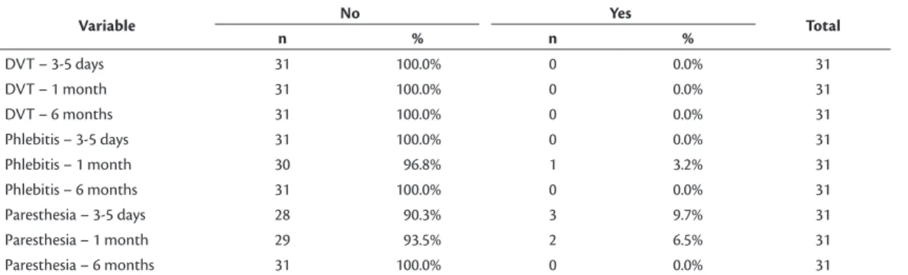
With regard to procedure-related complications, no patients had deep vein thrombosis, one had thrombophlebitis of the varicose vein at the level of the knee, and three reported symptoms of paresthesia in one of the limbs at the irst follow-up examination. In two of these patients, symptoms persisted over the irst month, but by 180 days symptoms had resolved in all three patients (Table 4).
Table 1. Saphenous vein diameters.
Variable Time of assessment n Mean Median Minimum Maximum SD p-value*
SFJ Preoperative 31 8.2 7.6 5.5 12.1 2.09
3-5 days 31 8.6 8.0 5.0 15.4 2.47 <0.001
1 month 31 7.5 6.7 1.3 14.1 2.45
6 months 31 6.6 6.3 3.7 13.0 1.85
high Preoperative 31 5.6 5.3 2.4 11.0 1.88
3-5 days 31 5.6 4.8 3.0 12.3 2.09 <0.001
1 month 31 4.6 4.0 0.0 8.8 2.08
6 months 31 2.3 2.9 0.0 6.5 2.16
Knee Preoperative 31 4.3 4.3 0.0 7.3 1.70
3-5 days 31 4.3 4.1 0.0 7.7 1.55 <0.001
1 month 31 3.9 3.8 0.0 7.0 1.55
6 months 31 3.0 3.1 0.0 6.4 1.67
Leg Preoperative 31 3.0 2.9 0.0 6.0 1.11
3-5 days 31 2.8 2.8 0.0 4.9 0.87 0.635
1 month 31 2.7 2.7 0.0 4.2 0.83
6 months 31 2.9 2.9 0.0 5.0 1.00
Ankle Preoperative 31 2.8 2.8 0.0 4.8 0.96
3-5 days 31 2.8 2.8 0.0 5.6 0.99 0.991
1 month 31 2.7 2.9 0.0 4.2 0.91
6 months 31 2.6 2.8 0.0 3.8 0.99
SD: standard deviation; SFJ: saphenofemoral junction. *Nonparametric Friedman test; p<0.05.
Figure 3. Duplex scan showing recanalization with great saphenous vein relux at 6-month follow-up.
DISCUSSION
Varicose veins of the lower extremities are a common medical problem. Recently, EVLA has been shown to be a minimally invasive treatment with excellent results in terms of a high technical success rate and low complication rate.8 However, there is still
no consensus on the optimal energy level that will provide the highest rate of technical success and the lowest rate of complications when performing EVLA.
EVLA.9,10 However, use of higher LEED signiicantly
increases the incidence of paresthesia. It therefore seems appropriate to use LEED below 100 J/cm in future trials.
The efficacy of energy delivered at different ranges of energy density has also been evaluated. Timperman et al.11 conducted a study using 810-nm
and 940-nm laser treatment and reported a signiicant difference in energy delivery between the “success” and “failure” groups (63.4 J/cm vs. 46.6 J/cm, p<0.0001). They also concluded that there was no signiicant difference in outcomes between patients treated with different wavelengths.11
Bueno et al.12 published a case series report
showing that use of 1470-nm laser is a good method for treating saphenous veins, with results similar to those obtained with 980-nm laser treatment, but with lower energy densities and power.
Park et al.13 concluded that EVLA conducted using
a1470-nm laser and low energy (LEED of 80 J/cm or lower) is an effective, safe, and technically successful option for the treatment of incompetent saphenous veins. In the current study, use of low-energy parameters with a mean LEED of 33 J/cm resulted in a GSV occlusion rate of 87.1% after 6 months.
Because recanalization can also occur after high-energy EVLA of saphenous veins, energy is probably not the only problem. If recanalization occurs, the ablated GSV may have incompetent collateral veins in the distal part of the reflux segment. If incompetent collateral veins are visualized preoperatively using duplex scanning, they should be removed by phlebectomy during the same procedure, thus preventing some cases of recanalization. In the current study, phlebectomy of incompetent collateral veins was performed concomitantly with EVLA of the saphenous vein to minimize recanalization rates related to this factor.
Figure 4. Venous Clinical Severity Scores (VCSS) over time.
Table 2. Pairwise comparison of GSV diameters measured at
the SFJ, thigh, and knee at diferent time points.
Time points compared SFJ high Knee
Preop × 3-5 days 0.330 0.902 0.476
Preop × 1 month 0.386 0.110 0.091
Preop × 6 months 0.001 <0.001 0.001
3-5 days × 1 month 0.068 0.140 0.017
3-5 days × 6 months <0.001 <0.001 <0.001
1 month × 6 months 0.014 <0.001 0.091
GSV: great saphenous vein; SFJ: saphenofemoral junction.
Table 3. Length of ablated GSV versus occlusion rate at 6 months.
Occlusion at 6 months
Length of ablated GSV
p-value*
n Mean Median Minimum Maximum SD
No 4 35 35 30 40 5.8 0.237
Yes 27 31 28 15 50 9.8
GSV: great saphenous vein; SD: standard deviation. *Nonparametric Mann-Whitney; p<0.05.
Table 4. Procedure-related complications.
Variable No Yes Total
n % n %
DVT – 3-5 days 31 100.0% 0 0.0% 31
DVT – 1 month 31 100.0% 0 0.0% 31
DVT – 6 months 31 100.0% 0 0.0% 31
Phlebitis – 3-5 days 31 100.0% 0 0.0% 31
Phlebitis – 1 month 30 96.8% 1 3.2% 31
Phlebitis – 6 months 31 100.0% 0 0.0% 31
Paresthesia – 3-5 days 28 90.3% 3 9.7% 31
Paresthesia – 1 month 29 93.5% 2 6.5% 31
Paresthesia – 6 months 31 100.0% 0 0.0% 31
Moreover, in this study recanalization of the GSV occurred in four limbs at 6 months, three of which exhibited reductions in VCSS. Only one limb exhibited an increase in VCSS, from 2 (preoperative, 3-5 days, and 1 month) to 4 at 6-month follow-up. It is of note that patients with excess BMI appear to be among those who have higher recanalization rates. However, it was not possible to perform a multivariate analysis taking into account the incidence of recanalization and BMI in this study.
The Trendelenburg position should always be used when performing EVLA, especially for veins with diameters greater than 8 mm, in which the amount of intravenous blood is very important. In such cases laser irradiation is not suficient to heat the vessel wall because the light energy is almost entirely absorbed by the blood. The initial success rate is therefore mainly the result of a thrombotic effect that, after thrombus dissolution, leads to recanalization of the GSV.14 Therefore, in the present study all patients
were placed in the Trendelenburg position for ELVA, in order to reduce the diameter of the GSV and, consequently, prolong vein contact with the laser iber, thereby avoiding thrombotic effects that could lead to subsequent recanalization.
The bare iber used for EVLA in this study has some technical shortcomings of its own, mainly because it is a rigid iber that emits the laser beam in a forward direction straight out of the tip, rather than emitting the laser energy in 360° around the tip, as is the case with radial ibers. When the bare iber is introduced into a GSV, which normally has some small tortuosities, it always has a tendency to stretch. As a consequence of stretching, the iber tip often occupies an eccentric position, with the tip touching the vein wall. In an ex vivo model, the use of bare
ibers was found to result in uneven application of light energy, requiring greater energy to ensure the destruction of the vein wall and prevent recanalization. Additionally, if the vein wall is carbonized by direct contact, a great amount of energy is emitted, increasing the likelihood of vein perforation.15
Hirokawa and Kurihara16 conducted a study using
1470-nm laser treatment, reporting that the incidence rates of bruising and pain were signiicantly reduced by using a radial iber, when compared with a conventional bare-tip iber. In the current study, we decided to use the bare iber because of the lower per-unit cost and the possibility of reuse by simply trimming the tip of the iber, which can be tested for effectiveness in the device and then sterilized. Notwithstanding, precautions were taken to minimize the disadvantages of the bare iber, adopting measures to produce more
uniform contact between the iber tip and the vessel wall, such as the Trendelenburg position, perivenous tumescence, and local compression as thermal ablation was occurring. These measures resulted in higher occlusion rates at 6-month follow-up.
In general, EVLA is a well-tolerated technique with a low incidence of complications and side effects, and most cases of such effects are transient and resolve within a few days without affecting normal walking or return to work. The main complications are bruising, saphenous nerve injury, skin burns, infection, and deep vein thrombosis/pulmonary embolism.
With the increasing popularity of endovenous GSV ablation, the possibility of endovenous heat-induced thrombosis (EHIT) of the saphenous vein extending to the femoral vein in the perioperative period has become a clinical reality. Although this occurrence has not been commonly reported in previous studies, all patients should be evaluated for this condition and treatment options should be discussed with them. Factors that may increase the risk of EHIT include patient age, unknown hypercoagulable states, and the severity of chronic venous disease.17
Suian et al.18 conducted a prospective study on the
incidence and factors contributing to the occurrence of EHIT after EVLA (1470 nm) and concluded that the following risk factors were associated with EHIT after EVLA: vein size, age, and multiple concomitant phlebectomies. EHIT usually resolves within 2 to 4 weeks in most patients, but it may worsen in some cases.18 In the current study, there was one case
of EHIT with minimal thrombus protrusion through the SFJ into the common femoral vein (involving 25% of the vein lumen). The patient was treated with anticoagulation. Duplex scanning was repeated after 4 weeks and showed that the thrombus had regressed and been resolved, and anticoagulation was then withdrawn. The risk of EHIT-related pulmonary embolism remains to be deined, but it is unlikely to occur after EVLA.
The risk of nerve injury during EVLA is directly related to the proximity of several nerves to the ablated veins. The site associated with the greatest risk of saphenous nerve injury is the middle/distal third of the leg. At the distal leg, the saphenous nerve may be directly damaged by the puncture needle or burned by laser energy transfer causing skin paresthesia, which is usually transient. Most nerve injuries can be prevented by performing ultrasound-guided puncture and avoiding EVLA on sites at high risk for nerve injury.19 In the current study, three patients reported
irst month in two patients, but by 6 months symptoms had resolved in all patients.
One limitation of this study is the lack of long-term follow-up. However, the mean follow-up of 6 months is consistent with that reported in previous studies.20
Min et al.21 reported that most failures in their study
occurred within the irst 3 months of follow-up, and all had occurred by 9 months. It therefore seems reasonable to assume that most failures that occurred in our study would have been detected within our follow-up period. Furthermore, a relatively small number of failures were observed in our sample, thereby precluding a multivariate analysis. Nevertheless, we only used nonparametric statistical tests, which are suitable for evaluation of variables that are not normally distributed or when the sample size is small. The sample size (31 cases) was considered adequate, and nonparametric methods were adopted mainly because the data did not it the normal distribution. An additional factor to be considered is that, when signiicant differences are detected for a particular sample, we can conclude that the sample size is suficient to support the results (differences) obtained in the analysis.
CONCLUSION
The thermal ablation therapy investigated in this study, using a 1470-nm water-speciic laser, allowed ablation at lower laser luence, leading to a reduction in the energy required for successful treatment. Our data support the possibility that, by using low energy density, the incidence of complications can be reduced without signiicantly affecting the clinical outcomes in the whole study group. However, this appears to be at the cost of reduced eficacy in terms of GSV occlusion rates.
REFERENCES
1. Evans CJ, Fowkes FG, Ruckley CV, Lee AJ. Prevalence of varicose veins and chronic venous insufficiency in men and women in the general population: Edinburgh Vein Study. J Epidemiol Community Health. 1999;53(3):149-53. http://dx.doi.org/10.1136/jech.53.3.149. PMid:10396491.
2. Andreozzi GM, Cordova RM, Scomparin A, et al. Quality of life in chronic venous insufficiency. An Italian pilot study of the Triveneto Region. Int Angiol. 2005;24(3):272-7. PMid:16158038.
3. van den Bos R, Arends L, Kockaert M, Neumann M, Nijsten T. Endovenous therapies of lower extremity varicosities: a meta-analysis. J Vasc Surg. 2009;49(1):230-9. http://dx.doi.org/10.1016/j. jvs.2008.06.030. PMid:18692348.
4. Boné Salat CB. Tratamiento endoluminal de las varices con laser de diodo: estudio preliminar. Rev Patol Vasc. 1999;5:35-46. 5. Navarro L, Min RJ, Bone C. Endovenous laser: a new minimally
invasive method of treatment for varicose veins--preliminary
observations using an 810 nm diode laser. Dermatol Surg. 2001;27(2):117-22. PMid:11207682.
6. Pannier F, Rabe E, Maurins U. First results with a new 1470-nm diode laser for endovenous ablation of incompetent saphenous veins. Phlebology. 2009;24(1):26-30. http://dx.doi.org/10.1258/ phleb.2008.008038. PMid:19155338.
7. Almeida J, Mackay E, Javier J, Mauriello J, Raines J. Saphenous laser ablation at 1470 nm targets the vein wall, not blood. Vasc Endovascular Surg. 2009;43(5):467-72. http://dx.doi. org/10.1177/1538574409335916. PMid:19628516.
8. Proebstle TM, Gül D, Lehr HA, Kargl A, Knop J. Infrequent early recanalization of greater saphenous vein after endovenous laser treatment. J Vasc Surg. 2003;38(3):511-6. http://dx.doi.org/10.1016/ S0741-5214(03)00420-8. PMid:12947269.
9. Proebstle TM, Moehler T, Gul D, Herdemann S. Endovenous treatment of the great saphenous vein using a 1,320 nm Nd:YAG laser causes fewer side effects than using a 940 nm diode laser. Dermatol Surg. 2005;31:1678-83.
10. Timperman PE. Prospective evaluation of higher energy great saphenous vein endovenous laser treatment. J Vasc Interv Radiol. 2005;16(6):791-4. http://dx.doi.org/10.1097/01.RVI.0000165044.41012. C8. PMid:15947042.
11. Timperman PE, Sichlau M, Ryu RK. Greater energy delivery improves treatment success of endovenous laser treatment of incompetent saphenous veins. J Vasc Interv Radiol. 2004;15(10):1061-3. http:// dx.doi.org/10.1097/01.RVI.0000130382.62141.AE. PMid:15466791. 12. Bueno KS, Albernaz DT, Albernaz LF, Zignani FR. Endolaser venoso: estudo série de casos. J Vasc Bras. 2012;11(4):286-8. http://dx.doi. org/10.1590/S1677-54492012000400006.
13. Park JA, Park SW, Chang IS, et al. The 1,470-nm bare-fiber diode laser ablation of the great saphenous vein and small saphenous vein at 1-year follow-up using 8-12 W and a mean linear endovenous energy density of 72 J/cm. J Vasc Interv Radiol. 2014;25(11):1795-800. http://dx.doi.org/10.1016/j.jvir.2014.07.009. PMid:25156646.
14. Proebstle TM, Lehr HA, Kargl A, et al. Endovenous treatment of the greater saphenous vein with a 940-nm diode laser: thrombotic occlusion after endoluminal thermal damage by laser-generated steam bubbles. J Vasc Surg. 2002;35(4):729-36. http://dx.doi. org/10.1067/mva.2002.121132. PMid:11932671.
15. Schmedt CG, Sroka R, Steckmeier S, et al. Investigation on radiofrequency and laser (980 nm) effects after endoluminal treatment of saphenous vein insufficiency in an ex-vivo model. Eur J Vasc Endovasc Surg. 2006;32(3):318-25. http://dx.doi. org/10.1016/j.ejvs.2006.04.013. PMid:16781172.
16. Hirokawa M, Kurihara N. Comparison of bare-tip and radial fiber in endovenous laser ablation with 1470 nm diode laser. Ann Vasc Dis. 2014;7(3):239-45. http://dx.doi.org/10.3400/avd.oa.14-00081. PMid:25298824.
17. Puggioni A, Kalra M, Carmo M, Mozes G, Gloviczki P. Endovenous laser therapy and radiofrequency ablation of the great saphenous vein: analysis of early efficacy and complications. J Vasc Surg. 2005;42(3):488-93. http://dx.doi.org/10.1016/j.jvs.2005.05.014. PMid:16171593.
18. Sufian S, Arnez A, Labropoulos N, Lakhanpal S. Endovenous heat-induced thrombosis after ablation with 1470 nm laser: Incidence, progression, and risk factors. Phlebology. 2014;30(5):325-30. PMid:24609619.
19. Dexter D, Kabnick L, Berland T, et al. Complications of endovenous lasers. Phlebology. 2012;27(Suppl 1):40-5. http://dx.doi.org/10.1258/ phleb.2012.012S18. PMid:22312066.
with increased energy dosing: definition of a threshold for the endovenous fluence equivalent. J Vasc Surg. 2006;44(4):834-9. http://dx.doi.org/10.1016/j.jvs.2006.05.052. PMid:16945499. 21. Min RJ, Khilnani N, Zimmet SE. Endovenous laser treatment of
saphenous vein reflux: long-term results. J Vasc Interv Radiol. 2003;14(8):991-6. http://dx.doi.org/10.1097/01.RVI.0000082864.05622. E4. PMid:12902556.
*
Correspondence
Walter Junior Boim Araujo Instituto da Circulação Avenida Sete de Setembro, 5348, Conjunto 905 – Batel CEP 80240-000 – Curitiba (PR), Brazil Tel.: +55 (41) 3244-5000 E-mail: [email protected]
Author information
WJBA - Vascular and endovascular surgeon; Vascular sonographer, Instituto da Circulação; MSc, PhD candidate in Surgical Medicine, Universidade Federal do Paraná (UFPR). JRRT - Vascular and endovascular surgeon, Instituto da Circulação; Adjunct professor, Universidade Federal do Paraná (UFPR). CSNJ - Vascular surgeon; MSc, PhD in Surgical Medicine, Universidade Federal do Paraná (UFPR). FCC - Vascular surgeon, Instituto da Circulação; MSc candidate in Surgical Medicine, Universidade Federal do Paraná (UFPR).
Author contributions
Conception and design: WJBA, JRRT, CSNJ Analysis and interpretation: WJBA Data collection: WJBA, CSNJ, FCC Writing the article: WJBA Critical revision of the article: JRRT Final approval of the article*: WJBA, JRRT, CSNJ, FCC Statistical analysis: WJBA Overall responsibility: WJBA