www.jped.com.br
ORIGINAL ARTICLE
Oxidant-antioxidant status in Egyptian children with sickle cell
anemia: a single center based study
夽
Mona Kamal El-Ghamrawy
a,∗, Wagdi Maurice Hanna
b, Amina Abdel-Salam
a,
Marwa M. El-Sonbaty
b, Eman R. Youness
c, Ahmed Adel
baDepartment of Pediatrics, New Children’s Hospital, Cairo University, Cairo, Egypt
bDepartment of Child Health, National Research Center, Cairo, Egypt
cDepartment of Medical Biochemistry, National Research Center, Cairo, Egypt
Received 1 July 2013; accepted 4 September 2013 Available online 5 February 2014
KEYWORDS
Antioxidants; Nitrite; Paraoxonase; Malondialdehyde; Sickle cell anemia; Children
Abstract
Objective: the present study was conducted to investigate the oxidant-antioxidant status in Egyptian children with sickle cell anemia.
Methods: the serum levels of total antioxidant capacity (TAO), paraoxonase (PON), vitamin E, nitrite, and malondialdehyde (MDA) were measured in 40 steady state children with homozygous sickle cell anemia (24 males and 16 females) and 20 apparently healthy age- and gender-matched controls.
Results: mean serum TAO, PON, vitamin E, and nitrite levels were significantly lower in the group with sickle cell anemia, whereas mean serum MDA was significantly higher in these chil-dren compared to controls. No significant differences in mean levels of TAO, PON, nitrite, vitamin E, and MDA were found in sickle cell anemia patients receiving hydroxyurea when com-pared with those not receiving hydroxyurea. A significant negative correlation between serum nitrite and the occurrence of vaso-occlusive crises (VOC) was observed (r =−0.3, p = 0.04). PON level was found to be positively correlated with patients’ weight and BMI (r =−0.4, p = 0.01; r =−0.7, p < 0.001, respectively), but not with frequency of VOC. The area under the curve of serum nitrite in predicting occurrence of VOC was 0.782,versus0.701 for PON, and 0.650 for TAO (p = 0.006). Serum MDA was not correlated with nitrite, PON, TAO, or vitamin E levels. No significant correlations were detected between serum nitrite and hemoglobin or antioxidant enzymes.
Conclusion: children with sickle cell anemia have chronic oxidative stress that may result in increased VOC, and decreased serum nitrite may be associated with increases in VOC frequency. A novel finding in this study is the decrease in PON level in these patients, which is an interesting subject for further research.
© 2014 Sociedade Brasileira de Pediatria. Published by Elsevier Editora Ltda. All rights reserved.
夽 Please cite this article as: El-Ghamrawy MK, Hanna WM, Abdel-Salam A, El-Sonbaty MM, Youness ER, Adel A. Oxidant-antioxidant status
in Egyptian children with sickle cell anemia: a single center based study. J Pediatr (Rio J). 2014;90:286---92. ∗Corresponding author.
E-mail:[email protected] (M.K. El-Ghamrawy).
PALAVRAS-CHAVE
Antioxidantes; Nitrito; Paraoxonase; Malondialdeído; Anemia falciforme; Crianc¸as
Estado oxidante-antioxidante em crianc¸as egípcias com anemia falciforme: estudo baseado em um único centro
Resumo
Objetivo: o presente estudo foi realizado com o objetivo de investigar o estado oxidante-antioxidante em crianc¸as egípcias com anemia falciforme.
Métodos: dosamos os níveis séricos da capacidade antioxidante total (CAT), paraoxonase (PON), vitamina E, nitrito e malondialdeído (MDA) em 40 crianc¸as estáveis com anemia falciforme homozigótica (24 meninos e 16 meninas), e 20 controles pareados por idade/sexo aparente-mente saudáveis.
Resultados: os níveis séricos médios da CAT, PON, vitamina E e nitrito foram significativa-mente menores, ao passo que o nível sérico médio de MDA foi significativasignificativa-mente maior em crianc¸as com anemia falciforme (AF), em comparac¸ão aos controles. Não foram encontradas diferenc¸as significativas nos níveis médios de CAT, PON, nitrito, vitamina E e MDA em pacientes com AF em tratamento com hidroxiureia, em comparac¸ão aos que receberam hidroxiureia. Encontramos uma correlac¸ão negativa significativa entre o nitrito sérico e a ocorrência de crises vaso-oclusivas agudas (CVO) (r =−0,3, p = 0,04). Descobrimos que o nível de PON está correlacionado positivamente com o peso e o IMC dos pacientes (r =−0,4; p = 0,01; r =−0,7; p<0,001, respectivamente), porém não com a frequência de CVO. A área sob a curva (ASC) do nitrito sérico na previsão da ocorrência de CVO foi 0,782, em comparac¸ão a 0,701 para PON e 0,650 para CAT (p = 0,006). O MDA não está correlacionado a nitrito, PON, CAT ou vitamina E. Não foram detectadas correlac¸ões significativas entre nitrito sérico e hemoglobina ou enzimas antioxidantes.
Conclusão: crianc¸as com AF apresentam estresse oxidativo crônico que pode resultar em aumento das CVO. Em crianc¸as com AF, a reduc¸ão nos níveis de nitrito sérico pode estar asso-ciada a aumentos da frequência de CVO. Um novo achado neste estudo é a reduc¸ão no nível de PON em pacientes com AF, que é um campo interessante de novas pesquisas.
© 2014 Sociedade Brasileira de Pediatria. Publicado por Elsevier Editora Ltda. Todos os direitos reservados.
Introduction
Sickle cell anemia (SCA) is one of the most common mono-genic disorders in the world, predominantly observed in Africa and Southeast Asia. It is a multi-system disease, asso-ciated with episodes of acute illness and progressive organ damage.1 SCA results from a p mutation in the genetic
code such that glutamic acid is replaced by valine in the globin chain of hemoglobin. This substitution transforms nor-mal adult hemoglobin (HbA) into sickle hemoglobin (HbS). When deoxygenated, HbS polymerizes, and when a crit-ical amount of HbS polymer accumulates within a sickle erythrocyte, cellular injury occurs. A sufficient number of damaged erythrocytes cause the phenotype of sickle cell disease (SCD), characterized by hemolytic anemia and vasoocclusion.2
SCD is emerging as an important model of oxidative stress. Since red blood cells (RBCs) carry oxygen to the body tissues, they are already rich in oxidative fuel. Their distinc-tive structural features make them susceptible to an oxidant assault. Chronic oxidative stress resulting from an imbal-ance between the production of reactive oxidant species (ROS) and antioxidant enzymes constitutes a critical fac-tor in endothelial dysfunction, inflammation, and multiple organ damage in SCD. In addition, the disease is character-ized by damage to the cell membrane due to increased lipid peroxidation products, such as malondialdehyde (MDA) and the increased consumption of nitric oxide (NO).3,4
Increased ROS production is caused by intrinsic mecha-nisms of disease, such as increased activity of several oxi-dases (NADPH oxidase and endothelial xanthine oxidase),5
auto-oxidation of HbS, release of heme iron, increased asymmetric dimethylarginine,6 uncoupling of NO synthase
activity, and decreased NO levels.7This enhanced
produc-tion of free radicals in SCA and subsequent decreased NO bioavailability inactivate NO-mediated vascular relaxation.8
Impaired vascular relaxation and increased endothelial adherence contribute to the vaso-occlusive phenomena.9
Several reports indicate that SCA patients have lower levels of antioxidants such as NO, total antioxidant capac-ity (TAO), and vitamin E as compared to normal healthy controls.10---13 Moreover, one study showed a significantly
enhanced lipid peroxidation in SCA patients when compared to controls.14 However, limited studies have evaluated the
role of oxidants and antioxidant status in children with SCA. To the authors’ knowledge, none have been conducted in patients with SCD in Egypt. The present study aimed to eval-uate the oxidant-antioxidant status in Egyptian children with SCA in a steady state through the estimation of serum levels of the lipid peroxidation product MDA, nitrite, PON, vitamin E, and TAO.
Material and methods
the Child Health and Medical Biochemistry Departments of the National Research Center, Cairo, Egypt. Forty chil-dren with established diagnosis of homozygous (HbSS) SCA (24 males and 16 females aged 10.6±4.5 years) and 20 healthy subjects (age- and gender-matched controls, 12 males and 8 females aged 10.0±2.8 years [p>0.05]) were
enrolled in the study after their legal guardians signed the informed consent. All recruited patients were in a steady state attending routine follow-up during the study period (from December 1, 2011 to June 30, 2012). Patients aged > 18 years, those with acute febrile illness within 72 hours, or acute vaso-occlusive crises (VOC) within three months prior to enrollment, serious concurrent illness, and those assigned to a regular blood transfusion program were excluded. None of recruited subjects received supplemental antioxidants or vitaminse.g.vitamin E. The study protocol was approved by the Ethics Committee of the Cairo University and by the Ethics Committee of the National Research Center, Cairo, Egypt, according to the Institutional Committee for the Pro-tection of Human Subjects, and adopted by the 18th World Medical Assembly, Helsinki, Finland.
Detailed history-taking and thorough clinical examina-tions were performed. At enrollment, the number of severe painful episodes in the preceding 12 months was recorded (frequency of VOC per year), with a working definition of a VOC as pain in the extremities, back, abdomen, chest, or head that led to an unscheduled clinic or emergency room visit and required hospitalization, and that could only be explained by SCD, with exclusion of hand-foot syndrome, chest syndrome, osteomyelitis, and any episode of pain that was treated entirely at home.15
Thirty-one patients were on hydroxyurea (HU) therapy with a mean dose of 19.8±3.4 (range 15---30 mg/kg/day, given orally once a day). The mean duration of HU was 2.12±1.49 years. Dose escalation was guided by clinical and hematological response with no attempt to reach the maxi-mum tolerated dose (MTD);163 subjects were at MTD at time
of enrollment.
Blood samples for determination of MDA, nitrite, PON, TAO, and vitamin E were collected and processed as follows: 5 mL of blood were collected into plain tubes and allowed to clot for 30 min at 25◦C; it was then centrifuged at 3,000 rpm
for 15 min at 4◦C, and the serum was separated into clean,
properly labeled tubes for analysis.
Determination of lipid peroxidation:Lipid peroxidation
was assayed by measuring the level of MDA. It was deter-mined by measuring thiobarbituric reactive species using the method of Ruiz-Larrea et al.,17 in which the
thiobarbi-turic acid-reactive substances react with thiobarbithiobarbi-turic acid to produce a red colored complex with peak absorbance at 532 nm.
Determination of serum nitrite:Serum nitrite (NO2−), as
an acceptable surrogate marker to serum NO, was measured using Griess reagent, following the Moshage et al. method;18
nitrite, a stable end-product of NO radical, is commonly used as indicator for the production of NO.19
Determination of PON activity:Arylesterase activity of
PON was measured spectrophotometrically in supernatants using phenylacetate as a substrate.20
Measurement of serum TAO levels: Serum TAO levels
were determined using an automated measurement method, which is based on the bleaching of the characteristic color
of a more stable 2, 2-azino-bis (3-ethylbenz-thiazoline-6-sulfonic acid, [ABTS]) radical cation by antioxidants (Beckman Coulter - Fullerton, CA, USA).21 The ABTS
radi-cal cation is decolorized by antioxidants according to their concentrations and antioxidant capacities. The results are expressed in mmol Trolox equivalents/L.
Measurement of vitamin E:Vitamin E as tocopherol was
measured by HPLC.22 Freshly-obtained erythrocytes were
stored in 2% pyrogallol in ethanol at -70◦C. All samples were
analyzed within one month of storage using a reverse-phase C-18 column (Waters - Milford, MA, USA), a 95% methanol solvent system, and a UV/VIS detector set at 292 nm.22
Statistical analysis
Patients’ data were analyzed using the Statistical Package for Social Sciences (SPSS), version 17.0 for Windows. Quantitative variables were expressed by mean±standard deviation (SD), and compared using Student’s t-test for unpaired samples and Mann---Whitney’s test. Spearman’s rank-order test was used for correlating quantitative variables. Qualitative variables were expressed as num-bers (frequency) and percentages, and compared between groups using the chi-squared test. Logistic regression anal-ysis was performed, and accuracy, sensitivity, specificity, positive predictive value (PPV), and negative predictive value (NPV) were calculated. A receiver operating charac-teristic (ROC) curve was made, and area the under the curve (AUC) was calculated. The optimal cut-off was determined for the variables required. p-value was considered to be significant if < 0.05.
Results
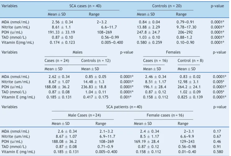
Table 1 illustrates a comparison of the tested variables between SCA patients and the control group. Mean values of nitrite, PON, TAO, and vitamin E were significantly lower, while the MDA level was significantly higher in SCA patients than in control group. This statistically significant difference in all measured variables was also observed when com-parison was made between patients and gender-matched controls. Gender did not appear to affect the oxidant-antioxidant status of SCA patients; there were no significant differences in the mean levels of nitrite, PON, TAO, and MDA in SCD males and females (Table 1).
No significant differences in the mean levels of nitrite, PON, TAO, and MDA were observed between SCD patients on HU therapy and those not receiving HU (Table 2). Com-pared to the mean vitamin E level in studied controls, the prevalence of vitamin E deficiency among SCA patients was 100%.
No significant correlations were detected between the frequency of VOC and levels of MDA, vitamin E, PON, or TAO (p > 0.05). However, serum nitrite correlated negatively with the frequency of VOC (r = -0.3, p = 0.04), but did not correlate with the levels of hemoglobin, MDA, PON, TAO, or vitamin E (r = 0.19, -0.3, 0.08, 0.03, and 0.05, respec-tively, p > 0.05). MDA did not correlate with any of the tested variables, including PON, TAO, and vitamin E (p > 0.05).
Table 1 Comparison of serum malondialdehyde, nitrite, paraoxonase, total antioxidant capacity, and vitamin E in sickle cell anemia patients and controls.
Variables SCA cases (n = 40) Controls (n = 20) p-value
Mean±SD Range Mean±SD Range
MDA (nmol/mL) 2.56±0.34 2---3.2 0.84±0.04 0.79---0.91 0.0001a
Nitrite (um/mL) 8.61±1.1 6.6---11.7 13.88±2.29 9.78---17.30 0.0001a
PON (u/mL) 191.33±33.19 108---269 247.8±24.7 206---292 0.0001a
TAO (mmol/L) 0.87±0.10 0.56---0.99 1.03±0.10 0.88---1.2 0.0001a
Vitamin E(mg/mL) 0.174±0.123 0.005---0.400 0.580±0.259 0.10---0.90 0.0001a
Variables Males p-value Females p-value
Cases (n = 24) Controls (n = 12) Cases (n = 16) Control (n = 8)
Mean±SD Mean±SD Mean±SD Mean±SD
MDA (nmol/mL) 2.62±0.34 0.85±0.05 0.0001a 2.46±0.34 0.83±0.02 0.0001a
Nitrite (um/mL) 8.67±1.07 14.48±1.3 0.0001a 8.51
±1.17 12.98±3.1 0.005a
PON (u/mL) 188.08±36.2 236.83±18.8 0.0001a 196.1±28.4 264.2±24.1 0.0001a
TAO (mmol/L) 0.87±0.08 1.04±0.11 0.0001a 0.87
±0.12 1.02±0.09 0.005a
Vitamin E (mg/mL) 0.185±0.131 0.417±0.175 0.001a 0.158±0.112 0.825±0.139 0.0001a
Variables SCA patients (n = 40) p-value
Male Cases (n = 24) Female cases (n = 16)
Mean±SD Range Mean±SD Range
MDA (nmol/mL) 2.6±0.34 2.1---3.2 2.4±0.34 2---3.1 0.17
Nitrite (um/mL) 8.67±1.07 6.9---11.7 8.5±1.17 6.6---9.9 0.67
PON (u/mL) 188.08±36.2 108---269 169.19±28.4 129---243 0.46
TAO (mmol/L) 0.87±0.08 0.71---0.9 0.87±0.12 0.56---0.98 0.91
Vitamin E (mg/mL) 0.185±0.131 0.005---0.400 0.158±0.112 0.01---0.40 0.580
MDA, malondialdehyde; PON, paraoxonase; SCA, sickle cell anemia; SD, standard deviation; TAO, total antioxidant capacity,
a Statistically significant value.
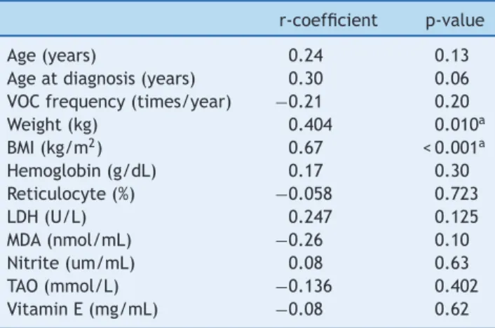
respectively). No significant correlations were observed between serum PON and frequency of VOC; laboratory indices of hemolysis including hemoglobin, reticulocyte count, or lactate dehydrogenase; or with levels of MDA, nitrite, vitamin E, or TAO (p > 0.05) (Table 3).
Figure 1 presents the sensitivity and specificity of nitrite, PON, and TAO in predicting the occurrence of VOC at differ-ent cut-off values. As there is no gold standard to compare with, nitrite, PON, and TAO were compared by ROC curve. The AUC of nitrite (0.782) was significantly higher when com-pared to that of PON (0.701) and TAO (0.650) (p = 0.006),
indicating that the overall predictability of nitrite is signif-icantly higher than that of to PON or TAO. However, when fixing the sensitivity or specificity of nitrite, it was found that either its sensitivity or specificity became unsatisfac-tory; this makes its adoption as a good predictor of the occurrence of VOC unlikely.
Discussion
The present data showed that there were decreases in serum nitrite, PON, TAO, and vitamin E levels in SCA children, as
Table 2 Comparison of serum malondialdehyde, nitrite, paraoxonase, total antioxidant capacity, and vitamin E in sickle cell anemia patients receiving HU and those not receiving HU.
Variables SCA cases receiving HU (n = 31) SCA cases not receiving HU (n = 9) p-value
Mean±SD Range Mea±SD Range
MDA (nmol/mL) 2.55±0.34 2---3.2 2.57±0.37 2.1---3.1 0.91
Nitrite (um/mL) 8.67±0.94 6.6---10.1 8.39±1.57 6.9---11.7 0.51
PON (u/mL) 186.6±30.9 108---240 207.3±37.6 148---269 0.10
TAO (mmol/L) 0.88±0.09 0.63---0.9 0.82±0.12 0.56---0.93 0.08
Vitamin E (mg/mL) 0.17±0.127 0.005---0.4 0.19±0.12 0.01---0.4 0.583
Table 3 Correlations of PON level and patients’ clinical and laboratory variables.
r-coefficient p-value
Age (years) 0.24 0.13
Age at diagnosis (years) 0.30 0.06 VOC frequency (times/year) −0.21 0.20
Weight (kg) 0.404 0.010a
BMI (kg/m2) 0.67 < 0.001a
Hemoglobin (g/dL) 0.17 0.30
Reticulocyte (%) −0.058 0.723
LDH (U/L) 0.247 0.125
MDA (nmol/mL) −0.26 0.10
Nitrite (um/mL) 0.08 0.63
TAO (mmol/L) −0.136 0.402
Vitamin E (mg/mL) −0.08 0.62
BMI, body mass index; LDH, lactate dehydrogenase; MDA, malon-dialdehyde; PON, paraoxonase; TAO, total antioxidant capacity; VOC, vaso-occlusive crises.
aStatistically significant values.
well as an increase in oxidative stress represented by MDA level. Gender and HU therapy did not appear to affect the oxidant-antioxidant status of SCA children. Serum nitrite was the only marker that correlated negatively with the fre-quency of VOC. The overall predictability of nitrite in VOC occurrence was significantly higher when compared to PON and TAO, but an increase in its sensitivity was invariably accompanied by a concomitant decrease in specificity.
Several studies have observed decreases in the activity levels of nitrite,10---12 PON, TAO,11,12and vitamin E13 in SCD
adult patients in steady state. Additionally, increased values of MDA as a lipid peroxidation product were reported as an index of the generation of ROS and oxidative stress in several disorders, including SCD.23To the authors’ knowledge, this
study was the first to investigate the oxidant-antioxidant status in Egyptian SCA children and to report increased oxidative stress in children with SCD.
One of the main findings of the present study is that the nitrite level was comparable in males and females with SCA. This contradicts previous studies reporting reductions of basal and stimulated nitrite production and responses to exogenous NO in male patients with SCA when compared to
Roc curve 1-specificity 1.0 0.8 0.6 0.4 0.2 0.0
0.0 0.2 0.4 0.6 0.8 1.0
No um/mL PON u/mL TAO mmoL/L Reference line
Sensitivity
Source of the curve
Fig. 1 ROC curve of nitrite, PON, and TAO for prediction of occurrence of vaso-occlusive crisis.
NO, nitrite; PON, paraoxonase; ROC curve, receiver operating characteristic curve; TAO, total antioxidant capacity.
females.24 Nevertheless, these differences disappear when
only children are considered. Gender differences in NO bioavailability are probably caused in part by the protec-tive effects of ovarian estrogen on NO synthase expression and activity in pubertal females.24
HU is described as an inducer of fetal hemoglobin expres-sion, which reduces HbS polymerization in SCA patients, reducing mortality and VOC.25 In vitroand animal studies
have demonstrated that HU may play an additional role as a NO donor.26 The activation of fetal hemoglobin
expres-sion by HU may occur through this NO pathway.27 Clinical
studies suggested an antioxidant effect of HU on SCA by measuring glutathione levels and other antioxidants in SCA patients treated with HU and those who were not given this medication.28 However, the small number of patients who
were not receiving HU in the present study makes it dif-ficult to draw any conclusions regarding the effects of HU (whether direct or indirect) on the measured oxidant and antioxidant markers.
The present study observed a weak association between the decreases in serum nitrite level and increased frequency of VOC. However, no correlations of serum nitrite level with laboratory indices, such as total hemoglobin level, other antioxidants, or MDA were observed.
SCD is an extremely heterogeneous disease for many reasons, and the occurrence of VOC appears to be multi-factorial. Even if low nitrite is one of these factors, it is impossible to conclude that serum nitrite level may provide specific prognostic or clinical information beyond that given by the simple, conventional measurement of hemoglobin concentration. However, it may be reserved as a simple biomarker of oxidative stress in children with SCA in steady state to help in selection and follow-up of those in need for antioxidant supplements.
In the present study, TAO was measured, as its value is more informative than the knowledge of individual antioxidant.12Its level reflects the collective contribution to
the reducing property of non-protein individual antioxidant or electron donating components. However, no correlation was found between TAO and increased frequency of VOC.
An important new observation in the present study was the decrease in PON level in SCA children. PON level was found to increase with body weight and BMI, but it did not correlate with the frequency of VOC or indicators of hemol-ysis. However, and to the authors’ knowledge, there are no reports on PON level or its impact on the phenotype in SCD patients. PON is a calcium-dependent serum esterase that is synthesized by the liver and is released into the circulation, where it associates mainly with high-density lipoproteins and protects low-density lipoproteins and cellular mem-branes against lipid peroxidation.29It is largely believed to
have a role in protection against oxidative stress.30Patients
with coronary heart disease showed increased lipid peroxi-dation and decreased PON activity.31This may suggest that
patients with SCA who showed decreased PON and increased lipid peroxidation (MDA) may be at risk of other forms of vas-culopathy, including coronary heart disease, especially with growing up indicating future studies.
antioxidant capacity among these children, and whether this may improve their clinical course.
Children with SCA have chronic oxidative stress that may result in increased VOC. In children with SCA, decreased serum nitrite may be associated with increases in VOC fre-quency. A novel finding in this study was the decrease in PON levels in SCA patients, which is an interesting field for further research.
Funding
Equipment from the Pediatric Hematology Clinic, Cairo University and from the Biochemistry department at the National Research Center were used. This work received no financial assistance from any funding agency in the public, commercial, or non-profit sectors.
Conflicts of interest
The authors declare no conflicts of interest.
Acknowledgements
The authors would like to thank all patients who participated in this study. They would like to express their appreciation to their colleagues and nurses at the Pediatric Hematology and BMT Unit who facilitated this work.
References
1. Weatherall D, Hofman K, Rodgers G, Ruffin J, Hrynkow S. A case for developing North-South partnerships for research in sickle cell disease. Blood. 2005;105:921---3.
2. Steinberg MH. Sickle cell anemia, the first molecular disease: overview of molecular etiology, pathophysiology, and therapeu-tic approaches. ScientificWorldJournal. 2008;8:1295---324. 3. Ray D, Deshmukh P, Goswami K, Garg N. Antioxidant vitamin
levels in sickle cell disorders. Natl Med J India. 2007;20:11---3. 4. Wood KC, Granger DN. Sickle cell disease: role of reactive
oxygen and nitrogen metabolites. Clin Exp Pharmacol Physiol. 2007;34:926---32.
5. Wood KC, Hebbel RP, Granger DN. Endothelial cell NADPH oxi-dase mediates the cerebral microvascular dysfunction in sickle cell transgenic mice. FASEB J. 2005;19:989---91.
6. Landburg PP, Teerlink T, Biemond BJ, Brandjes DP, Muskiet FA, Duits AJ, et al. Plasma asymmetric dimethylarginine concen-trations in sickle cell disease are related to the hemolytic phenotype. Blood Cells Mol Dis. 2010;44:229---32.
7. Morris CR, Kato GJ, Poljakovic M, Wang X, Blackwelder WC, Sachdev V, et al. Dysregulated arginine metabolism, hemolysis-associated pulmonary hypertension, and mortality in sickle cell disease. JAMA. 2005;294:81---90.
8. Kaul DK, Liu XD, Zhang X, Ma L, Hsia CJ, Nagel RL. Inhibition of sickle red cell adhesion and vasoocclusion in the micro-circulation by antioxidants. Am J Physiol Heart Circ Physiol. 2006;291:H167---75.
9. Francis Jr RB, Johnson CS. Vascular occlusion in sickle cell disease: current concepts and unanswered questions. Blood. 1991;77:1405---14.
10. Emokpae AM, Ojiefo UP, Aisha KG. Antioxidant enzymes and acute phase proteins correlate with marker of lipid peroxide in adult Nigerian sickle cell disease patients. Iran J Basic Med Sci. 2010;13:177---82.
11. Arinola OG, Olaniyisa SA, Akibinu MO. Evaluation of antioxi-dant levels and trace elements status in Nigerian sickle cell disease patients with plasmodium parasitae-mia. Pak J Nut. 2008;7:766---9.
12. Foluke F, Kayode A, Johan A, Modupe K. Total anti-oxidant status in sickle cell disease patients in steady state. J Natl Med Assoc. 2008;100:891---4.
13. Hasanato RM. Zinc and antioxidant vitamin deficiency in patients with severe sickle cell anemia. Ann Saudi Med. 2006;26:17---21.
14. Hundekar PS, Suryakar AN, Karnik AC, Valvi R, Ghone RA, Bhagat SS. The effect of antioxidant supplementation on the oxidant and antioxidant status in sickle cell anemia. J Clin Diag Res. 2011;5:1339---42.
15. Darbari DS, Onyekwere O, Nouraie M, Minniti CP, Luchtman-Jones L, Rana S, et al. Markers of severe vaso-occlusive painful episode frequency in children and adolescents with sickle cell anemia. J Pediatr. 2012;160:286---90.
16. Kinney TR, Helms RW, O’Branski EE, Ohene-Frempong K, Wang W, Daeschner C, et al. Safety of hydroxyurea in children with sickle cell anemia: results of the HUG-KIDS study, a phase I/II trial Pediatric Hydroxyurea Group. Blood. 1999;94:1550---4. 17. Ruiz-Larrea MB, Leal AM, Liza M, Lacort M, de Groot H.
Antioxidant effects of estradiol and 2-hydroxyestradiol on iron-induced lipid peroxidation of rat liver microsomes. Steroids. 1994;59:383---8.
18. Moshage H, Kok B, Huizenga JR, Jansen PL. Nitrite and nitrate determinations in plasma: a critical evaluation. Clin Chem. 1995;41:892---6.
19. Guevara I, Iwanejko J, Dembi´nska-Kie´c A, Pankiewicz J, Wanat A, Anna P, et al. Determination of nitrite/nitrate in human bio-logical material by the simple Griess reaction. Clin Chim Acta. 1998;274:177---88.
20. Watson AD, Berliner JA, Hama SY, La Du BN, Faull KF, Fogel-man AM, et al. Protective effect of high density lipoprotein associated paraoxonase. Inhibition of the biological activity of minimally oxidized low density lipoprotein. J Clin Invest. 1995;96:2882---91.
21. Erel O. A novel automated direct measurement method for total antioxidant capacity using a new generation, more stable ABTS radical cation. Clin Biochem. 2004;37:277---85.
22. Jain SK, Levine SN, Duett J, Hollier B. Reduced vitamin E and increased lipofuscin products in erythrocytes of diabetic rats. Diabetes. 1991;40:1241---4.
23. Yıldırım F, Sermetow K, Aycicek A, Kocyigit A, Erel O. Increased oxidative stress in preschool children exposed to passive smok-ing. J Pediatr (Rio J). 2011;87:523---8.
24. Reiter CD, Gladwin MT. An emerging role for nitric oxide in sickle cell disease vascular homeostasis and therapy. Curr Opin Hematol. 2003;10:99---107.
25. Steinberg MH, Barton F, Castro O, Pegelow CH, Ballas SK, Kut-lar A, et al. Effect of hydroxyurea on mortality and morbidity in adult sickle cell anemia: risks and benefits up to 9 years of treatment. JAMA. 2003;289:1645---51.
26. King SB. Nitric oxide production from hydroxyurea. Free Radic Biol Med. 2004;37:737---44.
27. Cokic VP, Smith RD, Beleslin-Cokic BB, Njoroge JM, Miller JL, Gladwin MT, et al. Hydroxyurea induces fetal hemoglobin by the nitric oxide-dependent activation of soluble guanylyl cyclase. J Clin Invest. 2003;111:231---9.
28. Teixeira Neto PF, Gonc¸alves RP, Elias DB, de Araújo CP, Magalhães HI. Analysis of oxidative status and biochemical parameters in adult patients with sickle cell anemia treated with hydroxyurea, Ceará, Brazil. Rev Bras Hematol Hemoter. 2011;33:207---10.
30. Amengual-Cladera E, Nadal-Casellas A, Gómez-Pérez Y, Gomila I, Prieto RM, Proenza AM, et al. Phytotherapy in a rat model of hyperoxaluria: the antioxidant effects of quercetin involve serum paraoxonase 1 activation. Exp Biol Med (Maywood). 2011;236:1133---8.