Bo1m Inst. oceanogr., S Paulo, 33(2):201-212, 1985
NOTES ON ELECTROPHEROGRAMS OF EYE-LENS
JMUSCLE PROTEINS AND ZYMOGRAMS
OF MUSCLE ESTERASES OF FISH COLLECTED DURING THE FIRST BRAZILIAN
EXPEDITION TO THE ANTARCTICA
PHAN Van Ngan; Hana SUZUKI*; Vicente GOMES*
&Maria José de Arruda Campos Rocha
PASSOS**
Instituto Oceanográfico da Universidade de são Paulo (Caixa Postal, 9075, 01000 são Paulo, SP)
Synopsis
A preZiminary study
wasoarried out on eZeotropherograms of eye-Zens, muscle
proteins and zymograrns of muscZe esterases of ten
No-to-the.ru..a.
.tilJL6erti.,six
Notothe.ru..a.
nu~6~on&and one Zanternfish,
E.te.~onaantaftctica.
The fish were
coUected by the R/V "nof. W. Besnard" of the Institute of Oceanography.,
University of
são
PauZo, during the
First
BraziZian Expedition to Antaratica.
Eye-Zens proteins were anaZysed onoellulose aoetate membrane, musole proteins and
esterases on gel of polyaarylamide. · Eye-Zens proteins showed three types of
eZectropherograms for
N. laJu,e.n.L
and
ruo types for
N.
nud..t6~on&.One of the
electropherograms of N.
laJu,e.n.Laan be readily distinguished from those of N.
nu~6~ol'L6.
Electropherograms of musale proteins of
N. laJu,e.n.L
and
N.
nud..t6~on6are
very similar and consist of sixteen to seventeen fractions.
EZectropherograrns of
muscZe proteins of
N.
~e.n.Lare severely affected by the aonservation of the
extracts overnight under -20°C. AZl
N.
nudi6~on&were of the same zymograms of
esterases while those of
N •
.t~e.n.Lvaried. Eleotropherograms of eye-Zens and
muscZe proteins
asweZZ as zymograms of esterases of the Zanternfish are different
from those of nototheniids.
Descr i ptors: Marine fishes, E1ectrophoresis, Esterases, Proteins, Muscle, Eye-lens,
No-to-the.nia
~e.n.L,Notothe.n.La
nud..t6~ol'L6,E.te.ct4ona
ant~ctica,R/V "Prof. W. Besnard", Bransfield Strait, Antarctica.
Descritores: Peixes marinhos, Eletroforese, Esterases, Proteínas, Músculo,
Cristalino,
Notathe.n.La
~e.n.L,Notothe.n.La
nud..t6~an6, E.te.c~onaa.rr.-taJtc.Uca, N/Oc. "Prof. W. Besnard", Estreito de Bransfield,
Antártica.
Introduction
Electrophoretic, techniques and
histochemical Jtaining methods (Smithies, 1959; Hunter
&
Markert, 1957) have been widely used for studies of genetic variation in species (Ayala, 1976; Selander&
Johnson, 1973). In many areas of the world's seas, the fish(*) Graduate Student, Institute of Oceanography, Departament of
Biological Oceanography, Fellow of CNPq.
(>~>~) CIRM - IOUSP
Pubi. n.
637 do InJ.d.oce.anogJt. da
U.6p.fauna has been a subject of this kind of investigation (de Ligny, 1969; 1972) but there is very few information in this field on fish of the region south of the Antarctic Convergence.
During the First Brazilian Expedition to Antarctica, a small number of fish belonging to several species was caught in experimental fishing with drag net and Isaacs-Kidd mid-water trawl realized by the research vessel "Prof. W. Besnard" of the Institute of Oceanography,
University of são Paulo. Eye-lens and musc1e samples of some os these fish were collected for this preliminary
202 Bolm Inst. oceanogr., 5 Paulo, 33(2), 1985
possible use of these samples for more detailed investigations in the future.
Material and lIlethods
MateriaIs used in this study were eye-lenses and musele samples of ten
Notothenia
l~eni (Lonnberg, 1905), S1XN. n.u.cü6Jt0n6
(Lonnberg, 1905) of the family Nototheniidae and one lanternfishEleetJton.a
an.taJte~ea (Gunther, 1878) of the family Myetophidae. Sexes of the fishes were unknown. The nototheniids, ranging from 129 to 158 rnrn in total length,were eaught by the drag net at station no. 24 (062°43'.0 S;054°24'.0 w) and the lanternfish, 80 rnrn in total lenght, was caught by lsaacs-Kidd mid-water trawl at station no. 25 (062°25'.0 S; 054°24'.0 w) in the Bransfield Strait, Austral Summer 1982-1983.Eye-lenses and muscle samples were eollected from alive specimens on board of the vessel and kept in the freezer at -20°C until use. Eye-lenses were
rnaeerated in solution of 0.9% NaCl by means of an electrie homogenizer. Two proportions, namely 1:2 and 1:4 (tissue weight/volume of extraetion fluid) were tested. The extracts were centrifuged at 3500 rpm for 30 mino Muscles were macerated in solution of glycerin-EDTA--Tris pH 8.7 (Scopes, 1968) in the proportion of 1:2 (weight/volume).
Maeerated material were spun at 3500 rpm for 15 min, supernatants were colleeted and submitted again to another
eentrifugation at 10 000 rpm for 15 mino Total protein eontents were determined by Biuret method (Gornall
et
al.,
1949). Eye-lens proteins were analysed byelectrophoresis on cellulose acetate membranes (Cellogel, Chemetron, 5.7x14 em). Two systems of buffers, Tris-glyeine pH 8.3 and Tris-glyeine pH 9.5 were tested. Electrophoresis was performed at 300V across the membrane during 25 mino Origin was at 1.5 cm from the cathode. Electropherograms were visualized by Ponceau S.
Densitometrie readings were taken on an Atago densitometer using filter of 500 nm. Musele proteins were analysed by vertical polyacrylamide flat gel eleetrophoresis. The method used was a modification of the method by Akroyd
(1967) as described by Phan (1980). The membrane consists of two layers of gels of different concentrations in different
buffers (Brewer
et
al.,
1974).Tris-Glycine buffer adjusted to pH 8.9 with IN NaOH was used as electrode buffer. For general proteins analysis, 5 ~l, and for esterases, 40 ~l of
extract containing 10% of saccharose were used. Electrophoresis was carried out, passing firstly by a current of 20 mA for about 5 min until the
Bromophenol Blue used as tracer reached the interface of the upper and the lower gel then passing a current of 45 mA for about 30 min for the analysis in the lower gel until the tracer dye reached the determined point. Electropherograms of muscle proteins were visualized by Coomassie Blue. Zyrnograms of esterases were obtained by the modified techniques of Gomori as described by Flowerdew
&
Cr i s p (197 5) •Results
The proportion between weight of eye-lenses and vglume of extraction fluid is important for the quality of the electropherograms of eye-lens proteins. Under the same conditions extracts obtained by homogenizing eye-lens in the proportion 1:2
(weight/volume) resulted in electropherograms with fractions sharper than those obtained with samples homoge~ized in the proportion 1:4. Total protein concentrations were 8.71 ± 1.80 g/dl (n=8) and 5.82 ± 0.47 g/dl (n=8) for extracts obtained by homogenizing in the proportion of 1:2 and 1:4 respectively. Electrophoresis of the same samples realized with Tris-glyeine pH 8.3 and Tris-Glycine pH 9.5 buffers resulted in
electropherograms of the same number of fractions. Electropherograms obtained with Tris-g1yeine pH 9.5, however, showed sharper separation and higher migration of fraetions, specia11y the cathodic ones (Fig. 1). Electropherograms
of
N.
l~en.~ comprised from ten toeleven fractions and those of N. n.ucü6!ton6 from nine to ten fraetions. The
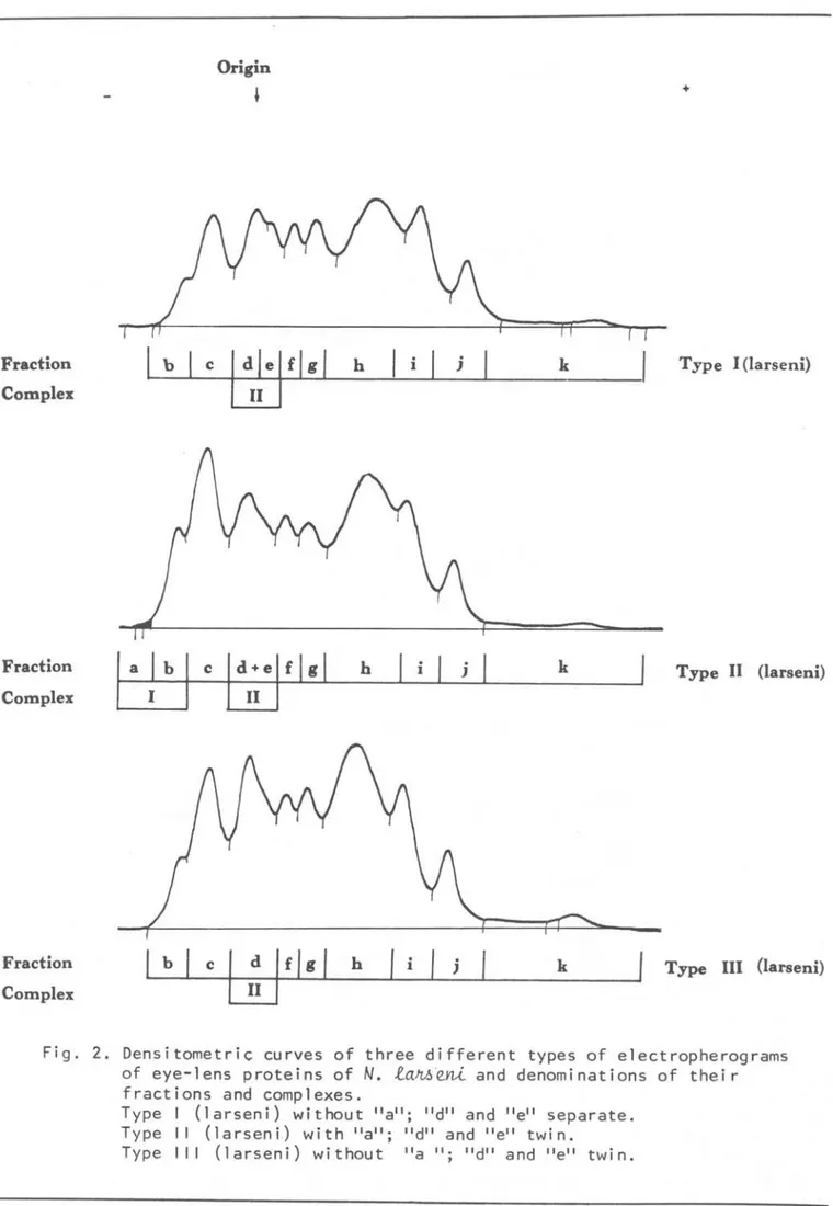
fractions were denominated sucessively by alphabetic letters. Two eonsecutive or twin fraetions may be classified as a complex (Figs 2-3). Three types of electropherograms of eye-lens proteins
of
N.
l~eni were recognized. Theywere denominated Type-l (larseni),
PHAN ,
et
al.:
Electrophoresis: Eye-lens: Muscle 203Origin
+
A
Length of electropherogram
---+l
8
Length of electropherogram
Fig. 1. Densitometric curves of the electropherograms of eye-lens proteins of the same
N.
l~eni obtained in cellulose acetate membrane with different buffers.A. Tris-Glycine pH
8.3
buffer B. Tris-Glycine pH9.5
buffer(larseni) and Type-III (larseni) by the Complex-II. In Type-I (larseni) this complex is IDade up by two separate fractions "d" and "e" while in Type-II (larseni) and.Type-III (larseni) it is made up by two twin fractions "d +
e".
Type-II (larseni) differs from Type-I(larseni) and Type-III (larseni) hy the fraction "a". In electropherograms this fraction appears as a weak stain at the cathode side and in densitometric curves as a tail of fraction "b" (Fig. 2). Relative concentration of this fraction, whenever possible, was computed together
with that of fraction "h" as relative concentration of Complex-I. Of the ten examined
N.
~eni eight were Type-I(larseni), one was Type-II (larseni) and one Type-III (larseni).
Two types of electropherograms of eye-lens proteins of
N.
nudi6~on~,namely Type-I (nudifrons) and Type-II (nudifrons), can he recognized. These types differ from each other by fraction "a" (Fig. 3). Two out of the six
N.
nudi6~o~ examined were Type-I
204
Fraction
Complex
Fraction
Complex
Fraction
Complex
Origin
b h
Bolm Inst. oceanogr., S Paulo,
33(2), 1985
+
Type I (larseni)
Type 11 (larseni)
i j k Type lU (Iarseni)
Fig. 2. Densitometriç curves of three different types of electropherograms of eye-lens proteins of
N.
la~eni and denominations of their fractions and complexes.Type I (larseni) without "a"; "d" and "e" separate. Type II (larseni) with "a"; "d" and "e" twin.
PHAN
e..t
(ti.: Electrophoresis: Eye-lens: Muscle 205Fraction
Fraction
Complex
Origin
c d
I
fI
g h+
J k
Type I (nudifrons)
Type 11 (nudifrons)
Fig. 3. Densitometric curves of two different types of electropherograms of eye-lens proteins of
N.
nudin~on6 and denominations of their fractions and complexoType I (nudifrons) without lia"; "e" absent. Type II (nudifrons) with "a"; "e" absent.
eye-lens proteins of
N.
t~e.ni andN.
nudi6~oY1.6 differ qualitatively and quantitatively. Qual.itatively
electropherograms of eye-lens proteins of both fish differ by the Complex 11.
In
N.
~eni. this complex consists oftwo separate or twin fractions "d" and fie"; in
N.
nudi6~on6. however, in the position of this complex, there is only fraction "d" (Figs 2-3).Electropherograms of
I:.
[~e.ni and N.nudi6~on.6 also differ by fraction "k"
which in
N.
t~eni appeared as a small peak at the lnode extremity ofdensitometric curves (Fig. 3).
Quantitatively, both species differ by
the relative concentration of fraction "c" which is significantly higher in N. nudi6~on6 (Table 1).
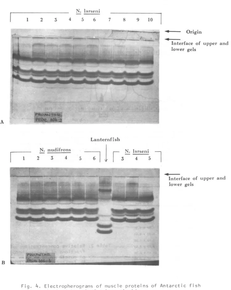
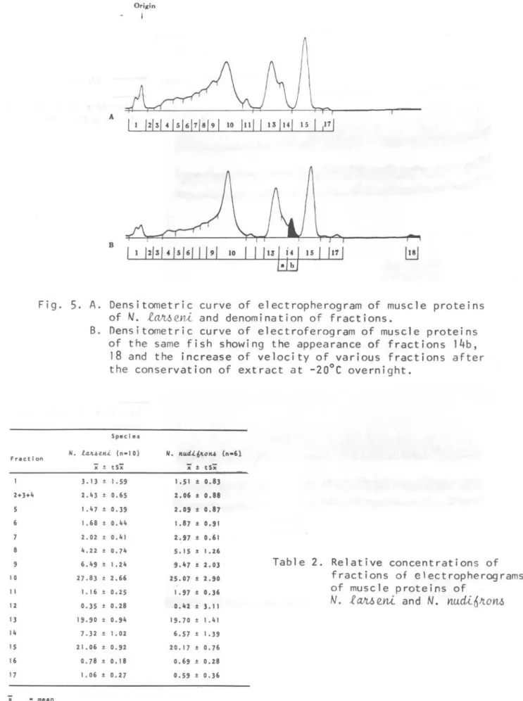
Electropherograrns of muscle proteins
of N. taM
eni
and N. nudi6~on6 are verysimilar. Qualitatively, only .a slight difference in sharpness of several fine and faint fractions ' in the cathode side of electropherograrns can be recognized
(Fig. 4).
206 Bolm Inst. oceanogr., S Paulo, 33(2), 1985
Table 1. Relative concentrations
(%)
of fractions and comolexes of electropherograms of eye-lens proteins ofN.
tan~e~A and N.Ylu.cü..
fi
Jto
YlJ.JSpecies
N.
Fractions and Complexes
x ±
b 3.55
a + b (Complex I ) 6.81
c 14.88
d 11 .29
e 6.44
d + e (Complex I I ) 17.58
f 7.54
9 9.35
h 28.40
1 1 .94
j 5.41
k 1 .69
x mean
txS-;Z confidence interval ( leve 1 of
n number of individuals
Quantitatively, only the difference in relative concentration of fraction "11" of both species is significant (Table 2).
Extracts of three
N.
t~eni were kept overnight at -20°C and their electropherograms were compared with those obtained before the conservation.It was noticed that the conservation affected not only the number but also the migration of the fractions. Fraction "14" was divided in to "14a." and "146"; fraction "18" a new and faint fraction, appeared near the anode of
electropherograms of conserved extracts. Migration velocities of various
fractions, especially those of fractions
faft.6e.Y!-t N. I1Udft-t6Jton.6
t
±
±
±
±
±
±
±
±
±
±
±
-
S-;Z(n)x S-;Z(n) x ± t x
O . 41 ( 9 ) 4. O G ± 8.51 ( 2 )
( 1 ) 6.03 ± 0.88 ( 4 )
1 .12 ( 1 O ) 17.77 ± 1 .75 ( 6 )
1 . 1 O (8 ) 12.37 ± 0.49 (6 )
1 .21 (8 )
0.49 ( 1 O )
0.27 ( 1 O ) 8.01 ± 0.64 ( 6 )
0.54 ( 1 O ) 10. 16 ± 0.74 (6 )
0.85 ( 1 O ) 28.60 ± 0.73 (6 )
O . 7 1 ( 1 O ) 12. O O ± 0.77 (6 )
0.46 ( 1 O) 4 . 91 ± 0.74 ( 6 )
0.82 (6 ) 1 . 14 ± 1 .72 (5 )
significance 5 %)
"11" onward, become more rapid than their correspondents before the conservation (Fig. 5).
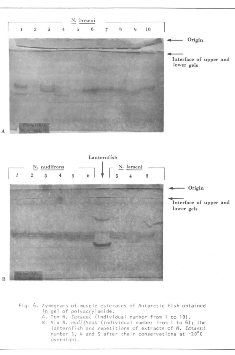
Zymograms of muscle esterases are shown in Figure 6. AlI
N.
Ylu.cü..6JtoYl~ are of the same zymograms while those ofN.
l~eni varied. Four out of the ten
N.
l~eYli showed the same zymograms as
those of
N.
Ylu.cü..6JtoYl~. Apparently zymograms of muscle esterases were not affected by the conservation of extracts.Electropherograms of eye-lens, muscle proteins and zymograms of muscle
PHAN
et
al.:
Electrophoresis: Eye-lens: Muscle 207A
B
N. larseni
1 2 4 5 6 7
Lanternfish N. nudifrons
2 3 4
li,
i'
8 9 10
1
• Origin
•
Interface of upper and lower gels
Interface of upper and lower gels
Fig.
4.
Electropherograms of muscle proteins of Antarctic fish obtained in gel of polyacrylamide.A. Ten
N.
fah~eni (individual number from 1 to la). B. SixN.
nu~i6no~ (individual number from 1 to6).
the lanternfish and repetitions of extracts of
208 Bolm Inst. oceanogr., S Paulo,
33(2),
1985A
B
Fig.
5. A.
Densitometric curve of electropherogram of muscle proteinsof N. l~eni and denomination of fractions.
Fractlon
2+3+4
8
9
10 11 12 13 14 15 16 17
x
•
me.n11.
B. Densitometric curve of electroferogram of muscle proteins of the same fish showing the appearance of fractions 14b, 18 and the increase of velocity of various fractions after the conservation of extract at -20°C overnight.
Specles
la.Jt<lell.i. (n-l0)
'i ± tSX'
3.13 1. 59 2.43 ± 0.65
1.47 ± 0.39
1. 68 0.44
2.02 0.41
4.22 ± 0.74
6.49 ± 1.24 27.83 2.66
1. 16 0.25 0.35 ± 0.28 19.90 ± 0.94
7.32 ± 1. 02
21.06 ± 0.92 0.78 ± 0.18
1. 06 ± 0.27
11. lIud.i.6JtOll4
X' ± tSX'
1. 51 ± 0.83 2.06 ± 0.88
2.09 ± 0.87 1.87 ± 0.91
2.97 ± 0.61 5.15 ± 1.26 9.47 ± 2.03 25.07 ± 2.90
1. 97 ± 0.36 0.42 ± 3.11
19.70 ± 1. 41 6.57 ± 1. 39
20.17 ± 0.76 0.69 ± 0.28 0.59 ± 0.36
(n-61
Table 2. Relative concentrations of fractions of electropherograms of muscle proteins of
N. R~eni and N.
Yl.udi6JtoYl-6
PHAN
et
ai.:
Electrophoresis: Eye-lens: Muscle 209A
B
N. l"lrseni
1 2 3 4 5 6 7
Lanternfísh N. nudifrons
2 3 4
8 9 10
larseni
4
.. Origin
..
Interface of upper and lower gels
• Origin
..
Interface of upper andlower gels
Fig.
6.
Zymograms of muscle esterases of Antarctic fish obtained in gel of polyacrylamide.A. Ten
N.
lc~e~ (individual number from 1 to 10). B. Six N. Ywd~6 , '1ol1!.l (individual nUfllber hom 1 to 6); the210 Bolrrt Inst. oceanogr., S Paulo,
33(2), 1985
A
B
Origin
Origin
,
+
+
Fig.
7.
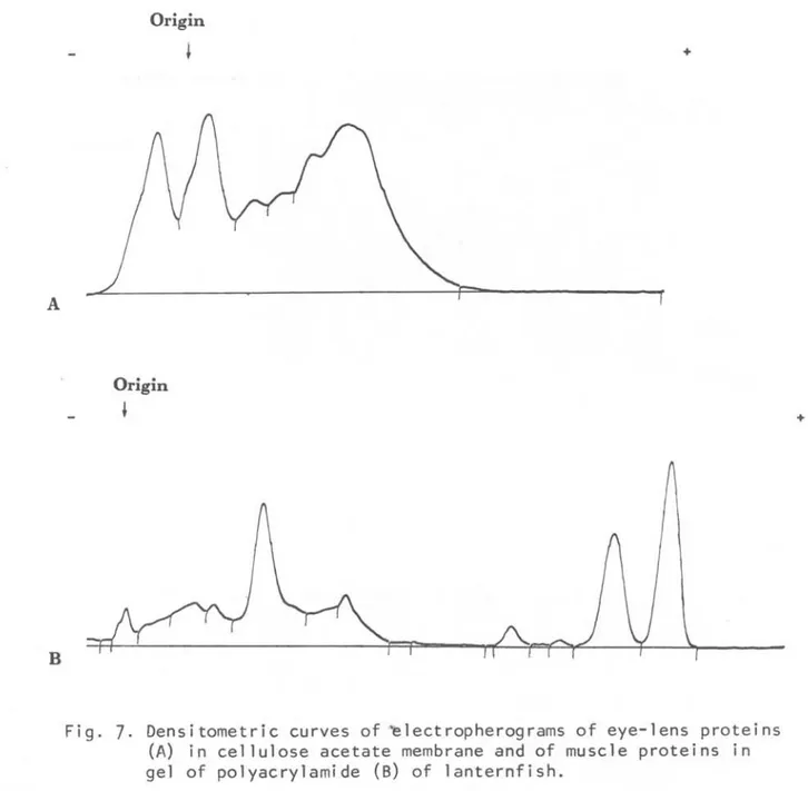
Densitometric curves of ~lectropherograms of eye-lens proteins (A) in cellulose acetate membrane and of muscle proteins in gel of polyacrylamide (8) of lanternfish.Discussion and cQnclusiQn
The so1ub1e proteins of the eye-1ens were considered to have great va1ue for taxonomic studies because they are synthesized by on1y one ce11 type present in the eye as a sing1e 1ayer
(O'Rourke, 1974). The use of eye-1ens proteins, however, has its drawback because they are known to be affected by ontogenetica1 and patho1ogica1 processes
(Bon, 1957; Haen
&
O'Rourke, 1969; Barrett&
Wi11iams, 1967; Peterson&
Shehadeh, 1971; Phanet
al.,
1977; Zigman & Yu10, 1979; Vazzo1er & Phan, 1981). Variation of e1ectropherograms of eye-1ens proteins of nototheniids in re1ation to growth was not studied dueto the sma11 number of samp1es co11ected in on1y one 1ocation and sma11 range of body 1ength of specimens. The
observation that one type of
e1ectropherograms of eye-1ens proteins
of
N.
l~eni can be readi1ydistinguished from those of
N.
~udi6~o~imp1ied that this tissue might be usefu1 for the identification of species.
Solub1e musc1e proteins of fish were considered a good material for the study of interspecific variations by means of e1ectrophoretic techniques (Tsuyuki
PHAN
et al.:
Electrophoresis: Eye-lens: Muscle 211Suzuki
et
al.,
1983). Electropherograms of muscle proteins of the twonototheniids investigated showed very little intra - and inter-specific differences. Electropherograms of one
species were severely affected by the conservation of extracts. Muscle esterases, on the other hand, showed a great degree of intra-specific
heterogeneity in
N.
t~en~ and a high percentage of inter-specific homogeneity betweenN.
t~e~ andN.
nudió~onó.These characteristics of muscle
esterases coupled with the observation that their zymograms were not affected by conservation of extract make these
isozymes a suitable material for the study of protein variations in
N.
t~e~ and
N.
nudió~onóResumo
Foi realizado um estudo preliminar sobre eletroferogramas de proteínas de crista-lino e de músculo esquelético, e zimo-gramas de esterases de músculo esquelé-tico de dez Notothe~a t~en~, seis
Notothen~a nudi6~on~ e de um
peixe-lan-terna. Ete~~ona ant~~~a. Os peixes foram,coletados pelo N/Oc. "Prof. W. Besnard" do Instituto Oceanográfico da Universidade de são Paulo durante a I Expedição Brasileira
ã
Antártica. As pr teínas do cristalino foram analisadas em membranas de acetato de celulose, en-quanto que as proteínas e esterases do músculo esquelético, em gel depoliacri-lamida.
As proteínas do cristalino apresentam três tipos distintos de eletroferogramas para
N.
t~e~ e dois paraN.
nudió~on~.Um dos eletroferogramas de
N.
t~en~pode ser prontamente distingüido dos de
N.
nudi6~on~. Eletroferogramas depro-teínas de músculo de N.
taJL6e~
e de t' N.nudió~onó
são muito semelhantes eco~
sistem de 16 a 17 frações. Os e1etro-fêrogramas de proteínas de músculo deN.
f~e~ são severamente afetados pelapreservação dos extratos por uma noite a -20°C.
Todos os N. nudi6~onó apresentam um mesmo zimograma de esterases enquanto que os de
N.
taJL6e~ variam.Tanto os e1etroferogramas de proteí-nas do cristalino e do músculo como os zimogramas de esterases do peixe-lan-terna são diferentes dos apresentados pelos nototeniídeos.
Acknowledgements
The authors wish to express their thanks to Dr Adolf Kellermann, Institut fur Polarokologie der Christian-Albrechts Universitat, and Mr. Uwe Rowedder, Küsten und Binnenfischerei, Kiel, West Germany, for identification of the nototheniids and the lanternfish,
respectively. The authors wish also to thank Dr Renato Amaral, Universidade Federal do Rio Grande do Sul, Brazil, for his excellent assistance in sampling during the Expedition, and to Mrs Maria Cecília Catunda for polishing the
English in the manuscript.
References
AKROYD, P. 1967. Acrylamide gel slab: electrophoresis in a simple glass cell for improved resolution and comparison of serum protein. Analytical Biochem., 79:399-410.
AYALA, F. J., ed. 1976. Molecular evolution. Sunderland, Mass., Sinauer Associates, 277p.
BARREIT, I. & WILLIAMS, A. A. 1967. Soluble lens proteins of some
scombroid fishes. Copeia, (2):468-471.
BON, W. F. 1957. Some physico-chemical data about the ontogenetic and
phylogenetic development of the eye lens proteins of fish. Pubbl. Staz. , Zool. Napoli, 30(3):373-380.
BREWER, J. M.; PESCE, A. J. & ASHWORTH, R. B. 1974. Experimental techniques in biochemistry. New Jersey,
Prentice-Hall, p.351.
De LIGNY, H. 1969. Serological and biochemica1 studies on fish
populations. Oceanogr. Mar. Biol. Ann. Rev., 7:411-513.
1972. Blood and
biochemical polymorphism in fishes. In: European Conference on Animal Blood Groups and Biochemical
Polymorphisms:12. The Hague, Dr. W. Junk, p.55-65.
FLOWERDEW, M. W. & CRISP, D. J. 1975. Esterase hetero gen e it y a nd an
212
in the cirriped Balan~ balano~d~
using acrylamide gel electrophoresis. Mar. Biol., 33:33-39.
GORNALL, A. C.; BARDAWILL, C. S. &
DAVID, M. M. 1949. Determination of serum proteins by means of biuret reaction. J. biol. Chem., 177:751-766.
HAEN, P. J.
&
O'ROURKE, F. J. 1969. Comparative electrophoretic studies on soluble eyelens proteins of some Irish freshwater fishes. Proc. R. Ir. Acad., 68(B4):67-75.HUNTER, R. L.
&
MARKERT, C. L. 1957. Histochemical demonstration of enzymes separated by zone electrophoresis in starch gelo Science, N.Y., 125:1294-1295.MOORE, G. S.; PETERS, H. A.
&
LEVIN, R. E. 1970. A1terations in theelectrophoretic patterns of
refrigerated fish. J. Fish. Res. Bd Can., 27: 31-38.
O'ROURKE, F. J. 1974. Fish. In:
Wright, C. A., ed. - Biochemica1 and innnuno10gica1 taxonomy of animals. London, Academic Press, p.243-302.
PETERSON, G. L.
&
SHEHADEH, Z. H. 1971. Subpopulations of the Hawaiianstriped mullet MU9~ eephal~:
analysis of variations of nuclear eye-1ens protein electropherograms and nuclear eye-lens weights. Mar. Biol.,
17(1):52-60.
PHAN, V. N. 1980. Estudo bioquímico e fisiológico sobre os bagres marinhos do Brasil. I. Sobre padrão eletrofo-retico do plasma em gel de poliacri-lamida dos bagres da região estuarino lagunar de Cananeia. Bolm Inst.
oceanogr., S Paulo, 29(2):301-303.
---; VAZZOLER, A. E. A. de M. &
PARDO, W. M. 1977. ~~opo90n 6~
~~. 11. Estudo dos padrões ele-troforeticos de proteínas totais de cristalinos da população I (Cabo Frio - Torres). Ciênc. Cult., S Paulo, 29(7, supl.):539. Abstract.
Bolm Inst. oceanogr.,
S
Paulo,33(2), 1985
SCOPES, R. K. 1968. Methods for starch ge1 electrophoresis of sarcop1asmic proteins. An investigation of the relative mobilities of the glycolytic enzymes from the muscle of a variety of species. Biochem J., 707:139-150.
SELANDER, R. K.
&
JOHNSON, W. E. 1973. Genetic variation among vertebrate species. Ann. Rev. Ecol. Syst., 4: 75-91.SMITHIES, O. 1959. An improved procedure for starch-gel
electrophoresis: further variation in the serum proteins of normal adu1ts. Biochem. J., 77:585-587.
SUZUKI, H.; VAZZOLER, A. E. A. de M.
&
PHAN, V. N. 1983. Estudo eletrofo-retico de proteínas do músculo esque-letico de ~~OP090~a..6 6~~(Desmarest, 1822) da costa SE-S do Brasil. I. Considerações tecnicas. Bo1m Inst. oceanogr., S Paulo, 32(2): 153-165.
TSUYVKI, H.; ROBERTS, E.
&
VANSTONE, W. E. 1965. Comparative zoneelectropherograms of muscle myogens and blood hemoglobins of marine and freshwater vertebrates and their application to biochemical
systematics. J. Fish. Res. Bd Can., 22 (1) :203-213.
VAZZOLER, A. E. A. de M.
&
PHAN, V. N. 1981. Ocorrência de'catarata emMie~opogo~ 6~~ (Desmarest,
1822), na área entre Cabo Frio e Torres (23°S - 29°S), Brasil: inves-tigação de causas e estudo eletrofo-retico das proteínas totais dos cris-talinos. Bolm Inst. oceanogr., S Paulo, 30(1):65-76.
ZIGMAN, S.
&
YULO, T. 1979. Eye lens ageing in the dogfish(MU6telU6
ea~). Comp. Biochem. Physiol.,
63B:379-385.