Correspondence: Prof. Dr Jesus D. Pécora, Departamento de Odontologia Restauradora, Faculdade de Odontologia de Ribeirão Preto, USP, 14040-904 Ribeirão Preto, SP, Brasil. Tel: +55-16-3602-3982. Fax: +55-16-633-0999. e-mail: [email protected]
Influence of Cervical Preflaring on Apical File Size
Determination in Maxillary Lateral Incisors
Guilherme Siqueira IBELLI Juliana Machado BARROSO
Alexandre CAPELLI Júlio César Emboava SPANÓ
Jesus Djalma PÉCORA
Department of Restorative Dentistry, Faculty of Dentistry of Ribeirão Preto, University of São Paulo,Ribeirão Preto, SP, Brazil
The purpose of this study was to investigate the influence of cervical preflaring on determination of the initial apical file in maxillary lateral incisors. Forty human lateral incisors with complete root formation were used. After standard access cavities, a size 06 K-file was inserted into each canal until the apical foramen was reached. The WL (WL) was set 1 mm short of the apical foramen. Four groups (n=10) were formed at random, according to the type of cervical preflaring performed. Group 1 received the initial apical instrument without previous preflaring of the cervical and middle root canal thirds. Group 2 had the cervical and middle root canal thirds enlarged with nickel-titanium Orifice Opener instruments. Group 3 had the cervical and middle root canal thirds enlarged with Gates-Glidden drills. Titanium-nitrite treated, stainless steel LA Axxess burs were used for preflaring the cervical and middle root canal thirds of group 4. Each canal was sized using manual K-files, starting with size 08 files with passive movements until the WL was reached. File sizes were increased until a binding sensation was felt at the WL, and the instrument size was recorded for each tooth. The apical region was then observed under a stereoscopic magnifier, images were recorded digitally and the differences between root canal and maximum file diameters were recorded (in mm) for each sample. Significant differences were found between the groups regarding the anatomical diameter at the WL and the first file to bind the canal (p = 0.01). The major discrepancy was found when no preflaring was performed (0.1882 mm average). Canals preflared with Orifice Opener instruments (0.0485 mm average) and Gates-Glidden drills (0.1074 mm average) also showed great discrepancy. The LA Axxess burs produced the smallest differences between anatomical diameter and first file to bind (0.0119 mm average). Instrument binding technique for determining anatomical diameter at WL was not accurate. Preflaring of the cervical and middle thirds of the root canal improved anatomical diameter determination; the instrument used for preflaring played a major role on determination of the anatomical diameter at the WL. Canals preflared with LA Axxess burs created a more accurate relationship between file size and anatomical diameter.
Key Words: cervical preflaring, initial apical file, anatomic diameter.
INTRODUCTION
Current standards in root canal treatment are based on cleaning and shaping the root canal prior to filling. Some authors suggest that the amount of apical enlargement to be achieved during canal shaping should be done according to the estimation of initial apical diameter and by three file sizes larger than the first file that fits at the apex (1-4).
Detection of apical constriction and determination of the size of the first file that binds at working length (WL) are based on the operator’s tactile sensitivity. This
premise relies on the assumptions that the root canal is narrower in the apical third and that the file would pass without interference until reaching this constriction, which offers resistance to further penetration (5).
These dentin projections at the coronal and middle root canal thirds should thus be removed during endodontic instrumentation by preflaring (7-13).
Studies on dental anatomy have shown that the anatomic diameter of the apical portion of mesiobuccal canals of maxillary molars corresponds to that of a #25 or #30 file (12). Therefore, it may be assumed that when a #25 file is last used for instrumentation, root canal cleaning is not efficient. If cleaning of the canals is not appropriate, especially in teeth with necrotic pulps with or without lesion, the use of too much interappointment endodontic dressing is required (14).
The purpose of this study was to evaluate the influence of cervical preflaring performed with different rotary instruments on determination of the apical diameter.
MATERIAL AND METHODS
Forty human maxillary lateral incisors with com-plete root formation, obtained from the stock of the Endodontics Research Laboratory of the School of Dentistry of Ribeirão Preto, University of São Paulo, Brazil, were used. The teeth were kept in 0.1% thymol solution at 9°C, and placed under running water to eliminate traces of thymol 48 h prior use.
Standard access to the pulp chamber was per-formed and the pulp tissue was removed with a barbed broach (Dentsply/Maillefer, Ballaigues, Switzerland), avoiding contact with the root canal walls. The root canal of each tooth was explored using a size 06 K-file (Dentsply/Maillefer) until the apical foramen was reached and the tip of the file was visible. The actual canal length was determined and WL was established by subtracting 1 mm this measurement. Thereafter, the teeth were randomly assigned to four groups (n=10), as follows. 1. In Group 1, the size of the initial apical file (instrumented that adjusted at the WL) was determined without previous cervical preflaring of the root canal. 2. Group 2 had the cervical and middle root canal thirds enlarged with nickel-titanium K3 Orifice Opener
instruments (Sybronendo, Glendora, CA, USA) in the following sequence: 25/.08; 25./10 and 25/.12, 3 mm short of the WL.
3. Group 3 had the cervical and middle root canal thirds enlarged with Gates-Glidden drills sizes 90, 110 and 130 (Dentsply/Maillefer). The length of this preflaring was determined by the resistance felt at the middle portion of the canal.
4. Titanium-nitrite treated, stainless steel LA Axxess burs (Sybronendo, Glendora, Ca, USA) sizes 20/.06, 35/.06, 45/.06 were used for preflaring the cervical and middle root canal thirds in Group 4, 3 mm short of the WL. Copious irrigation with 10 mL of 1% hypochlorite was done during preflaring of all canals.
Each canal was sized using manual K-files (Dentsply/Maillefer), starting with size 08 files until the WL was reached. File sizes were increased until a binding sensation was felt at the WL, and the instrument size was recorded for each tooth. The handles of the files were painted in black in order to avoid identifica-tion, thus the operator was unaware of the file size used until a binding sensation at WL was felt.
After apical file size determination for each tooth, the binding instruments were fixed into the canals at the WL with methylcyanoacrylate. The teeth were then sectioned transversally 1 mm from the apex, with the binding file in position. Cross-sections of the WL region were observed under a stereoscopic magnifier (30X magnification, Wild, Heerbrugg, Switzerland) and im-ages recorded digitally. A metal ring (1.35 mm diameter) was used around the area of interest in order to stan-dardize the area for analysis.
The analysis of the images obtained was per-formed on a computer using the free UTHSCSA ImageTool software (developed at the University of Texas Health Science Center at San Antonio, Texas, USA and available from the Internet by anonymous FTP from ftp://maxrad6.uthscsa.edu). Root canal and file maximum diameters were recorded for each sample. The diameter of the root canal at the WL and the diameter of the initial apical file were measured for each specimen. The discrepancy between these diameters was calculated (in mm) and the results of each group were submitted to statistical analysis.
Data were submitted to statistical analysis using the Kruskall-Wallis test to assess the effect of the four preflaring techniques on the discrepancies between the diameter of the binding instruments and the anatomic diameter of root canals.
RESULTS
and validates the experimental model.
Differences between canal size and the diameter of the initial apical file are given on Table 1.
There were statistically significant differences (p=0.01) among all groups with respect to discrepancies between anatomical diameter and the size of the first file to bind at the WL.
The greatest discrepancy was found in Group 1 (nonflared canals) (Fig. 1). Canals preflared with Ori-fice Opener instruments (Fig. 2) and Gates-Glidden drills (Fig. 3) also showed great discrepancies, the latter presenting greater discrepancy than former. LA Axxess burs produced the smallest discrepancies between ana-tomical diameter and the first apical file (Fig 4.).
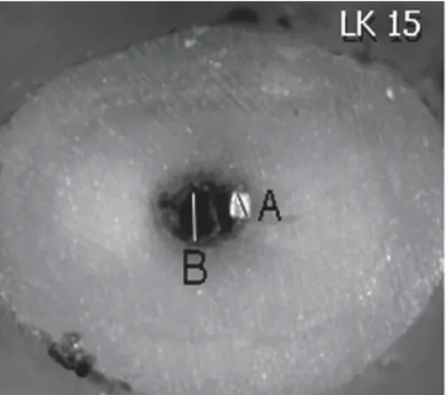
Figure 1. Group 1 (without cervical and middle preflaring). Transverse section at WL. A: instrument diameter; B: canal diameter.
Figure 4. Group 4 (cervical and middle preflaring with LA Axxess burs). Transverse section at WL. A: Instrument diameter; B: canal diameter.
Figure 3. Group 3 (cervical and middle preflaring with Gates-Glidden drills). Transverse section at WL. A: Instrument diameter; B: canal diameter.
Figure 2. Group 2 (cervical and middle preflaring with Orifice Opener instruments). Transverse section at WL: Instrument diameter; B: canal diameter.
Table 1. Discrepancies (mm) between the diameters of the binding files and canals at the working length, for the different groups.
Diameter
Means (±SD) Range 99% CI
No
preflaring 0.1882 (±0.0818) 0.3355-0.0703 0.2548, 0.1216
Gattes
Glidden 0.1074 (±0.0379) 0.1784-0.0453 0.1383, 0.0765
Orifice
Opener 0.0485 (±0.0184) 0.0729-0.0172 0.0635, 0.0335
DISCUSSION
Apical access by cervical flaring has been in-creasingly investigated. This procedure aims to remove cervical interferences from the root canal entrances, which represent an obstacle to free access of endodon-tic instruments to the apical portion of the root canal. Removal of these anatomical interferences enhances canal shaping at the apical third (8,10,11,13). There-fore, preflaring of the coronal and middle root canal thirds has been recommended prior to determining the first file that binds at the WL (7) in order to establish the correct final diameter required for complete apical enlargement.
The findings of this study revealed that when the cervical third was not preflared, the determination of the initial apical file did not reflect the real apical anatomic diameter. The group without cervical preflaring pre-sented the greatest discrepancies between the canal size and the diameter of the file that fit the WL, compared to the other experimental groups.
From all specimens evaluated, the root canals preflared with LA Axxess system presented the least discrepancies between the canal size and the diameter of the first file that adjusted at the WL. This may possibly be attributed to characteristics of the LA Axxess sys-tem, which include the configuration, metal alloy prop-erties and mode of operation. Additionally, the taper (0.06), safe-end and flute design of LA Axxess instru-ments have been shown to yield complete removal of cervical dentin projections without occurrence of canal transportation or perforations (10).
The Gates-Glidden drills provided direct access to both cervical and middle thirds of root canals, reducing the contact area of the instrument in these regions. Nevertheless, these instruments, as well as the Orifice Opener did not allow for accurate determination of the initial apical file. These findings are in agreement with those of previous studies (8-12).
The parameter generally adopted for enlarge-ment of the apical portion at the WL consists of determination of the initial apical file and instrumentation of this region using three file sizes larger than the first file that binds at the WL. The binding sensation is based on the operator’s tactile sensation. However, this has been claimed to be an unreliable and empiric method for accomplishment of such an important step of the biomechanical preparation (8).
In this study, a size 35 K-file represented the diameter of the maxillary lateral incisors. This file size was obtained by determination of the initial apical file after cervical flaring with LA Axxess burs. Kerekes and Tronstad (15) have stated that instrumentation of nar-rowed canals up to sizes 25 or 30 K-files does not provide an accurate apical cleaning. Pécora et al. (10) have postulated that canals of the maxillary central incisors should be enlarged with NiTi files up to a size 45 K-file. These findings are in agreement with those of Wu et al. (16). Although a definite criterion for apical instrumentation in canals has not yet been established, the literature has agreed that the minimal final diameter should correspond to a size 25 K-file (3).
The concept of preparing the canal using three successively larger instruments than the binding file needs to be reviewed, as it is ineffective and may leave uninstrumented canal walls when no preflaring is done. According to the methodology proposed and based on the findings of this study, the following conclusions may be drawn: the instrument binding technique for determining anatomical diameter at WL was not precise; preflaring of the cervical and middle root canal thirds improved the determination of the anatomical diameter; the instrument used for preflaring may play a role in determining the anatomical diameter at the WL; canals preflared with LA Axxess burs presented the least discrepancy between file size and anatomical diameter.
RESUMO
do canal e o diâmetro do instrumento apical inicial foi calculada para cada amostra (em mm). A análise estatística indicou diferença estatisticamente significante entre os grupos experimentais (p=0.01). A maior discrepância foi representada pelo grupo que não recebeu o pré-alargamento (média: 0,1882 mm). O grupo no qual o pré-alargamento foi realizado com instrumentos Orifice Opener também apresentou elevada discrepância entre o diâmetro anatômico e o instrumento apical inicial (média: 0,0485 mm), seguido pelo grupo que se utilizou Gates-Glidden (média: 0,1074 mm). As brocas LA Axxess promoveram a menor diferença entre o diâmetro anatômico no comprimento de trabalho e o instrumento apical inicial (média: 0,0119 mm). Pode-se concluir que o pré-alargamento dos terços cervical e médio permitiu uma melhor determinação do instrumento apical inicial. O grupo no qual foram utilizados instrumentos LA Axxess refletiu com maior precisão o diâmetro anatômico no comprimento de trabalho de incisivos laterais superiores.
REFERENCES
1 . Grossman LI, Oliet S, Del Rio CE. Preparation of the root canal: equipment and technique for cleaning, shaping and irrigation. In: Endodontic Practice. Grossman LI, Oliet S, Del Río CE (Editors). 11th ed. Philadelphia: Lea & Febiger; 1988. p. 179-227.
2 . Ingle JI, Bakland LK, Peters DL, Buchanan LS. Endodontic cavity preparation. In: Endodontics. Ingle JI, Bakland LK (Editors). 5th ed. Malvern: Williams & Wilkins; 1994. p. 92-228.
3 . Torabinejad M. Passive step-back technique. Oral Surg Oral Med Oral Pathol 1994;77:398-401.
4 . Walton RE, Rivera EM. Cleaning and shaping. In: Principles and Practice of Endodontics. Walton RE, Torabinejad M (Editors). 2nd ed. Philadelphia: W.B. Saunders; 1996. p. 201-233.
5 . Leeb J. Canal orifice enlargement as related to biomechanical
preparation. J Endod 1983;9:463-470.
6 . Philippas GG. Influence of occlusal wear and age on forma-tion of dentin and size of pulp chamber. J Dent Res 1961;40:1186-1198.
7 . Contreras MAL, Zinman EH, Kaplan SK. Comparison of the first file that fits at the apex, before and after early flaring. J Endod 2001;27:113-116.
8 . Wu MK, Barkis D, Roris A, Wesselink PR. Does the first file to bind correspond to the diameter of the canal in the apical region? Int Endod J 2002;35:264-267.
9 . Tan BT, Messer H. The effect of instrument type and preflaring on apical file size determination. Int Endod J 2002;35:752-758.
10. Pécora JD, Capelli A, Guerisoli DMZ, Spanó JCE, Estrela C. Influence of cervical preflaring on the apical file size determi-nation. Int Endod J 2005;38:430-436.
11. Barroso JM, Guerisoli DMZ, Capelli A, Saquy, PC, Pécora JD. Influence of cervical preflaring on determination of apical file size in maxillary premolars: SEM analysis. Braz Dent J 2005;16:30-34.
12. Vanni JR, Santos R, Limongi O, Guerisoli DM, Capelli A, Pecora JD. Influence of cervical preflaring on determination of apical file size in maxillary molars: SEM analysis. Braz Dent J 2005;16:181-186.
13. Weiger R, Bartha T, Kalwitzki M, Lost C. A clinical method to determine the optimal apical preparation size. Part I. Oral Surg Oral Med Oral Pathol Oral Radiol Endod 2006;5:686-691.
14. Pécora JD, Capelli, A. Shock of paradigms on the instrumen-tation of curved root canals. Braz Dent J 2006;17:3-5. 15. Kerekes K, Tronstad L. Morphometric observations on the
root canals of human molars. J Endod 1977;3:114-118. 16. Wu MK, Roris A, Barkis D, Wesselink PR. Prevalence and
extent of long oval canals in the apical third. Oral Surg Oral Med Oral Pathol Oral Radiol Endod 2000;89:739-743.