Influence of diltiazem in combination
with a sucrose-rich diet on gingival
alterations in rats
Abstract: The aim of this study was to evaluate the inl uence of diltiazem in combination with a sucrose-rich diet on gingival alterations in rats. One hundred and twenty male Holtzman rats were randomly assigned to 10 groups (n = 12), being 2 control groups treated with saline and 8 test groups treated with diltiazem in daily doses of 5, 25, 50 and 100 mg/kg during 40 or 60 days. Afterwards, the mandibles were removed for mac-roscopic, histologic and histometric analyses of the buccal gingiva of the mandibular right i rst molar. No macroscopic characteristic of gingival overgrowth was observed in any of the groups. The microscopic analy-sis showed characteristics of normality with inl ammatory cells only ad-jacent to the crevicular epithelium in all groups for both periods. The histometric analysis showed signii cant differences only for the epithelial tissue area in the 40-day period (Kruskal-Wallis; P = 0.032). Comparing the periods, signii cant differences regarding the connective and epithe-lial tissue areas were observed only in the group treated with a 25 mg/kg dose (Mann-Whitney; P = 0.004 and P = 0.007, respectively). Oral ad-ministration of diltiazem in combination with a sucrose-rich diet did not induce gingival alterations in rats.
Descriptors: Diltiazem; Sucrose; Gingiva; Rats.
Fernanda Oliveira Bello Corrêa(a)
Lucas Moura Faria(b)
Romeu Belon Fernandes-Filho(c)
Luís Carlos Spolidorio(d)
Silvana Regina Perez Orrico(e)
(a) MSc, PhD Student in Periodontics; (b)DDS; (c)DDS, MSc Student in Periodontics; (e)PhD, Associate Professor of Periodontics – Department of Diagnostics and Surgery, School of Dentistry at Araraquara, São Paulo State University.
(d) PhD, Associate Professor of Oral Pathology, Department of Physiology and Pathology, School of Dentistry at Araraquara, São Paulo State University.
Corresponding author:
Silvana Regina Perez Orrico
Depto. de Diagnóstico e Cirurgia, Faculdade de Odontologia – UNESP
Rua Humaitá, 1680 – Centro Araraquara - SP - Brazil CEP: 14801-903
E-mail: [email protected]
Introduction
Calcium channel blockers have been widely used in the treatment of cardiovascular diseases and may be classii ed into four structural subclasses accord-ing to their chemical and pharmacological char-acteristics: phenylalkylamines (e.g., verapamil), dihydropiridines (e.g., nifedipine, nicardipine, isra-dipine, amloisra-dipine, nitrenisra-dipine, felodipine), benzo-thiazepines (e.g., diltiazem) and T-channel antago-nists (e.g., mibefradil).1
The side-effects of calcium channel blockers in-clude headache, facial redness, dizziness, peripheral edema and gingival overgrowth.2 The development of gingival overgrowth in patients treated with nife-dipine, associated or not with other drugs such as cyclosporin-A, has been extensively reported.2,3 On the other hand, there are few reports of diltiazem-induced gingival overgrowth in humans.4-7 The i nd-ings of a recent study8 showed that in rats with non-inl amed gingival tissue, diltiazem did not induce gingival overgrowth. Nevertheless, the pathogenesis of drug-induced gingival enlargement is uncertain and conl icting and has been related to several fac-tors, namely age, gender, treatment duration, plas-matic and salivary drug concentration, presence of bacterial bioi lm, gingival tissue inl ammation, ge-netics and synergism with other medicaments.9,10
Considering that these factors are important de-terminants of gingival overgrowth, the purpose of this study was to evaluate the inl uence of diltiazem, in different doses and periods, in combination with a sucrose-rich diet on gingival alterations in rats.
Material and Methods
The study protocol was approved by the Ethics in Animal Research Committee, São Paulo State University, Araraquara, SP, Brazil (Process No. 04/2004).
One hundred and twenty male Holtzman rats (Norvegicus albinus) weighing approximately 70 g were randomly assigned to 10 groups of 12 ani-mals each. The room temperature was thermostati-cally regulated to 22ºC ± 1ºC and the humidity was maintained at 60% ± 5%. The time of exposure to light was automatically controlled (12h30min/day).
Two groups were used as control and received
saline (JP Indústria Farmacêutica, Ribeirão Preto, SP, Brazil) orally during the experimental periods. The other 8 groups received diltiazem (Alcon Biosci-ences Pvt. Ltd., Munbai, India) therapy. Diltiazem was administered orally in daily doses of 5, 25, 50 or 100 mg/kg body weight during 40 or 60 days, us-ing a hypodermic needle and an injector device as-sembled manually.
The animals were fed daily a sucrose-rich diet composed of 56% rei ned sugar, 30% integral pow-dered milk and 14% standard ground chow,11,12 which were weighed in an electronic balance and mixed manually. The food was prepared daily by adding a small amount of water to obtain a pasty consistence.2,13 The goal of this diet was to prevent self cleaning during mastication and facilitate bac-terial bioi lm accumulation in order to induce an inl ammatory process.14 During the experimental period the body weight gain of all animals was eval-uated. Afterwards, the animals were sacrii ced and the mandibles were carefully removed together with the gingival tissue surrounding the teeth and stored in 10% buffered formalin solution (Synth, Diadema, SP, Brazil).
Macroscopic analysis
The presence of characteristics of gingival over-growth such as volume increase of the gingival margin and interdental papilla and clinical inl am-matory alterations, namely alterations in gingival contouring, color and brightness, were examined by a single calibrated blind examiner using a magnify-ing glass.
Histological analysis
Microscopic analysis
The microscopic analysis was performed on the buccal gingiva of the mandibular right i rst molar by a single calibrated blind examiner using a light transmission microscope BX51 (Olympus, Melville, NY, USA). Cell adhesion, number of cell layers and presence/absence of deep papillary interdigitations were examined in the epithelial tissue whereas the morphology of collagen i bers and the number of cells and blood vessels were examined in the con-nective tissue.
The inl ammatory response was evaluated ac-cording to a score scale:
presence of inl ammatory cells only adjacent to the epithelium;
presence of inl ammatory cells throughout the connective tissue;
presence of inl ammatory cells close to the alveo-lar bone.
Histometric analysis
For the histometric analysis, four glass micro-scope slides were examined per animal, using a previously described technique.8 Briel y, the slides were selected in a standardized manner, represent-ing the initial, intermediate and i nal portions of the gingival margin, thus totalizing 10 readings of his-tological sections at 60-μm intervals for each ani-mal. The histometric analysis was performed using a DIASTAR optical microscope (Leica Reichert & Jung Products, Wetzlar, Germany) with an objective lens 10/0.25 adapted to a video camera (Sony DXC - 107A, Sony Electronics Inc., Shinagawa-ku, Tokyo, Japan) and connected to a computer. Gingival epi-thelium and connective tissue areas were measured using image-analysis software (Sigma Scan®; Mo-cha; Jandel Scientii c, San Rafael, CA, USA). The lower limit of the connective and epithelial tissue areas measured with the software was the end of the junctional epithelium. Statistical analysis was based on the average obtained for each animal.
Statistical analysis
Statistical analysis of the histometric data was performed by the Mann-Whitney test using dose as a factor and by the Kruskal-Wallis test using admin-1.
2.
3.
istration period as a factor. The signii cance level was set at 5%.
Results
Macroscopic and microscopic data
All rats survived the experimental periods. Com-pared to the control animals, no animal treated with diltiazem had its body weight gain affected by any of the diltiazem doses for either the 40-day (P = 0.271) or the 60-day (P = 0.186) administration periods.
No macroscopic characteristic of gingival over-growth was observed in any of the groups, either con-trol or experimental, for both evaluations periods.
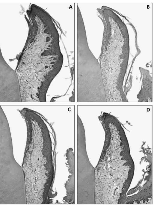
In addition, no microscopic alteration suggestive of gingival overgrowth was observed, regardless of the treatment protocol. In all groups, the epithelium had 3-5 cell layers without hyperplasia or deep pap-illary interdigitations. The connective tissue had a normal number of collagen i bers and blood vessels and few macrophages (Figure 1).
The application of the score scale to evaluate the inl ammatory response showed the presence of inl ammatory cells only adjacent to the crevicular epithelium (score 1) in all groups for both evalua-tion periods.
Histometric analysis
Table 1 shows the results of the Kruskal-Wallis test used to evaluate the effect of the different doses (5, 25, 50 and 100 mg/kg) of diltiazem on the con-nective and epithelial tissue areas of the marginal gingiva of the buccal face of mandibular right i rst molar. No statistically signii cant difference was ob-served in the connective tissue area for both peri-ods [40 days (P = 0.128) and 60 days (P = 0.278)]. Regarding the epithelial tissue area, there was signii cant difference only for the 40-day period (P = 0.032). The application of the multiple-compar-ison test (Dunn’s Method) did not show statistically signii cant difference between the comparisons. However, the results of the groups in which 25 mg/ kg and 100 mg/kg were administered were close to signii cance when compared to the control groups.
Figure 1 - Buccolingual sections of the buccal region of the first mandibular molar from the control groups after the 40- and 60-day periods (1A and 1C, respectively) and test groups, treated with 100 mg/kg of diltiazem, after the same periods (1B and 1D, respectively). No alteration in the gingival tissues was observed (Hematoxylin and Eosin stain, magnification x40).
D C
B A
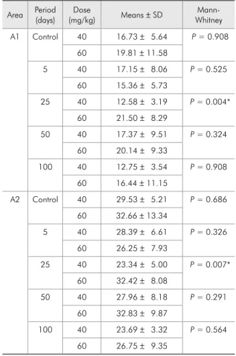
and epithelial tissue areas of the marginal gingiva of the buccal surface of mandibular right i rst molar. Statistically signii cant differences for the connec-tive and epithelial tissue areas between the 40- and 60-day periods were observed only in the group that received a 25 mg/kg dose (P = 0.004 and P = 0.007, respectively). For the other doses, the experimental period did not inl uence the induction of gingival al-teration in the animals treated with diltiazem.
Discussion
The hypothesis investigated in the present study
was raised from our recent i ndings, which showed that the treatment with increasing doses of diltia-zem in rats did not induce gingival alterations.8 At the same time, it has been suggested that drug-in-duced gingival overgrowth is related to factors such as treatment duration, presence of bacterial bioi lm and gingival inl ammation.9,10
Therefore, our hypothesis was to explore in rats treated with diltiazem whether gingival inl amma-tion, secondary to a sucrose-rich diet, would induce gingival alterations.
been widely employed to investigate the effect of drugs on the oral environment because it provides more uniform responses compared to humans and allows better control of several important variables including genetic predisposition, gender, age, dose and duration of drug administration.15
The diltiazem-treated rats did not present clinical evidence of toxicity that compromised their weight gain or survival. In fact, the i ndings of previous in-vestigations have shown that the administration of diltiazem in the doses used in the present study did not induce toxicity and that the treatment was well supported by the animals.8,13,16,17
Macroscopically, the sucrose-rich diet induced dental bioi lm accumulations in all animals though
none of them exhibited clinical characteristics of gingival overgrowth, which coni rms our previous i ndings.8 However, Morisaki et al.16 (2000) ob-served gingival overgrowth in rats after administra-tion of diltiazem in doses greater than those used in the present study during a similar experimental period. Microscopically, there was only a mild in-l ammation in the animain-ls (Figure 1), which is par-ticularly relevant because the diet followed by the animals was expected to cause gingivitis. Based on previous studies11,12,18 that described the sucrose-rich diet as a model to induce periodontal disease (gingivitis and periodontitis), we aimed to promote natural accumulation of supragingival plaque on the dentogingival area and consequently experimental
Table 1 - Histometric analysis of the influence of different administration doses of diltiazem on the connective tissue (A1) and epithelial tissue (A2) areas (mm²) of the buccal gin-giva of the mandibular right first molar of rats.
Area Period (days)
Dose
(mg/kg) Means ± SD
Kruskal-Wallis
A1 40 Control 16.73 ± 5.64 P = 0.128 5 17.15 ± 8.06
25 12.58 ± 3.19
50 17.37 ± 9.51
100 12.75 ± 3.54
A2 40 Control 29.53 ± 5.21 P = 0.032* 5 28.39 ± 6.61
25 23.34 ± 5.00
50 27.96 ± 8.18
100 23.69 ± 3.32
A1 60 Control 19.81 ± 11.58 P = 0.278 5 15.36 ± 5.73
25 21.50 ± 8.29
50 20.14 ± 9.33
100 16.44 ± 11.15
A2 60 Control 32.66 ± 13.34 P = 0.163 5 26.25 ± 7.93
25 32.42 ± 8.08
50 32.83 ± 9.87
100 26.75 ± 9.35
*Statistically significant difference.
Table 2 - Histometric analysis of the influence of different administration periods of diltiazem on the connective tissue (A1) and epithelial tissue (A2) areas (mm²) of the buccal gin-giva of the mandibular right first molar of rats.
Area Period (days)
Dose
(mg/kg) Means ± SD
Mann-Whitney
A1 Control 40 16.73 ± 5.64 P = 0.908 60 19.81 ± 11.58
5 40 17.15 ± 8.06 P = 0.525 60 15.36 ± 5.73
25 40 12.58 ± 3.19 P = 0.004* 60 21.50 ± 8.29
50 40 17.37 ± 9.51 P = 0.324 60 20.14 ± 9.33
100 40 12.75 ± 3.54 P = 0.908 60 16.44 ± 11.15
A2 Control 40 29.53 ± 5.21 P = 0.686 60 32.66 ± 13.34
5 40 28.39 ± 6.61 P = 0.326 60 26.25 ± 7.93
25 40 23.34 ± 5.00 P = 0.007* 60 32.42 ± 8.08
50 40 27.96 ± 8.18 P = 0.291 60 32.83 ± 9.87
100 40 23.69 ± 3.32 P = 0.564 60 26.75 ± 9.35
gingivitis. However, our result was similar to that of Galvão et al.19 (2003), who reported that a sucrose-rich diet was not capable of promoting more inl am-mation than that seen in rats treated with standard rat lab chow.
Another possible explanation for our results would be the inl uence of diltiazem on the bio-chemical composition of saliva. Dehpour et al.20 (1995) evaluated the effects of calcium channel blockers on the functions of the parotid and sub-mandibular glands of rats. They reported that the administration of 10 mg/kg of diltiazem decreased signii cantly the l ow and the concentration of cal-cium (both glands) and amylase (parotid gland) in the saliva of these animals. Moreover, Rudiger et al.21 (2002) have demonstrated that there is a great-er concentration of salivary amylase in the pres-ence of gingival inl ammation caused by a greater bioi lm accumulation. They have also shown that the concentration of proteins present in saliva may modify the bacterial adhesion and the initial com-position of dental bioi lm. Given the alterations relative to modii cation of the amount of salivary amylase that might be triggered by diltiazem, it may be speculated that there was less formation of dental bioi lm or an alteration of its composition in the present study.
In humans, the presence of inl ammation in the pathogenesis of gingival growth is debatable. Miranda et al.7 (2005) observed that individuals in therapy with diltiazem or nifedipine presented greater gingival enlargement when the use of these drugs was associated with the presence of gingivitis. On the other hand, Morisaki et al.17 (1993) reported that nifedipine induced gingival overgrowth in rats with or without gingival inl ammation and/or den-tal bioi lm, these factors being able to potentialize the effect of the drug.
In the present study, the histometric analysis showed no statistically signii cant difference among the groups for most part of the sample. Only the group that received a dose of 25 mg/kg of diltiazem presented a signii cant increase of the connective and epithelial tissue areas when comparing the 40- and 60-day periods (Table 2). Since this i nding was
not observed when the higher doses were adminis-tered, there is reason to believe that it might be a casual i nding.
The results of this study showed that differ-ent doses of diltiazem did not inl uence the devel-opment of gingival overgrowth (Table 1). Correa et al.8 (2005) had similar results, although using lower doses of diltiazem. Another study16 reported gingival overgrowth in rats after administration of diltiazem in doses > 1,000 mg/kg body weight for 40 days, which is much greater than the maximum dose used in our study (100 mg/kg). The rationale for choosing these doses of diltiazem was to simu-late as close as possible the doses used in humans based on the fact that, in rats, 5 mg/kg correspond to the average daily intake of a hypertensive adult patient.
Another factor taken into account was the ad-ministration route. In our previous study,8 the drug was administered subcutaneously while in the pres-ent investigation the drug was given orally. The rationale was to simulate the administration route of diltiazem in humans. Nevertheless, this factor was proved not to have a signii cant inl uence on the outcomes, given that the results of the present study were similar to those reported by Correa et al.8 (2005).
Most studies that evaluated diltiazem-induced gingival enlargement in humans4-7 had to deal with a heterogenous sample regarding age, duration of drug use, dose and other variables. Therefore, fur-ther research using animal models should be under-taken to evaluate the behavior of gingival tissue af-ter long-af-term use of diltiazem, in order to evaluate other mechanisms that might be involved in gingival overgrowth, not only gingival inl ammation.
Conclusion
References
1. Cummins DF. Newer calcium channel antagonists and the treatment of hypertension. Expert Opin Investig Drugs. 1999;8(7):1031-42.
2. Seymour RA. Calcium channel blockers and gingival over-growth. Br Dent J. 1991;170(10):376-9.
3. James JA, Marley JJ, Jamal S, Campbell BA, Short CD, John-son RW et al. The calcium channel blocker used with cyclospo-rin has an effect on gingival overgrowth. J Clin Periodontol. 2000;27(2):109-15.
4. Bullon P, Machuca G, Armas JR, Rojas JL, Jimenez G. The gingival inflammatory infiltrate in cardiac patients treated with calcium antagonists. J Clin Periodontol. 2001;28(10):897-903.
5. Bullon P, Pugnaloni A, Gallardo I, Machuca G, Hevia A, Battino M. Ultrastructure of the gingiva in cardiac patients treated with or without calcium channel blockers. J Clin Peri-odontol. 2003;30(8):682-90.
6. Ellis JS, Seymour RA, Steele JG, Robertson P, Butles TJ, Thomason JM. Prevalence of gingival overgrowth induced by calcium channel blockers: a community based study. J Periodontol. 1999;70(1):63-7.
7. Miranda J, Brunet L, Roset P, Berini L, Farre M, Mendieta C. Prevalence and risk of gingival overgrowth in patients treated with diltiazem or verapamil. J Clin Periodontol. 2005;32(3):294-8.
8. Correa FOB, Giro G, Gonçalves D, Spolidorio LC, Orrico SRP. Diltiazem did not induce gingival overgrowth in rats: a clinical, histological and histometric analysis. Braz Oral Res. 2005;19(3):163-8.
9. Nishikawa S, Nagata T, Morisaki I, Oka T, Ishida H. Patho-genesis of drug-induced gingival overgrowth. A review of studies in the rat model. J Periodontol. 1996;67(5):463-71. 10. Seymour RA, Ellis JS, Thomason JN. Risk factors for
drug-induced gingival overgrowth. J Clin Periodontol. 2000;27(4):217-23.
11. Offenbacher S, Jared HL, O’Reilly PG, Wells SR, Salvi GE, Lawrence HP et al. Potential pathogenic mechanisms of
peri-odontitis-associated pregnancy complications. Ann Periodon-tol. 1998;3(1):233-50.
12. Pilatti GL, Sampaio JEC. The influence of chlorhexidine on the severity of cyclosporine A-induced gingival overgrowth. J Periodontol. 1997;68(9):900-4.
13. Ishida H, Kondoh T, Kataoka M, Nishikawa T, Morisaki I, Kido J et al. Factors influencing nifedipine-induced gingival overgrowth in rats. J Periodontol. 1995;66(5):345-50. 14. Adachi C, Kitamura K, Kato K, Yoshida M, Morisaki I,
So-bue S. [Cyclosporin-A induced gingival overgrowth – strain differences in the rats] [Article in Japanese]. Shoni Shikagaku Zasshi. 1991;29(1):24-31.
15. McKevitt KMB, Irwin CR. Phenotypic differences in growth, matrix synthesis and response to nifedipine between fibro-blasts derived from clinically healthy and overgrown gingival tissue. J Oral Pathol Med. 1995;24(2);66-71.
16. Morisaki I, Fukui N, Fujimori Y, Murakami J, Daikoku H, Amano A. Effects of combined oral treatments with cyclospo-rine A and nifedipine or diltiazem on drug-induced gingival overgrowth in rats. J Periodontol. 2000;71(3):438-43. 17. Morisaki I, Kato K, Loyola-Rodriguez JP, Nagata T, Ishida
H. Nifedipine-induced gingival overgrowth in the presence or absence of gingival inflammation in rats. J Periodontal Res. 1993;28(6):396-403.
18. Hemmerle J, Frank RM. Short-term effect of cyclosporine injections on experimental gingival inflammation in the rat. J Periodontol. 1993:64(1):24-8.
19. Galvão MP, Chapper A, Rosing CK, Ferreira MB, de Souza MA. Methodological considerations on descriptive studies of induced periodontal diseases in rats. Pesqui Odontol Bras. 2003;17(1):56-62.
20. Dehpour AR, Ghafourifar P, Massoudi S, Abdollahi M, Mousavizadeh K. On the relation of calcium channel block-ers to rat parotid and submandibular glands function in vivo.
Gen Pharmacol. 1995;26(3):619-22.