ABSTRACT: Anti-inlammatory agents have been reported to regulate bone healing. The aim of this study was to in -vestigate the effect of a selective cyclooxygenase-2 inhibitor (meloxicam) on bone healing in calvarial defects in rats. Thirty-six adult male Wistar rats were included. After anesthesia, a linear incision was made through the skin of the scalp, a full-thickness lap was relected and a 4 mm round defect was made with a trephine drill. The animals were randomly assigned to one of the following 4 treatment groups (9 animals each), including daily subcutaneous injections: A: saline solution for 15 days; B: saline solution for 45 days; C: 3 mg/kg of meloxicam for 15 days and D: 3 mg/kg of meloxicam for 45 days. The animals were sacriiced and the specimens, routinely processed. The bone illing was histometrically measured and statistical analysis, performed. Intergroup comparisons demonstrat -ed that the meloxicam groups present-ed a signiicant r-eduction in bone healing when compar-ed to their respective controls (group A, 44.5 ± 5.75%, against group C, 57.5 ± 7.25%, p < 0.05; group B, 40.25 ± 13.75%, against group D, 52.25 ± 17.25%). Within the limits of the present study, it can be concluded that selective cyclooxygenase-2 inhibitors may reduce bone healing in calvarial defects in rats after continuous administration.
DESCRIPTORS: Anti-inlammatory agents, non-steroidal; Cyclooxygenase inhibitors; Wound healing; Rats.
RESUMO: Agentes antiinlamatórios têm sido descritos como reguladores do reparo ósseo. O objetivo deste estu -do foi investigar o efeito de um inibi-dor seletivo da cicloxigenase-2 (meloxicam) no reparo ósseo em defeitos de calvárias de ratos. Trinta e seis ratos machos Wistar foram incluídos no estudo. Após anestesia, incisão linear e rebatimento de retalho de espessura total, um defeito de 4 mm de diâmetro foi criado na calvária dos animais com broca treina. Os mesmos foram aleatoriamente distribuídos em um dos 4 grupos de tratamento (9 animais por grupo), recebendo injeções subcutâneas diárias de: A: soro isiológico por 15 dias; B: soro isiológico por 45 dias; C: 3 mg/kg de meloxicam por 15 dias; D: 3 mg/kg de meloxicam por 45 dias. Os animais foram sacriicados e os espécimes rotineiramente processados. Medidas do preenchimento ósseo foram histometricamente realizadas e analisadas estatisticamente. Comparações intergrupos demonstraram que os grupos com meloxicam apresenta-ram uma redução signiicativa no reparo ósseo quando comparados com os respectivos grupos-controle (grupo A, 44,5 ± 5,75%, contra grupo C, 57,5 ± 7,25%, p < 0,05; grupo B, 40,25 ± 13,75%, contra grupo D, 52,25 ± 17,25%). Dentro dos limites do presente estudo, pode ser concluído que os inibidores seletivos da cicloxigenase-2 podem reduzir o reparo ósseo em defeitos em calvárias de ratos após sua administração contínua.
DESCRITORES: Antiinlamatórios não esteróides; Inibidores de ciclooxigenase; Cicatrização de feridas; Ratos.
INTRODUCTION
The effect of anti-inflammatory drugs on bone healing has been evaluated in several studies1,3,11.
Non-steroidal anti-inflammatory drugs (NSAIDS) interfere with aracdonic acid metabolism by block-ing prostaglandin synthesis through
cyclooxygen-ase pathway inhibition, which has a fundamental role in bone healing22,24.
Selective COX-2 inhibitors have emerged with
the objective of reducing stomach and renal toxic -ity. Additionally, they can also promote effects on
* PhD Student, Department of Prosthodontics and Periodontics, Division of Periodontics; **Graduate Student, Department of Prosthodontics and Periodontics, Division of Periodontics; ***PhD Student, Department of Histology, Division of Morphology; ****Professors, Department of Prosthodontics and Periodontics, Division of Periodontics; *****Professor and Chairman, Depart-ment of Prosthodontics and Periodontics, Division of Periodontics; ******Visiting Professor, DepartDepart-ment of Prosthodontics and Periodontics, Division of Periodontics; *******Assistant Professor, Department of Prosthodontics and Periodontics, Division of Periodontics – School of Dentistry at Piracicaba, State University of Campinas.
Selective COX-2 inhibitor reduces bone healing in bone defects
Inibidor seletivo de COX-2 reduz reparo em defeitos ósseos
Bruno César de Vasconcelos Gurgel* Fernanda Vieira Ribeiro**
Marco Antônio Dias da Silva*** Francisco Humberto Nociti Júnior**** Antonio Wilson Sallum****
Enilson Antônio Sallum***** Sérgio de Toledo******
bone healing inhibiting prostaglandins23. It has
been shown that the COX-2 enzyme participates in early phases of osteogenesis and that it is more related to osteoblast maturation in later stages20.
COX-2 inhibitors reduce the osteoblastogenesis process and alter genes activities responsible for osteoblastic differentiation19,23.
In order to clarify the role of NSAIDS on bone healing after surgical procedures, the purpose of the present study was to evaluate the influence of meloxicam, a selective COX-2 inhibitor, on bone healing in calvarial defects in rats.
MATERIALS AND METHODS
AnimalsThirty-six male Wistar rats (250-350 g) were included in the study. The animals were kept in plastic cages with access to food and water ad libi-tum. Prior to surgical creation of the defects, all an-imals were allowed to acclimatize to the laboratory environment for a period of 7 days. The protocol was approved by the State University of Campinas Institutional Animal Care and Use Committee.
Experimental design
General anesthesia was obtained by intra-muscular administration of ketamine (0.5 ml/kg) (Dopalen, Agribrands Brasil Ltda., Paulínia, SP, Brazil). After cranial dorsal tricotomiae, the surgi-cal site was scrubbed with iodine (Geyer Medica-mentos S.A., Porto Alegre, RS, Brazil) and a 15 mm mid-sagittal linear incision was made through the skin of the scalp. A full-thickness flap, including periosteum, was then reflected, exposing the cal-varial bone. After dissection and bone exposure, a standardized, circular defect with 4 mm in diameter was made in one side of the parietal bone with the use of a trephine drill (3i Implants Innovations do Brasil Ltda., São Paulo, SP, Brazil) and abundant ir-rigation with sterile saline solution (Fresenius Kabi Brasil Ltda., Campinas, SP, Brazil), exposing the dura mater membrane. The soft tissues and perios-teum were then repositioned for total coverage and sutured with 4.0 mono-nylon suture (Shalon, B.A. da B. Vista, S.L.M. Belos, GO, Brazil). The animals were then randomly assigned to one of 4 groups (9
animals/group) that were subjected to daily subcu
-taneous injections. Groups A and B received con -tinuous administration of 1 ml/kg of saline solution for 15 and 45 days, respectively; and groups C and D received continuous administration of 3 mg/kg of meloxicam (Movatec - Boehringer Ingelheim do
Brasil Química e Farmacêutica Ltda., Itapecerica da Serra, SP, Brazil) for 15 and 45 days, respectively.
The animals were sacrificed by deep anesthe-sia, and the calvarial bones, including the surgical sites, were removed and fixed in 4% neutral formalin (Dinâmica Reagentes Analíticos, São Paulo, SP, Brazil) for 48 hours. The specimens were deminer-alized in a solution with equal parts of 50% formic acid (Dinâmica Reagentes Analíticos, São Paulo, SP, Brazil) and 20% sodium citrate (Chemco In-dústria e Comércio Ltda., Campinas, SP, Brazil) for 40 days. Paraffin semi-serial sections (6 µm) were obtained in an anteroposterior direction and stained with hematoxilin and eosin (Dinâmica Reagentes Analíticos, São Paulo, SP, Brazil). Twenty sections were uniformly selected from the beginning to the end of the defect and analyzed with a light micro-scope (Zeiss, Jena, Germany) under a 2.5 X magnifi-cation. Using an image analysis system(Image-Pro; Media Cybernetics, Silver Spring, MD, USA), linear measurements of the healing remaining defect were calculated from one side to the other side of the bone by the same examiner. A blind examiner was trained and calibrated in performing the measurements.
Measurements of the twenty sections were averaged to allow intra- and intergroup analysis related to time. Comparisons between groups were made by Bifactorial Variance Analysis with a sig-nificance level of 5%.
RESULTS
Histologic analysis
Two weeks postsurgically, the presence of a thin connective tissue membrane was observed over the defect sites in both control and treated samples. The connective tissue layer of test animals presented an advanced process of angiogenesis, and a decreased number of hemorrhagic and inflammatory sites, when compared to the samples of the control ani-mals. However, new bone formation, originated from the margins and/or as bone isles throughout the defect, was greater in the control groups than in the test groups (p < 0.05). In addition, cubical or cylindri-cal osteoblasts were observed in the control groups (Figure 1) while in the test group, osteoblasts were squamous (surface or bone lining cells) (Figure 2).
formed bone in the control groups, whereas the test groups presented a pattern similar to that of the 15 days observations, marked by squamous osteoblasts originating small amounts of osteoid matrix and reduced bone formation.
Histomorphometric analysis
Data from linear measurements of remaining osseous defect were subtracted from the original defect (4 mm) and mean percentages and standard deviations were also subtracted from the original size defect (Table 1) (Figura 3).
Statistically significant differences (p < 0.05) were observed between group A and C and between B and D. However, no significant difference was observed between experimental periods (A to B and C to D).
DISCUSSION
The present study demonstrates, histologi-cally, that COX-2 inhibitors may significantly decrease the bone healing in calvarial defects in rats. This is in accordance with other studies that have suggested that NSAIDS also inhibit bone
re-pair and bone formation12,21. This fact could be
explained by prostaglandin synthesis inhibition, due to the blockade of cyclooxygenase, which has an important role in osteogenesis and bone heal-ing, such as endochondral and intramembranous ossification21,23.
Sato et al.20 (1997) showed that COX-2 and
COX-1 have a fundamental role in early phases of osteogenesis. In addition, COX-2, in later stages, is more related to osteoblast maturation. Zhang
et al.23 (2002) demonstrated that COX-2 regulates
genes, such as cbfa1 and osterix, which are re-quired for osteoblast differentiation and bone for-mation. In this process, COX-1 does not have a critical role. Decreases in the expression of these genes may contribute to incomplete bone repair19,
as observed in the present study. Therefore, since COX-2 regulates genes activities in osteoblastic dif-ferentiation, a reduction in the osteoblastogenesis
TABLE 1 - Mean percentages and standard deviations of linear measurements from the remaining defect of the control and test groups at 15 and 45 days.
Period (days) Control Group Test Group 15 (A-C) 44.5 ± 5.75% a 57.5 ± 7.25% b
45 (B-D) 40.25 ± 13.75% a 52.25 ± 17.25% b
Means followed by different letters differ statistically by Bifacto-rial Variance Analysis (p < 0.05).
FIGURE 2 - In the test groups, osteoblasts were
squa-mous (bone lining cells). Magniication 50 X. H. E.
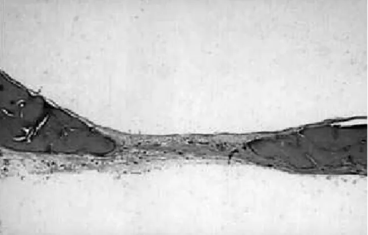
FIGURE 3 - Healing remaining defect between bone
margins. Original magniication 2.5 X. H. E. FIGURE 1 - In control groups, osteoblasts surfacing the
bone were cubical or cylindrical. Magniication 50 X.
process could be the main hypothesis to explain the remaining bone defects in the test groups ob-served in the present study.
Additionally, studies have shown that pros-taglandins may increase fracture repair rates in experimental models, improving bone healing14
and also stimulating bone formation22,24. Zhang
et al.23 (2002)has suggested that prostaglandins
also participate in osteoblast recruitment and dif-ferentiation, and induce cbfa-1 and osterix gene expression.
Previous studies have shown that COX-2 spe-cific inhibitors may not have any effect on frac-ture healing in animal models5 and do not inhibit
bone fusion following spinal arthrodesis16. On the
other hand, studies also demonstrate that COX-2 inhibitors can present radiographic,histological, morphological, and biomechanical evidence of de-layed fracture healing at four weeks12,21, even when
administered before injury11,21.
As observed in the present study, the amount of bridging boneformation is reduced in the test groups (p < 0.05), as compared to the control group, at 2 weeks. Although intragroup analysis did not show any significant difference in bone healing, a numerical difference could be observed favoring the latter control group.
In this study, the animals were treated for 15 or 45 days. Although studies have suggested that bone healing is increased during the first 15 days7,8, the present study investigated if a longer
period (45 days) of bone repair could reduce the remaining calvarial defect more than 15 days, even in the control group.
The apparent stabilization of the healing process observed in the present study at 45 days postoperatively could be the result of using a criti-cal size defect. The regenerative capacity of the bone tissue can be limited by the size of the de-fect, where large bone defects cannot regenerate spontaneously6. The critical size defect depends on
animal species, age, size and place4. In calvaria of
rats, this size can vary from 2 mm18 to 8 mm15. In
the present study, a defect of 4 mm was not able to regenerate spontaneously, as shown in the lat-ter control group.
Probably, the apparent high quantity of prosta-glandins encountered in the early phase of healing, as observed in previous studies7,23, might justify
the results of the present study, where differences in defect closure were not noticed between 15 and 45 days. This observation suggests that the high peak of osteogenesis activity may occur during the first 15 days. Dekel et al.7 (1981) showed that high
prostaglandin levels (PGE and PGF) were increased between the 3rd and 14th days of repair in fractures
of rat tibiae. According to the same authors, the mRNA expression of COX-2 was also higher dur-ing the first 14 days, demonstratdur-ing the role of COX and its metabolites during the early phases of healing. Gerstenfeld et al.9 (2003) also showed
that COX-1 mRNA levels were constant during the first 21 days, while COX-2 mRNA levels presented peaks of elevation during the first 14 days of heal-ing, and returned to basal levels after 21 days. These findings indicate that in the first two weeks of bone repair, the osteogenic activity is more in-tense because of the high rate of COX-2 transcrip-tion and, consequently, high levels of prostaglan-dins found in this period, since these metabolites stimulate bone formation22,24. These findings are in
accordance with those of the present study, where increased bone healing was observed at 15 days compared to 45 days in non-treated groups, even without significant differences (p > 0.05).
The histological analysis showed that, during the early phases, the control group presented cu-boidal and cylindrical osteoblasts, characterizing a high activity of osteoid production (non calci-fied matrix). These findings could not be found in treated groups, where most of the osteoblasts were squamous, which demonstrates lower synthesis of osseous matrix. According to Marks et al.17 (1996),
actively secreting mature osteoblasts are, histo-logically, cuboidal or tall cells. When they start to secrete bone, the secretion is polarized towards the bone surface. This could be explained by the role of both COX-1 and COX-2 enzymes during the early phases of osteogenesis20. In addition, COX-2,
selectively inhibited by meloxicam, may also be as-sociated with osteoblast maturation in later stages, which explains the morphological distinction and, consequently, the difference in activity observed in the osteoblasts on new bone surface (bone lining cells), when compared to the control group. This histological evidence, associated with the lack of hemorrhagic features, few inflammatory cells and normal tissue vascularization, characterize ad-vanced bone and tissue healing processes2.
Ho et al.13 (1995) demonstrated that NSAIDS
effects are dose-dependent and that, after with-drawal, their effects may be reverted1. Apparently,
the use of NSAIDS seems to delay the healing proc-ess and their use should be avoided immediately postoperatively or should only be used during a short-term administration8, because early events
analy-ses in animal models and humans are required to clarify the mechanisms of the effect of nonste-roidal anti-inflammatory drugs such as COX-2 inhibitors, as well as the role of prostaglandins and cyclooxygenases in bone healing. As clinical stud-ies are limited, as are retrospectives3,10, additional
information about dosage and time of administra-tion also need investigaadministra-tion in the future due to concerns emerging in the literature.
CONCLUSION
Within the limits of the present study, it can be concluded that meloxicam, a selective
cyclo-oxygenase-2 inhibitor, may reduce bone healing in calvarial defects in rats after continuous ad-ministration.
ACKNOWLEDGMENTS
This study was supported by Fundação de Am-paro à Pesquisa do Estado de São Paulo – FAPESP, process 02/13416-6. We would also like to thank the histological technician, Mariana Piovezzan Fug-golin, who contributed to this study, and Gláucia Maria Bovi Ambrosano for the statistical analysis support.
REFERENCES
1. Altman RD, Latta LL, Keer R, Renfree K, Hornicek FJ, Banovac K. Effect of nonsteroidal antiinflammatory drugs on fracture healing: a laboratory study in rats. J Orthop Trauma 1995;9(5):392-400.
2. Applegate EJ. The Anatomy and Physiology Learning System: Textbook. W.B. Philadelphia: Saunders Company; 1995. 3. Bichara J, Greenwell H, Drisko C, Wittwer JW, Vest TM,
Yancey J et al. The effect of postsurgical Naproxen and a bioabsorbable membrane on osseous healing in intrabony defects. J Periodontol 1999;70(8):869-77.
4. Bosch C, Melsen B, Vargervik K. Importance of the Criti-cal-Size bone defect in testing bone regenerating material. J Craniofac Surg 1998;9(4):310-6.
5. Brown KM, Saunders MM, Kirsch T, Donahue HJ, Reid JS. Effect of COX-2-specific inhibition on fracture-healing in the rat femur. J Bone Joint Surg Am 2004;86-A(1):116-23. 6. Cormack DH. Ham - Histologia. 9ª ed. Rio de Janeiro:
Guanabara Koogan; 1991.
7. Dekel S, Lenthall G, Francis MJ. Release of prostaglandins from bone and muscle after tibiae fracture. An experimental study in rabbits. J Bone Joint Surg Br 1981;63-B(2):185-9. 8. Gerstenfeld LC, Einhorn TA. COX inhibitors and their effects
on bone healing. Expert Opin Drug Saf 2004;3(2):131-6. 9. Gerstenfeld LC, Thiede M, Seibert K, Mielke C, Phippard D,
Svagr B et al. Differential inhibition of fracture healing by non-selective and cyclooxygenase-2 selective non-steroidal anti-inflammatory drugs. J Orthop Res 2003;21(4):670-5. 10. Giannoudis PV, Macdonald D, Matthews SJ, Smith
RM, Furlong J, De Bôer P. Nonunion of the femoral diaphy-sis. The influence of reaming and non-steroidal anti-inflam-matory drugs. J Bone Joint Surg Br 2000;83(2):308. 11. Giordano V, Giordano M, Knackfuss IG, Apfel MI,
Gomes RD. Effect of tenoxicam on fracture healing in rat tibiae. Injury 2003;34(2):85-94.
12. Goodman S, Ma T, Trindade M, Ikenoue T, Matsuura I, Wong N et al. COX-2 selective NSAID decreases bone ingrowth in vivo. J Orthop Res 2002;20(6):1164-9. 13. Ho ML, Chang JK, Wang GJ. Antiinflammatory drug
effects on bone repair and remodeling in rabbits. Clin Or-thop Relat Res 1995;(313):270-8.
14. Keller J. Effects of indomethacin and local prosta-glandin E2 on fracture healing in rabbits. Dan Med Bull 1996;43(4):317-29.
15. Kleinschmidt JC. The critical size defects as an ex-perimental model to test bone repair materials. J Craniofac Surg 1990;1(1):60-8.
16. Long J, Lewis S, Kuklo T, Zhu Y, Riew KD. The effect of cyclooxygenase-2 inhibitors on spinal fusion [abstract]. J Bone Joint Surg Am 2002;84:1763-8.
17. Marks SC Jr, Cielinski MJ, Sundquist KT. Bone sur-face morphology reflects local skeletal metabolism. Microsc Res Tech 1996;33(2):121-7.
18. Mulliken JB, Glowacki J. Induced osteogenesis for repair and construction in the craniofacial region. Plast Reconstr Surg 1980;65(5):553-60.
19. Nakashima K, Zhou X, Kunkel G, Zhang Z, Deng JM, Behringer RR et al. The novel zinc finger-containing transcription factor osterix is required for osteoblast dif-ferentiation and bone formation. Cell 2002;108:17-29. 20. Sato Y, Arai N, Negishi A, Ohya K. Expression of
cy-clooxygenase genes and involvement of endogenous pros-taglandin during osteogenesis in the rat tibial bone marrow cavity. J Med Dent Sci 1997;44(4):81-92.
21. Simon AM, Manigrasso MB, O’ Connor JP. Cyclooxy-genase 2 function is essential for bone fracture healing. J Bone Miner Res 2002;17(6):977-8.
22. Suponitzky I, Weinreb M. Differential effects of sys-temic prostaglandin E2 on bone mass in rat long bones and calvariae. J Endocrinol 1998;156(1):51-7.
23. Zhang X, Schqarz EM, Young DA, Puzas JE, Rosier RN, O’Keefe RJ. Cyclooxygenase-2 regulates mesenchymal cell differentiation into the osteoblast lineage and is critically involved in bone repair. J Clin Invest 2002;110(8):1211. 24. Weinreb M, Suponitzky I, Keila S. Systemic
admistration of an anabolic dose of PGE2 in young rats in-creases the osteogenic capacity of bone marrow. Bone 1997;20(6):521-6.
25. Yazdi M, Cheung DT, Cobble S, Nimni ME, Schonfeld SE. Effects of non-steroidal anti-inflammatory drugs on demineralized bone-induced bone formation. J Periodontal Res 1992;27(1):28-33.