340
Radiol Bras. 2017 Set/Out;50(5):338–348 Letters to the Editorhttp://dx.doi.org/10.1590/0100-3984.2015.0222
Chordoma of the posterior mediastinum accompanied by synchronous lesion
Carla Lorena Vasques Mendes de Miranda1, Camila Soares Moreira de Sousa1, Nathalie Gonçalves Nascimento Pinheiro Cordão1, Breno Braga Bastos2, Francisco Edward Mont’Alverne Filho1
1. Med Imagem – Radiologia, Teresina, PI, Brazil. 2. UDI 24 horas – Radiologia, Teresina, PI, Brazil. Mailing address: Dra. Camila Soares Moreira de Sousa. Med Imagem – Radiologia. Rua Paissandu, 1862, Centro. Teresina, PI, Brazil, 64001-120. E-mail: [email protected].
Colorectal perforation is a serious complication of a barium enema. Although its exact occurrence is dificult to establish, some studies indicate a mean incidence of 0.02–0.23% among the exams performed, with a mortality rate of up to 50%(1,2). The sites most commonly affected are the sigmoid colon and the rectum.
Etiologically, colorectal perforations cause by enema ad-ministration can be divided into those that are iatrogenic and those that are secondary to weakness of the colorectal wall. Iat-rogenic perforations can occur as a result of forced introduction of the catheter into the anterior rectum wall, balloon hyperin-lation, or excessive hydrostatic pressure during contrast injec-tion. Perforations secondary to colorectal wall weakness occur in patients with a history of inlammatory bowel disease, acute diverticulitis, or obstructive colorectal processes, as well as in those who have recently undergone a surgical procedure, are of advanced age, or are on corticosteroid therapy, any of which make these patients more susceptible to perforation during the administration of the enema(3). In such high-risk cases, the use of water-soluble contrast should be considered.
The symptoms of colorectal perforation are variable, de-pending on the location and size of the lesion, and can initially manifest as abdominal pain progressing to peritonitis, sepsis, and shock. However, in fewer than 10% of cases, patients are asymptomatic in the irst days after the examination, and the ra-diologist can be the irst to suggest perforation, as was the case in the patient described here(3,4).
In cases of colorectal perforation in which the patient is stable, the puncture is small, and there is no fecal matter in
the gastrointestinal tract or retroperitoneum, conservative treat-ment is adopted. Otherwise, exploratory laparotomy is neces-sary(5).
Although barium enema is a routine examination, it should be performed with caution. In cases of perforation resulting from the examination, treatment should be initiated early and should be tailored to the type of injury, as well as to the clinical status of the patient, thus reducing the morbidity and mortality associated with the condition.
REFERENCES
1. Batista RR, Castro CAT, Pincinato A, et al. Perfuração retal incompleta após enema opaco: relato de caso. Rev Bras Coloproctol. 2010;30:347–51. 2. de Feiter PW, Soeters PB, Dejong CHC. Rectal perforations after barium
enema: a review. Dis Colon Rectum. 2006;49:261–71.
3. Gayer G, Zissin R, Apter S, et al. Perforations of the rectosigmoid colon induced by cleansing enema: CT indings in 14 patients. Abdom Imaging. 2002;27:453–7.
4. Paran H, Butnaru G, Neufeld D, et al. Enema-induced perforation of the rectum in chronically constipated patients. Dis Colon Rectum. 1999;42:1609–12.
5. Madhala O, Greif F, Cohen M, et al. Major rectal perforations caused by enema: is surgery mandatory? Dig Surg. 1998;15:270–2.
Dear Editor,
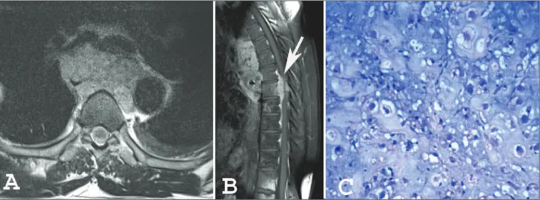
A 53-year-old male patient with a 3-month history of back pain presented with progressive paraparesis, although without loss of sphincter control. Magnetic resonance imaging (MRI) of the dorsal spine (Figures 1A and 1B) showed an expansile lesion with lobulated contours, involving the posterior medias-tinum and extending to the vertebral canal, thus reducing the amplitude of the vertebral canal and compressing the medulla.
A synchronic lesion of similar appearance, affecting the 12th dorsal vertebra, was observed. The histopathological study re-vealed large cells with vacuolated cytoplasm and partially vesic-ular nuclei (some demonstrating prominent nucleoli), with the appearance of physaliferous cells (from the Greek physallis, or bubble), consistent with a diagnosis of chordoma (Figure 1C).
Recent studies in the radiology literature of Brazil have highlighted the importance of imaging methods in improving the diagnosis of intrathoracic alterations(1–5). Chordomas are slow-growing malignancies derived from primitive remnants of the notochord. They typically occur in the ifth and sixth
341
Radiol Bras. 2017 Set/Out;50(5):338–348Letters to the Editor
http://dx.doi.org/10.1590/0100-3984.2016.0059
Bruno Niemeyer de Freitas Ribeiro1, Edson Marchiori2
1. Instituto Estadual do Cérebro Paulo Niemeyer, Rio de Janeiro RJ, Brazil. 2. Universidade Federal do Rio de Janeiro (UFRJ), Rio de Janeiro, RJ, Brazil. Mailing address: Dr. Bruno Niemeyer de Freitas Ribeiro. Instituto Estadual do Cérebro Paulo Niemeyer – Departamento de Radiologia. Rua do Rezende, 156, Centro. Rio de Janeiro, RJ, Brazil, 20231-092. E-mail: bruno.niemeyer@ hotmail.com.
cades of life(6,7), with a slight predilection for males and prefer-ential involvement of the sacrococcygeal region (50%), followed by the spheno-occipital region (35%), cervical spine, and lumbar spine, occurring only rarely in the dorsal spine and posterior mediastinum(6–8). Symptoms often appear only after the lesion has reached large proportions, with local invasion affecting neu-rovascular structures. Local recurrence is common when com-plete resection was not possible.
The differential diagnoses of chordoma include metastases, chondrosarcoma, multiple myeloma, neurogenic tumors, among others. Although imaging methods help delineate the lesion, the diagnosis is made on the basis of the histopathological analysis(7).
On MRI, most chordomas show isointense or hypointense signals in T1-weighted sequences, whereas they show hyperin-tense signals in T2-weighted and short-tau inversion-recovery sequences, relecting their high water content, some lesions con-taining ibrous septa and therefore showing low signal intensity in T2-weighted sequences(6–8). Gadolinium contrast enhance-ment tends to be moderate and heterogeneous(6,8). Lesions are often accompanied by bone erosion, which was not observed in the case reported here. Recent studies have highlighted the use of diffusion-weighted imaging in the differentiation between chordomas and chondrosarcomas, reporting that the latter show higher apparent diffusion coeficients(9,10).
In addition to an unusual site of involvement, our patient presented the peculiarity of a synchronous lesion. Although some authors have reported similar cases(7,8,11,12), there is no speciic criterion for differentiating between a multicentric chordoma and metastatic dissemination. We believe that our case could represent dissemination to the cerebrospinal luid, because there was involvement of the vertebral canal.
The treatment of choice for chordoma is surgical resection with adjuvant radiotherapy, resulting in a disease-free period approximately 2.5 years longer than that achieved after surgi-cal treatment alone(7). Because chordoma is resistant to con-ventional radiotherapy, other modalities, such as stereotactic radiosurgery, are used. Chordoma does not respond well to che-motherapy, antitumor activity having been observed, in small studies, only with the use of imatinib mesylate(13).
Albeit rare, a diagnosis of chordoma should be considered in patients with lesions affecting the posterior mediastinum. In
addition, the possibility of synchronous lesions should be inves-tigated in such patients.
REFERENCES
1. Guimaraes MD, Hochhegger B, Koenigkam-Santos M, et al. Magnetic resonance imaging of the chest in the evaluation of cancer patients: state of the art. Radiol Bras. 2015;48:33–42.
2. Pessanha LB, Melo AMF, Braga FS, et al. Acute post-tonsillectomy nega-tive pressure pulmonary edema. Radiol Bras. 2015;48:197–8.
3. Barbosa BC, Marchiori E, Zanetti G, et al. Catamenial pneumothorax. Radiol Bras. 2015;48:128–9.
4. Nishiyama KH, Falcão EAA, Kay FU, et al. Acute tracheobronchitis caused by Aspergillus: case report and imaging indings. Radiol Bras. 2014;47:317–9.
5. Fernandes GL, Teixeira AA, Antón AGS, et al. Churg-Strauss syndrome: a case report. Radiol Bras. 2014;47:259–61.
6. Rodallec MH, Feydy A, Larousserie F, et al. Diagnostic imaging of solitary tumors of the spine: what to do and say. Radiographics. 2008;28:1019–41. 7. Aydin AL, Sasani M, Oktenoglu T, et al. A case of chordoma invading
multiple neuroaxial bones: report of ten years follow up. Turk Neuro-surg. 2013;23:551–6.
8. Lim JJ, Kim SH, Cho KH, et al. Chordomas involving multiple neuraxial bones. J Korean Neurosurg Soc. 2009;45:35–8.
9. Yeom KW, Lober RM, Mobley BC, et al. Diffusion-weighted MRI: dis-tinction of skull base chordoma from chondrosarcoma. AJNR Am J Neu-roradiol. 2013;34:1056–61.
10. Freeze BS, Glastonbury CM. Differentiation of skull base chordomas from chondrosarcomas by diffusion-weighted MRI. AJNR Am J Neuro-radiol. 2013;34:E113.
11. Badwal S, Pal L, Basu A, et al. Multiple synchronous spinal extra-osseous intradural chordomas: is it a distinct entity? Br J Neurosurg. 2006;20:99–103.
12. Simon SL, Inneh IA, Mok CL, et al. Multiple epidural lumbar chordo-mas without bone involvement in a 17-year-old female: a case report. Spine J. 2011;11:e7–10.
13. Casali PG, Stacchiotti S, Sangalli C, et al. Chordoma. Curr Opin Oncol. 2007;19:367–70.
Esthesioneuroblastoma
Dear Editor,
A 64-year-old male presented with nasal obstruction, anos-mia, and a reduction in visual acuity over the last few months, together with weight loss and a two-year history of headache. Computed tomography (CT) of the brain (Figure 1A) showed an expansile lesion with poorly deined borders, occupying the ethmoid cells, sphenoid sinuses, and the anterior cranial fossa, accompanied by edema of the frontal lobes. On magnetic reso-nance imaging (MRI) scans (Figures 1B, 1C, and 1D), the le-sion showed restricted diffule-sion and intense enhancement after contrast administration. A biopsy was performed, and analysis of the biopsy sample revealed hyperchromatic cells organized around a ibrillar stroma, forming rosettes, consistent with a di-agnosis of olfactory neuroblastoma. The lesion was staged his-tologically as grade I in the Hyams grading system. There was no evidence of cervical involvement or distant metastases. The patient died 15 days after undergoing the examinations.
Olfactory neuroblastoma, also known as esthesioneurob-latoma, is a rare malignant neoplasm of neuroectodermal origin and accounts for 3–6% of all malignant tumors of the paranasal sinuses. It has a bimodal age distribution, being most common among adults in the second or ifth decades of life(1). It is believed that the neoplasm arises from the olfactory epithelium, origi-nating in the superior portion of the nasal cavities, ascending across the cribriform plate, and extending into the anterior cra-nial fossa(2).