Radiol Bras. 2016 Jan/Fev;49(1):26–34 26
Cardiac magnetic resonance imaging and computed tomography
in ischemic cardiomyopathy: an update
*
Ressonância magnética cardíaca e tomografia computadorizada na cardiomiopatia isquêmica: atualidades
Assunção FB, Oliveira DCL, Souza VF, Nacif MS. Cardiac magnetic resonance imaging and computed tomography in ischemic cardiomyopathy: an update. Radiol Bras. 2016 Jan/Fev;49(1):26–34.
Abstract
R e s u m o
Ischemic cardiomyopathy is one of the major health problems worldwide, representing a significant part of mortality in the general population nowadays. Cardiac magnetic resonance imaging (CMRI) and cardiac computed tomography (CCT) are noninvasive imaging methods that serve as useful tools in the diagnosis of coronary artery disease and may also help in screening individuals with risk factors for developing this illness. Technological developments of CMRI and CCT have contributed to the rise of several clinical indications of these imaging methods complementarily to other investigation methods, particularly in cases where they are inconclusive. In terms of accuracy, CMRI and CCT are similar to the other imaging methods, with few absolute contraindications and minimal risks of adverse side-effects. This fact strengthens these methods as powerful and safe tools in the management of patients. The present study is aimed at describing the role played by CMRI and CCT in the diagnosis of ischemic cardiomyopathies.
Keywords: Ischemic cardiomyopathy; Heart; Magnetic resonance imaging; Computed tomography.
A cardiomiopatia isquêmica é um dos principais problemas de saúde no mundo, representando significativa parcela da mortalidade. A ressonância magnética cardíaca (RMC) e a tomografia computadorizada cardíaca (TCC) são métodos de imagem não invasivos úteis no diagnóstico da doença arterial coronariana e também podem auxiliar no rastreamento de indivíduos com fatores de risco para o desen-volvimento de cardiomiopatia induzida por isquemia/infarto. Os avanços tecnológicos da RMC e da TCC contribuíram para o surgimento de diversas indicações clínicas para aplicação desses métodos de imagem de forma complementar a outros exames, principalmente quando estes se mostram inconclusivos. A RMC e a TCC apresentam acurácia semelhante aos demais métodos de imagem, poucas contraindicações absolutas e mínimos riscos de efeitos adversos, o que os fortalecem como ferramentas seguras no manejo dos pacien-tes. O presente estudo tem por objetivo descrever o papel da RMC e da TCC no diagnóstico das cardiomiopatias isquêmicas.
Unitermos: Cardiomiopatia isquêmica; Coração; Ressonância magnética; Tomografia computadorizada.
* Study developed at Department of Radiology of School of Medicine – Univer-sidade Federal Fluminense (UFF), Niterói, RJ, Brazil. Financial support: Fundação Carlos Chagas Filho de Amparo à Pesquisa do Estado do Rio de Janeiro (Faperj) and Conselho Nacional de Desenvolvimento Científico e Tecnológico (CNPq).
1. MDs, Trainees at School of Medicine – Universidade Federal Fluminense (UFF), Niterói, RJ, Brazil.
2. MD, Resident of Magnetic Resonance Imaging and Emergency Radiology at Complexo Hospitalar de Niterói (CHN), Niterói, RJ, Brazil.
3. Associate Professor and Vice-Chief of the Department of Radiology, School of Medicine – Universidade Federal Fluminense (UFF), Niterói, RJ, Brazil.
Mailing Address: Dr. Marcelo Souto Nacif. Rua Barão de Cocais, 324, Bosque Imperial. São José dos Campos, SP, Brazil, 12242-042. E-mail: [email protected] / www.msnacif.med.br.
Received June 19, 2014. Accepted after revision October 6, 2014.
ergism between the oxygen supply and demand is the main determining factor of ischemia in cases of stable chronic angina(1).
The clinical presentation of the coronary disease ranges from stable chronic angina to sudden death. This spectrum includes acute myocardial infarction with ST segment tion, acute myocardial infarction without ST segment eleva-tion, and unstable angina, currently catalogued as acute coro-nary syndrome(1). In about 50% to 70% of patients, acute myocardial infarction is the first manifestation of ischemic cardiomyopathy(2,3). The main etiopathogenic substrate of
ischemic cardiomyopathies is atherosclerosis(4). The risk
factors for development of atherosclerosis and subsequent ischemic cardiomyopathy include, besides age, systemic ar-terial hypertension, diabetes mellitus, smoking, dyslipidemia, sedentarism and obesity(5).
The diagnosis is based on the patient’s clinical history and presence of risk factors. Electrocardiography and chest radiography may also be useful to clarify chest pain etiol-ogy. Cardiac catheterization, however, is the gold standard for the diagnosis of coronary artery disease (CAD), despite its invasiveness and expensiveness(6). Currently, noninvasive
Fernanda Boldrini Assunção1, Diogo Costa Leandro de Oliveira1, Vitor Frauches Souza2, Marcelo Souto Nacif3
INTRODUCTION
syn-emphasis on the two latter methods(1).
CMRI is extremely useful in the evaluation of CAD, both in the acute and chronic phases. Because of its high spatial resolution, this method can currently be considered as a reference standard for evaluation of the global and re-gional myocardial function and for detection and quantifi-cation of myocardial infarction areas(7,8). CCT is a more
recent method with main clinical application focused on the diagnosis of CAD, determining the Agatston coronary cal-cium score (CCS) and the performance of noninvasive coro-nary angiography(7,8).
On the basis of the above considerations, the present study is aimed at describing the current concepts regarding the utilization of CMRI and CCT in ischemic cardiomyopa-thy, highlighting the diagnostic, therapeutic and prognostic impacts caused by the method. A systematic review was per-formed in the PubMed data basis sources (National Library of Medicine), utilizing the search terms “cardiac magnetic resonance in ischemic cardiomyopathy” and “computed to-mography in ischemic cardiomyopathy”, as well as in theme-correlated books, consensus and societies guidelines.
RESULTS
The authors found 1319 articles and, among those, 43 articles published in high-impact journals were selected by consensus of the authors, Additionally one societies consen-sus and three guidelines (I Diretriz de Ressonância e Tomo-grafia Cardiovascular da Sociedade Brasileira de Cardiolo-gia(8), III Diretriz Brasileira de Insuficiência Cardíaca
Crônica(9) and Diretriz de Doença Coronariana Crônica – Angina Estável(10)) were utilized. The present study was
de-veloped on the data collected in this review.
DISCUSSION
Cardiac magnetic resonance imaging (CMRI)
CMRI allows for evaluating the global and regional myo-cardial function, detecting and quantifying areas of myocar-dial infarction without utilizing ionizing radiation and neph-rotoxic contrast agents, being one of the most safe methods in cardiovascular diagnosis(8). Because of the obtention of
ventricular volumes and masses by means of a tridimensional approach, CMRI has high accuracy in the obtention of val-ues for both the left(11) and right(12) ventricles, and is
con-sidered to be the gold standard for such measurements(7).
Non contrast-enhanced echocardiography underestimates the ejection fraction values and volumes as compared with CMRI(13). Additionally, CMRI is useful to visualize and
Assessment of global and segmental function
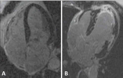
CMRI provides accurate and highly reproducible data regarding parameters of mass, volume, and global and re-gional contractility of right and left ventricles(8) (Figure 1).
The evaluation of the regional left ventricle function (seg-mental contractility) is performed both at rest and under pharmacological stress. The results of the segmental con-tractility analysis by means of CMRI are superior to those from echocardiography(16). The most utilized techniques for
investigating the presence of CAD involve the direct visual-ization of effects from ischemia, induced by pharmacologi-cal stress, and a multimodal analysis of the segmental con-tractility and myocardial perfusion. CMRI presents the unique characteristic of providing both types of information in a single procedure, combining the higher specificity in the evaluation of the regional function under stress with the higher sensitivity in the assessment of the myocardial perfu-sion(8).
Assessment of myocardial perfusion
The assessment of myocardial perfusion (Figure 2) is performed at rest and under pharmacological stress (dipy-ridamole or adenosine) and is analyzed together with the delayed enhancement images to identify necrotic or fibrotic areas(8). CMRI with myocardial perfusion has excellent
sen-sitivity and specificity as compared with cardiac catheteriza-tion(17,18). CMRI has already been validated utilizing the
methods currently available in the cardiological practice with comparative analyses and longitudinal evaluation for prog-nostic characterization of patients. Currently, the clinical utilization of this method is already quite consolidated(19).
As the myocardial perfusion evaluated by CMRI under stress is normal, the patient presents with a low rate of future car-diovascular events. On the other hand, in the presence of ischemia, the rate of future cardiovascular events is high, thus determining its prognostic capacity(20).
Evaluation of delayed myocardial enhancement
The protocols based on delayed myocardial gadolinium enhancement allow for accurate delimitation of the areas of myocardial necrosis or fibrosis in patients with chronic(8)
is a clear signal difference between the two tissues (black/ white)(8) (Figure 3).
In what concerns CAD, the presence and the pattern of delayed myocardial enhancement at CMRI in a patient with ventricular dysfunction allows for the diagnosis of ischemic or non-ischemic cardiomyopathy(7). The evaluation of
transmurality of the regions with myocardial necrosis or fi-brosis allows for predicting, with excellent accuracy, the probability of regional myocardial function recovery after revascularization, either surgical or percutaneous(21). As a
function of its excellent spatial resolution, CMRI can diag-nose, besides transmural infarction, small subendocardial infarctions(8). Cine CMRI allows for the same type of analysis
of segmental contractility than echocardiography. However, as such information is combined with the detailed evalua-tion of the infarcted region provided by the delayed myocar-dial enhancement technique, CMRI allows for accurately determining what is stunned myocardial tissue and what is irreversible necrosis(8).
Contraindications and limitations
CMRI has some relative contraindications, namely, pres-ence of pacemakers, implanted defibrillators, cerebral clips, cochlear implants and metal fragments in the eyes(8). Among
the method limitations, there is the fact that coronary MRI angiography, employing several acquisition techniques,
re-Figure 1. Ventricular function evaluation. Simpson’s technique (A,B) and 4D (C,D).
Figure 2. Evaluation of myocardial perfusion under pharmacological stress and at rest. ADCA, anterior descending coronary artery.
Perfusion – dipyridamole Perfusion – at rest Delayed enhancement Catheterization
veals moderate sensitivity (72–77%) and specificity (71–87%) values for detection of coronary stenosis as compared with invasive angiography(22,23). CMRI has modest spatial
reso-lution in the detection of coronary stenosis as compared with the high spatial resolution provided by CCT. It is possible that in the future coronary MRI becomes more usual, but currently, coronary CT angiography is a more robust method, with higher sensitivity and specificity as compared with CMRI(23).
The method limitations include systemic nephrogenic fibrosis, a disease that causes systemic tissue fibrosis and is associated with the use of gadolinium in patients with chronic renal failure stages 4 and 5 and in patients with hepatorenal syndrome. It is important to highlight that such warning does not apply to patients with normal renal function. The sys-temic nephrogenic fibrosis physiopathogenesis is still to be understood, but it is known that there is an association be-tween the development of the disease and use of gadolinium, which was demonstrated by the detection of gadolinium in biopsies of tissues of patients with systemic nephrogenic fi-brosis, and by the disease onset after a 2–12-week period after the use of gadolinium. Systemic nephrogenic fibrosis is a quick-onset and progressive condition, determining the on-set of symptoms such as muscle weakness, arthralgia, skin hardening and contractures, which, in conjunction, lead to the patient immobility. The risk for development of the dis-ease incrdis-eases at each exposure to gadolinium. For this rea-son, the Food and Drug Administration recommends the calculation of the creatinine clearance in the patients previ-ously to the gadolinium enhanced CMRI, and the indica-tion of this examinaindica-tion for patients at risk only if strictly necessary, followed by dialysis, despite the absence of scien-tific evidence that dialysis prevents the onset of systemic nephrogenic fibrosis(24).
In the clinical practice, all the imaging studies for in-vestigation of ischemia are performed with the patient
un-der pharmacological stress, which represents a partial limi-tation for the non-pharmacological evaluation, in spite of the fact that some studies have already demonstrated the possi-bility of performing MRI with exercise stress(25).
Cardiac computed tomography (CCT)
CCT is a method that utilizes ionizing radiation and iodinated contrast agent, with the main clinical application focused on the diagnosis of CAD(26). Such method presents
a high negative predictive value in the detection of CAD(9), and for this reason it may be utilized as an alternative to cardiac catheterization to rule out CAD(27).
Coronary calcium score
CCT detects and quantifies coronary artery calcium, a marker of the presence and extent of atherosclerotic dis-ease(8). The presence of calcium in the coronary arteries have
a strong predictive value for future cardiac events in asymp-tomatic patients(28–30), considering the high probability of
obstructive coronary disease associated with the increase in the amount of coronary calcium(30,31). Thus evaluation based
on the CCS allows for the differentiation between asymp-tomatic patients and those under risk to develop CAD over time(7). At the Bethesda Conference, it was concluded that
CCT and the CCS technique constitute the most accurate method currently available for early detection of coronary atherosclerosis(7).
Despite the wide applicability of the CCS, it is impor-tant to highlight that, in some situations, obstructive lesions might not contain calcium, and calcified lesions might not be obstructive(32). Such process is explained by the Gagov’s
phenomenon(23) that consists in the patency of the normal
volume of the vessel, despite the presence of an atheroscle-rotic process, which is called positive remodelling. The evalu-ation of the CCS (Figure 4) is complementarily added to the clinical risk stratification data, with possibility to add
clinical conducts, principally for patients considered to be at intermediate risk by the Framingham scores(33,34) and by the percentile stratified by the Multi-Ethnic Study of Ath-erosclerosis(35).
Coronary computed tomography angiography
Besides the coronary calcium evaluation, CCT may be used as a noninvasive modality of coronary angiography, which is called coronary computed tomography angiogra-phy (CCTA), with high sensitivity and specificity in the de-tection of coronary stenosis. It is indicated principally for patients at intermediate risk for CAD and with dubious tests
for ischemia, or in patients with low clinical probability of CAD, but with positive test for ischemia(8). CCTA is per-formed by means of multidetector CT, preferentially with 64 or more channels, under a single apnea. Routinely, the amount of contrast agent utilized is between 70 and 100 mL, which is considered to be low, therefore reducing the occur-rence of problems associated with nephrotoxicity. CCTA is capable of visualizing the vessel volume and walls, which allows for a noninvasive evaluation of the presence and size of noncalcified plaques(7) (Figure 5). In the evaluation of intra-stent restenosis in general, CCTA diagnostic accuracy is accepted as sufficient for clinical use in a noninvasive
Figure 4. Evaluation of coronary calcium score. Left coronary trunk, green (A). Proxi-mal third of the anterior descending coronary artery, yellow (B). Right coronary artery, red, and circumflex coronary artery, light blue (C).
method, depending on the utilized method(36,37) (Figure 6).
The method can still be utilized to evaluate the patency of surgical grafts or for differentiation between ischemic and non-ischemic cardiomyopathy(8) (Figures 7 and 8).
Contraindications and limitations
The main CCTA limitation occurs in the presence of dense calcification in the coronary arteries or the presence of a bare metal stent. In both situations, it will not possible to evaluate the degree of luminal obstruction. Another limi-tation is the necessity of a low heart rate (< 70 bpm), requir-ing the use of beta-blockers durrequir-ing the scan. The use of
sub-lingual nitrate, as indicated, might be considered as a limit-ing factor as it generates a tendency to the CCTA overesti-mate the degree of coronary stenosis(38,39), which frequently leads to the necessity for confirmation by means of a myocar-dial function test. The utilization of radiation is also consid-ered to represent a limitation of the method(7), but, with the
recent technological developments, radiation doses have been reduced. Additionally, iodine-based contrast agents utilized in CCTA are nephrotoxic, differently from the gadolinium-based ones utilized in CMRI. However, such a nephrotoxic-ity is usually self-limited and severe allergic reactions rarely occur. Preventive measures in relation to nephrotoxicity may
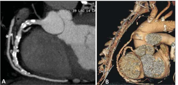
Figure 6. Evaluation of neointimal hyperplasia with intra-stent restenosis. Transversal axis (A) and longitudinal axis (B).
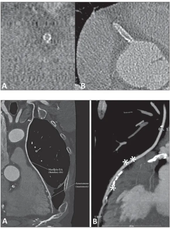
Figure 7. Evaluation of left internal mammary artery graft to anterior de-scending coronary artery (DA). Note the route of the graft in the mediasti-num (A) and the anastomosis of the permeable graft (double asterisks) with the native anterior descending artery (single asterisk) (B).
(Mammary–DA) ...
(Anastomosis)
. .
.. ..
.. ..
.. ..
.. ..
.. ..
.. ..
.. ..
be adopted, including a good hydration of the patient previ-ously to the scan and use of acetylcysteine in the previous and in the following days, although studies evaluating such measures still remain controversial(8).
Future prospects
CMRI as well as CCTA are diagnostic imaging meth-ods undergoing constant technological development.
Technological development of CMRI
The main future prospect for CMRI is related to the con-tinuous improvement of the images quality, allowing for a more detailed understanding of the myocardial anatomy, function and perfusion, of the tissues characterization and cardiac viability, besides representing an important tool for clinicians and cardiologists(40). Undoubtedly this will reduce
the acquisition time and will maintain or improve the diag-nostic potential of the method.
Technological development of CCT
The main prospects for CCT involve the reduction of the radiation dose affecting the patient, reduction of the ac-quisition time and, consequently, of the breath hold time, reduction of the volume of contrast agent required for the noninvasive acquisition of coronary arteries images, and reduction of the radiation dose. Another prospect involves the analysis of myocardial perfusion by means of dual en-ergy CT apparatuses(41–43). The high spatial and temporal
resolution of this method allows for images acquisition with a great anatomical detailing and noninvasive evaluation of the spatial relation between adjacent structures(26).
Anatomy versus ischemia / coronary flow reserve
Over the last four decades, the analysis of the coronary anatomy to predict the myocardial physiological status has ever been considered to be evidencing. However, it is cur-rently believed that the isolated anatomical analysis cannot
predict the physiological behavior of a single patient, since the biological variability is not taken into consideration. Anatomical parameters such as percentage of stenosis in the coronary diameter are not a very useful tool to understand the physiological behavior in general. In the future, the ten-dency is the adoption of parameters capable of more accu-rately and less invasively predicting the physiological status. Such physiological parameters can be evaluated by means of noninvasive imaging methods such as PET/CT or by in-vasive methods such as cardiac catheterization and, recently, noninvasively by CCTA. PET/CT, because of its high spa-tial resolution, is capable of quantifying myocardial perfu-sion at rest and stress and determining the coronary flow reserve. Cardiac catheterization allows for determining the fractional flow reserve that is defined as the quotient between the pressure distal to a stenosis and the proximal pressure. Both physiological parameters allow for more accurately predicting the degree of ischemia caused by the CAD, be-cause these parameters are superior to evaluate diffuse, multisegmental coronary diseases and those with heteroge-neous remodelling. As compared with each other, the coro-nary flow reserve is accurate to predict ischemia and supe-rior in the evaluation of diffuse diseases, so there is a future tendency towards the analysis of such parameter by PET/CT. The most relevant PET/CT clinical application in myocar-dial perfusion is the selection of patients with atherosclero-sis who will benefit more from undergoing myocardial revascularization(44). The calculation of the fractional flow
reserve has also been studied by means of CCTA. Although still undergoing improvements, it would be theoretically ideal for the management of patients with CAD, since a single scan would allow for the anatomical evaluation of a determined stenosis and its functional, demonstrating if, in fact, there was impairment of the myocardium. However, data about clinical validation and cost-effectiveness are still limited(45,46). Another approach under study is the evaluation of myo-cardial perfusion under stress by CT in association with
diagnostic accuracy when the techniques are utilized in com-bination as compared with CCTA alone. However, this tech-nique still lacks further studies to establish imaging proto-cols defining contrast agents and radiation doses(47–50).
CONCLUSION
CMRI and CCT are validated as highly sensitive and specific diagnostic tools, with few contraindications and mini-mal risks of adverse effects, and should be utilized by physi-cians as aid in the management of their patients.
REFERENCES
1. Carvalho ACC, Sousa JMA. Cardiopatia isquêmica. Rev Bras Hi-pertens. 2001;8:297–305.
2. Manfroi WC, Freitas FM, Gensini G, et al. Achados hemodinâmi-cos e cineangiocoronariográfihemodinâmi-cos em pacientes acometidos de in-farto do miocárdio: estudo comparativo entre uma população de Syracuse, NY, USA, com outra de Porto Alegre, RS, Brasil. Arq Bras Cardiol. 1980;34:359–62.
3. Behar S, Reicher-Reiss H, Abinader E, et al. The prognostic signifi-cance of angina pectoris preceding the occurrence of a first acute myocardial infarction in 4166 consecutive hospitalized patients. Am Heart J. 1992;123:1481–6.
4. Manfroi WC, Peukert C, Berti CB, et al. Infarto agudo do miocár-dio. Primeira manifestação da cardiopatia isquêmica e relação com fatores de risco. Arq Bras Cardiol. 2002;78:388–91.
5. Zaslavsky C, Gus I. Idoso. Doença cardíaca e comorbidades. Arq Bras Cardiol. 2002;79:635–9.
6. Yu PC, Caramelli B, Calderaro D. Performance diagnóstica de angiografia coronariana por tomografia computadorizada de 64 de-tectores (estudo CORE 64). Rev Assoc Med Bras. 2009;55:235–6. 7. Berman DS, Hachamovitch R, Shaw LJ, et al. Roles of nuclear car-diology, cardiac computed tomography, and cardiac magnetic reso-nance: assessment of patients with suspected coronary artery dis-ease. J Nucl Med. 2006;47:74–82.
8. Rochitte CE, Pinto IMF, Fernandes JL, et al. I Diretriz de Resso-nância e Tomografia Cardiovascular da Sociedade Brasileira de Car-diologia – Sumário executivo. Arq Bras Cardiol. 2006;87:e48–e59. 9. Schuijf JD, Bax JJ, Shaw LJ, et al. Meta-analysis of comparative diagnostic performance of magnetic resonance imaging and multi-slice computed tomography for noninvasive coronary angiography. Am Heart J. 2006;151:404–11.
10. Pletcher MJ, Tice JA, Pignone M, et al. Using the coronary artery calcium score to predict coronary heart disease events: a systematic review and meta-analysis. Arch Intern Med. 2004;164:1285–92. 11. Nayler GL, Firmin DN, Longmore DB. Blood flow imaging by cine
magnetic resonance. J Comput Assist Tomogr. 1986;10:715–22. 12. Helbing WA, Rebergen SA, Maliepaard C, et al. Quantification of
right ventricular function with magnetic resonance imaging in chil-dren with normal hearts and with congenital heart disease. Am Heart J. 1995;130:828–37.
13. Hoffmann R, von Bardeleben S, Kasprzak JD, et al. Analysis of re-gional left ventricular function by cineventriculography, cardiac magnetic resonance imaging, and unenhanced and
contrast-en-echocardiography. Circulation. 1999;99:763–70.
17. Nagel E, Klein C, Paetsch I, et al. Magnetic resonance perfusion measurements for the noninvasive detection of coronary artery dis-ease. Circulation. 2003;108:432–7.
18. Schwitter J, Nanz D, Kneifel S, et al. Assessment of myocardial per-fusion in coronary artery disease by magnetic resonance: a compari-son with positron emission tomography and coronary angiography. Circulation. 2001;103:2230–5.
19. Barranhas AD, Santos AASMD, Coelho-Filho OR, et al. Cardiac magnetic resonance imaging in clinical practice. Radiol Bras. 2014; 47:1–8.
20. Hundley WG, Morgan TM, Neagle CM, et al. Magnetic resonance imaging determination of cardiac prognosis. Circulation. 2002;106: 2328–33.
21. Kim RJ, Wu E, Rafael A, et al. The use of contrast-enhanced mag-netic resonance imaging to identify reversible myocardial dysfunc-tion. N Engl J Med. 2000;343:1445–53.
22. Budoff MJ, Achenbach S, Duerinckx A. Clinical utility of computed tomography and magnetic resonance techniques for noninvasive coronary angiography. J Am Coll Cardiol. 2003;42:1867–78. 23. Maintz D, Ozgun M, Hoffmeier A, et al. Whole-heart coronary
magnetic resonance angiography: value for the detection of coro-nary artery stenoses in comparison to multislice computed tomog-raphy angiogtomog-raphy. Acta Radiol. 2007;48:967–73.
24. Leite CC. Gadolínio e fibrose nefrogênica sistêmica: o que todo médico deve saber. Radiol Bras. 2007;40(4):iv–v.
25. Gusso S, Salvador C, Hofman P, et al. Design and testing of an MRI-compatible cycle ergometer for non-invasive cardiac assess-ments during exercise. Biomed Eng Online. 2012;11:13. 26. Shiozaki AA, Jasinowodolinski D, Sara L, et al. Ressonância
mag-nética cardiovascular e tomografia computadorizada cardiovascular nas doenças não-coronárias. Rev Soc Cardiol Estado de São Paulo. 2009;19:348–60.
27. Bocchi EA, Marcondes-Braga FG, Ayub-Ferreira SM, et al. Socie-dade Brasileira de Cardiologia. III Diretriz brasileira de insuficiên-cia cardíaca crônica. Arq Bras Cardiol. 2009;93(1 supl 1):3–70. 28. Greenland P, Bonow RO, Brundage BH, et al. ACCF/AHA 2007
clinical expert consensus document on coronary artery calcium scor-ing by computed tomography in global cardiovascular risk assess-ment and in evaluation of patients with chest pain: a report of the American College of Cardiology Foundation Clinical Expert Con-sensus Task Force (ACCF/AHA Writing Committee to Update the 2000 Expert Consensus Document on Electron Beam Com-puted Tomography). Circulation. 2007;115:402–26.
29. Taylor AJ, Bindeman J, Feuerstein I, et al. Coronary calcium inde-pendently predicts incident premature coronary heart disease over measured cardiovascular risk factors: mean three-year outcomes in the Prospective Army Coronary Calcium (PACC) project. J Am Coll Cardiol. 2005;46:807–14.
30. Kronmal RA, McClelland RL, Detrano R, et al. Risk factors for the progression of coronary artery calcification in asymptomatic sub-jects: results from the Multi-Ethnic Study of Atherosclerosis (MESA). Circulation. 2007;115:2722–30.
32. Wexler L, Brundage B, Crouse J, et al. Coronary artery calcification: pathophysiology, epidemiology, imaging methods, and clinical im-plications. A statement for health professionals from the American Heart Association. Writing Group. Circulation. 1996;94:1175–92. 33. Greenland P, LaBree L, Azen SP, et al. Coronary artery calcium score combined with Framingham score for risk prediction in as-ymptomatic individuals. JAMA. 2004;291:210–5.
34. Nasir K, Michos ED, Blumenthal RS, et al. Detection of high risk young adults and women by coronary calcium and National Cho-lesterol Education Program Panel III guidelines. J Am Coll Cardiol. 2005;46:1931–6.
35. McClelland RL, Chung H, Detrano R, et al. Distribution of coro-nary artery calcium by race, gender, and age: results from the Multi-Ethnic Study of Atherosclerosis (MESA). Circulation. 2006;113: 30–7.
36. Gaspar T, Halon DA, Lewis BS, et al. Diagnosis of coronary in-stent restenosis with multidetector row spiral computed tomogra-phy. J Am Coll Cardiol. 2005;46:1573–9.
37. Cademartiri F, Mollet N, Lemos PA, et al. Usefulness of multislice computed tomographic coronary angiography to assess in-stent restenosis. Am J Cardiol. 2005;96:799–802.
38. Cury RC, Pomerantsev EV, Ferencik M, et al. Comparison of the degree of coronary stenoses by multidetector computed tomogra-phy versus by quantitative coronary angiogratomogra-phy. Am J Cardiol. 2005;86:784–7.
39. Moselewski F, Ropers D, Pohle K, et al. Comparison of measure-ment of cross-sectional coronary atherosclerotic plaque and vessel areas by 16-slice multidetector computed tomography versus intra-vascular ultrasound. Am J Cardiol. 2004;94:1294–7.
40. Vitorino RR, Nacif MS. Ressonância magnética cardíaca na cardio-miopatia dilatada: atualidades. Rev Bras Clin Med. São Paulo. 2011;9:225–33.
41. Pinto IMF, Sousa AGMR, Ishikawa W, et al. Ressonância
magné-tica e tomografia computadorizada no diagnóstico de insuficiência coronária. Rev Bras Cardiol Invas. 2006;14:168–77.
42. Parga Filho JR, Lima CSLM, Lima FG, et al. Perfusão miocárdica dinâmica por tomografia computadorizada de dupla fonte de raio X. Arq Bras Cardiol. 2012;98,54–8.
43. Delgado C, Vázquez M, Oca R, et al. Myocardial ischemia evalua-tion with dual-source computed tomography: comparison with magnetic resonance imaging. Rev Esp Cardiol (Engl Ed). 2013;66: 864–70.
44. Gould KL, Johnson NP, Bateman TM, et al. Anatomic versus physi-ologic assessment of coronary artery disease. Role of coronary flow reserve, fractional flow reserve, and positron emission tomography imaging in revascularization decision-making. J Am Coll Cardiol. 2013;62:1639–53.
45. Yoon YE, Koo BK. Non-invasive functional assessment using com-puted tomography: when will they be ready for clinical use? Cardio-vasc Diagn Ther. 2012;2:106–12.
46. Nørgaard BL, Leipsic J, Gaur S, et al. Diagnostic performance of non-invasive fractional flow reserve derived from coronary CT an-giography in suspected coronary artery disease: The NXT trial. J Am Coll Cardiol. 2014;63:1145–55.
47. Rossi A, Merkus D, Klotz E, et al. Stress myocardial perfusion im-aging with multidetector CT. Radiology. 2014;270:25–46. 48. Johnson NP, Kirkeeide RL, Gould KL. Coronary anatomy to
pre-dict physiology: fundamental limits. Circ Cardiovasc Imaging. 2013;6:817–32.
49. Sociedade Brasileira de Cardiologia. Diretrizes de doença corona-riana crônica – angina estável. Arq Bras Cardiol. 2004;83(supl II):1– 43.