Radiol Bras. 2014 Jul/Ago;47(4):251–255 251
Imaging diagnosis of dural and direct cavernous carotid
fistulae
*
Diagnóstico por imagem das fístulas arteriovenosas da região do seio cavernoso
Santos D, Monsignore LM, Nakiri GS, Cruz AAV, Colli BO, Abud DG. Imaging diagnosis of dural and direct cavernous carotid fistulae. Radiol Bras. 2014 Jul/Ago;47(4):251–255.
Abstract
R e s u m o
Arteriovenous fistulae of the cavernous sinus are rare and difficult to diagnose. They are classified into dural cavernous sinus fistulae or direct carotid-cavernous fistulae. Despite the similarity of symptoms between both types, a precise diagnosis is essential since the treatment is specific for each type of fistula. Imaging findings are remarkably similar in both dural cavernous sinus fistulae and carotid-cavernous fistulae, but it is possible to differentiate one type from the other. Amongst the available imaging methods (Doppler ultrasonography, computed tomography, magnetic resonance imaging and digital subtraction angiography), angiography is considered the gold standard for the diagnosis and classification of cavernous sinus arteriovenous fistulae. The present essay is aimed at didactically presenting the classification and imaging findings of cavernous sinus arteriovenous fistulae.
Keywords: Arteriovenous fistula; Cavernous sinus; Ultrasonography; Computed tomography; Magnetic resonance imaging; Angiography.
As fístulas arteriovenosas da região do seio cavernoso são lesões raras e difíceis de diagnosticar. Elas são classificadas em fístulas durais do seio cavernoso e fístulas carótido-cavernosas diretas. Apesar de apresentarem sintomas semelhantes, o diagnóstico preciso é impor-tante, já que o tratamento é específico para cada uma delas. As alterações encontradas nos exames de imagem são muito semelhantes, tanto nas fístulas durais do seio cavernoso quanto nas fístulas carótido-cavernosas, contudo, é possível diferenciá-las. Dentre os exames de imagem disponíveis (ultrassonografia de órbita com Doppler, tomografia computadorizada, ressonância magnética e angiografia com subtração digital), a angiografia é considerada padrão ouro para o diagnóstico e classificação das fístulas arteriovenosas da região do seio cavernoso. O objetivo deste ensaio é mostrar de modo didático a classificação e o aspecto por imagem das fístulas arteriovenosas do seio cavernoso.
Unitermos: Fístula arteriovenosa; Seio cavernoso; Ultrassonografia; Tomografia computadorizada; Ressonância magnética; Angiografia.
* Study developed at Centro de Ciências das Imagens e Física Médica (CCIFM) – Hospital das Clínicas da Faculdade de Medicina de Ribeirão Preto de Universidade de São Paulo (HCFMRP-USP), Ribeirão Preto, SP, Brazil.
1. Masters, Physicians Assistants, Unit of Interventional Radiology and Therapeutic Neuroradiology, Hospital das Clínicas da Faculdade de Medicina de Ribeirão Preto da Universidade de São Paulo (HCFMRP-USP), Ribeirão Preto, SP, Brazil.
2. Fellow PhD degree, Physician Assistant, Unit of Interventional Radiology and Therapeutic Neuroradiology, Hospital das Clínicas da Faculdade de Medicina de Ri-beirão Preto da Universidade de São Paulo (HCFMRP-USP), RiRi-beirão Preto, SP, Brazil. 3. Full Professor, Teacher at Division of Ophthalmology, Department of Ophthal-mology, Otorhinolaryngology and Head and Neck Surgery – Faculdade de Medicina de Ribeirão Preto da Universidade de São Paulo (FMRP-USP), Ribeirão Preto, SP, Brazil. 4. Full Professor, Teacher at Division of Neurosurgery, Department of Surgery and Anatomy – Faculdade de Medicina de Ribeirão Preto da Universidade de São Paulo (FMRP-USP), Ribeirão Preto, SP, Brazil.
5. Professor Responsible for the Unit of Interventional Radiology and Therapeutic Neuroradiology – Hospital das Clínicas da Faculdade de Medicina de Ribeirão Preto da Universidade de São Paulo (HCFMRP-USP), Ribeirão Preto, SP, Brazil.
creased blood volume and stasis, increased venous pressure and, consequently, ophthalmic veins flow reversal and even reflux into cortical veins and other venous sinuses. This ex-plains the neuro-ophthalmic alterations found in such pa-tients(2).
The AVF of the CS are classified according to anatomy, etiology and hemodynamics, into dural cavernous sinus fis-tulae (DCSF) and direct carotid-cavernous fisfis-tulae (CCF). In spite of presenting with similar symptoms, an accurate diagnosis is important, since the treatment is specific for each one of the entities.
The present essay was aimed at didactically demonstrat-ing the classification and imagdemonstrat-ing finddemonstrat-ings of AVF of the cav-ernous sinus.
CLASSIFICATION
Dural cavernous sinus fistulae
Dural AVF are anomalous direct communications be-tween meningeal arterial branches and the dura mater or a
Daniela dos Santos1, Lucas Moretti Monsignore2, Guilherme Seizem Nakiri1, Antonio Augusto Velasco e Cruz3,
Benedicto Oscar Colli4, Daniel Giansante Abud5
INTRODUCTION
Arteriovenous fistulae (AVF) of the cavernous sinus (CS) region are rare and difficult to diagnose. The main signs and symptoms include proptosis; conjunctival hyperemia; chemo-sis; palsy of the II, IV, V and VI cranial nerves; ptochemo-sis; glau-coma; visual acuity reduction and headache(1). More severe cases such as amaurosis and intracranial bleeding are also described. The arteriovenous shunt into the CS leads to
in-Mailing Address: Dra. Daniela dos Santos. Centro de Ciências das Imagens e Física Médica – HCFMRP-USP. Avenida Bandeirantes, 3900, Monte Alegre. Ribeirão Preto, SP, Brazil, 14048-900. E-mail: [email protected].
Direct carotid-cavernous fistulae
In cases of CCF, there is a defect on the wall of the internal carotid artery (ICA) which communicates directly with the CS and, consequently, a large-volume arteriovenous shunt. Such type of fistulae may either occur after cranioencephalic trauma (CET) or may spontaneously de-velop after rupture of an aneurysm located in the cavernous portion of the ICA (post-traumatic or spontaneous CCF, respectively). Post-traumatic CCF represent 69-77% of cav-ernous sinus AVF, with a prevalence of only 0.2% in cases of CET(7). CCF predominate in male individuals in the age range between 20 and 30 years(8).
IMAGING DIAGNOSIS
Imaging findings are very similar for both DCSF and CCF, but it is possible to differentiate them. Among the available imaging methods – orbital Doppler ultrasonogra-phy (US), computed tomograultrasonogra-phy (CT), magnetic resonance imaging (MRI) and digital subtraction angiography (DSA) –, the latter is considered the gold-standard for the diagno-sis and classification of cavernous sinus AVF.
The patient’s clinical history is fundamental and a very important tool for a correct diagnosis. In patients with signs and more subtle and slow-progressing symptoms, the hypoth-esis of a DCSF should be considered. In a patient with a history of trauma, exuberant and fast-progressing neuro-oph-thalmic alterations, the hypothesis of a post-traumatic CCF should be initially considered. In patients with exuberant clinical presentation, without a history of trauma, spontane-ous CCF should be considered.
edema (Figures 2 and 3). Ectatic SOV is identified at the post-contrast phase in 86–100% of cases and it is one of the first signs of cavernous sinus AVF(10).
Because of the intensity of the arteriovenous shunt, gen-erally with low flow in the DCSF and high flow in CCF, the enhancement of the cavernous sinus is one of the signs that may be utilized in the differentiation between fistulas. In cases of DCSF, a slower contrast uptake by the cavern-ous sinus will occur, while in cases of CCF it will be faster and exuberant. Additionally, the increased flow of the direct CCF frequently leads to a CS dilatation. Such findings in association with the patient’s clinical history and presence of cranial fractures, allow for the differentiation of the fistu-las between DCSF and CCF.
Intracranial hemorrhage may occur as a complication of an AVF in the CS region, and CT is the method of choice for such an evaluation(11). Such a complication is mainly seen in cases of post traumatic direct CCF and is rarely observed in cases of DCSF. Generally, the bleeding is adjacent to the ectatic veins.
Magnetic resonance imaging
MRI is superior to CT in the detection of radiological signs. Minimum SOV dilation, subtle proptosis and small extraocular muscle thickening may be more easily identified by this method (Figures 4 and 5). The MRI sensitivity is enhanced by the utilization of venous injection of paramag-netic contrast, T2-weighted and SWI sequences. Thus, a better evaluation of the venous drainage and possible reflux into the dural sinuses and cortical veins is obtained.
Figure 3. Computed tomography, bone window. a: Female 73-year-old with a diagnosis of DCSF. Subtle proptosis at right (dashed line). b: Male eight-year-old patient with diagnosis of post-traumatic CCF. Subtle proptosis at right (dashed line) and sphenoid bone fracture (bold arrow). The presence of fracture in the skull base suggests a post-traumatic origin in the patient on the image b.
Figure 2. Contrast-enhanced computed tomography, arterial phase. Female 73-year-old patient with diagnosis of DCSF. a: Subtle proptosis (dashed line) and extraocular muscle thickening at right (fine arrows). b: Early contrast enhance-ment at the right SOV, which is slightly ectatic (bold arrow).
Ectatic SOV is detected by MRI both at T2-weighted sequences and at post-contrast phases, in 75–100% of the pa-tients(10). Early contrast-enhancement of the cavernous si-nus is shown even in low flow arteriovenous shunts, as it occurs in most DCSF patients. At T2-weighted sequences,
it is possible to identify the abnormal cortical venous drain-age into the leptomeningeal veins and possible throm-boses(12). Like at CT, in cases of DCSF there is a slower contrast enhancement of the CS, and in cases of CCF, there is cavernous sinus dilatation and contrast enhancement is Figure 4. Encephalon MRI. a,b: Axial, T2-weighted sequence. c: Contrast-enhanced axial T1-weighted sequence. Female 21-year-old patient with diagnosis of DCSF. Extraocular muscle thickening and periorbital fat edema (thin arrows). Proptosis at left (dashed line). Flow void in the SOV (dashed arrow) compatible with increased blood flow. Early contrast enhancement and SOV dilatation (bold arrow).
Figure 5. Encephalon MRI. a,b: Axial T2-weighted sequence. c: Axial FLAIR sequence. d: Contrast-enhanced T1-weighted sequence. Female 24-year-old patient with a diagnosis of spontaneous CCF. Flow void indicating high flow in the cavernous sinus and in the left SOV, which is ectatic (thin arrows). Also, observe is the increased sinus volume. Proptosis at left (dashed line). Early contrast enhancement and ectasia of both SOVs, predominant at left (bold arrows).
Digital subtraction cerebral angiography
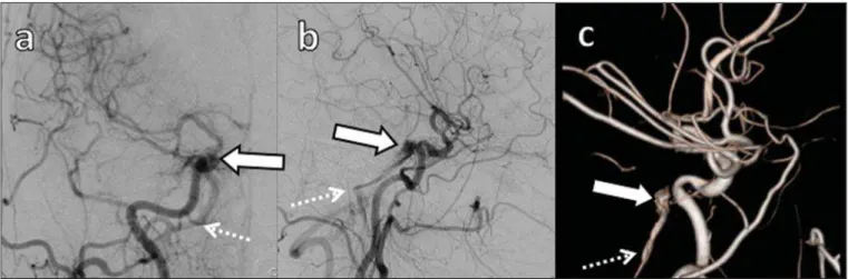
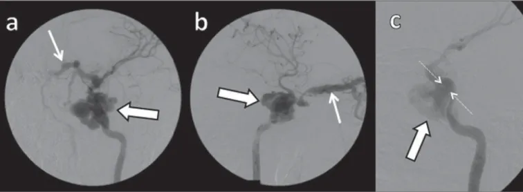
The utilization of angiography is essential for diagnos-tic confirmation, anatomical evaluation and classification of cavernous sinus AVF. Angiography detects the fistula and identifies the supplying meningeal branches in DCSF (Fig-ures 8 and 9) or ICA wall laceration in direct CCF (Figure 10). This method evaluates the flow in the cavernous sinus as well as in other venous sinuses, identifying possible throm-bosis or reflux into cortical veins and into other sinuses. It also identifies the venous drainage either into the SOV or into the IPS, and evaluates risk factors such as intracavernous pseudoaneurysms and reflux into cortical veins. The
thera-Figure 9. Digital subtraction cerebral angiography, arterial phase. a: Right anterior oblique view. b: Lateral view. c: 3D reconstruction. Female 31-year-old patient with a diagnosis of DCSF. Early contrast-enhancement of the cavernous sinus (bold arrows) which drains to the IPS (dashed arrows). One also observes that there is no drainage through the SOV, characterizing a DCSF with only posterior drainage.
Figure 6. Intracranial vessels MR angiography. a: Lateral view. b: Anteroposte-rior view. Female 73-year-old patient with a diagnosis of DCSF. Early enhancement of SOV (thin arrow) and facial vein (dashed arrow). Both are ectatic. Subtle early contrast enhancement of the right CS (bold arrow).
Figure 7. MR angiography of intracranial vessels. a: Lateral view. b: Anteropos-terior view. Female 24-year-old patient with diagnosis of spontaneous CCF. Con-trast enhancement of the left cavernous sinus which is dilated, with reflux into the right CS (bold arrows). Early contrast enhancement of the SOV (thin arrow) which is dilated.
Figure 8. Digital subtraction cerebral angiography, arterial phase. a: Lateral view.
the findings are the same at US, the analysis of CT and MRI findings allow for the classification between DCSF and CCF, while arteriography, besides being the gold-standard for the diagnosis, allows for the planning and performance of the therapeutic procedure.
REFERENCES
1. Vilela MAP. Fístula carotídeo-cavernosa. Rev Bras Oftalmol. 2013; 72:70–5.
2. Meyers PM, Halbach VV, Dowd CF, et al. Dural carotid cavernous fistula: definitive endovascular management and long-term follow-up. Am J Ophthalmol. 2002;134:85–92.
3. Szikora I. Dural arteriovenous malformations. In: Forsting M, Wanke I, editors. Intracranial vascular malformations and aneurysms. 2nd ed. Berlin: Springer; 2008. p. 101–41.
4. Satomi J, Satoh K. Epidemiology and etiology of dural arteriovenous fistula. Brain Nerve. 2008;60:883–6.
5. Woo HH, Masaryk TJ, Rasmussen PA. Treatment of dural arterio-venous malformations and fistulae. Neurosurg Clin N Am. 2005;16: 381–93.
6. Cognard C, Gobin YP, Pierot L, et al. Cerebral dural arteriovenous
fistulas: clinical and angiographic correlation with a revised classi-fication of venous drainage. Radiology. 1995;194:671–80. 7. Millman B, Giddings NA. Traumatic carotid-cavernous sinus
fis-tula with delayed epistaxis. Ear Nose Throat J. 1994;73:408–11. 8. Debrun G, Lacour P, Vinuela F, et al. Treatment of 54 traumatic
carotid-cavernous fistulas. J Neurosurg. 1981;55:678–92. 9. Flaharty PM, Lieb WE, Sergott RC, et al. Color Doppler imaging.
A new noninvasive technique to diagnose and monitor carotid cav-ernous sinus fistulas. Arch Ophthalmol. 1991;109:522–6. 10. Hirabuki N, Fujita N, Hashimoto T, et al. Follow-up MRI in dural
arteriovenous malformations involving the cavernous sinus: emphasis on detection of venous thrombosis. Neuroradiology. 1992;34:423– 7.
11. d’Angelo VA, Monte V, Scialfa G, et al. Intracerebral venous hem-orrhage in “high-risk” carotid-cavernous fistula. Surg Neurol. 1988; 30:387–90.
12. Chen JC, Tsuruda JS, Halbach VV. Suspected dural arteriovenous fistula: results with screening MR angiography in seven patients. Radiology. 1992;183:265–71.
13. Acierno MD, Trobe JD, Cornblath WT, et al. Painful oculomotor palsy caused by posterior-draining dural carotid cavernous fistulas. Arch Ophthalmol. 1995;113:1045–9.