Evaluation of the Metabolism of High Energy Phosphates in Patients
with Chagas’ Disease
Ana Maria Betim Paes Leme
1, Vera Maria Cury Salemi
1, José Rodrigues Parga
1, Bárbara Maria Ianni
1, Charles
Mady
1, Robert G. Weiss
2, Roberto Kalil-Filho
1Instituto do Coração (InCor) do Hospital das Clínicas da Faculdade de Medicina da Universidade de São Paulo1, São Paulo, SP - Brazil;
Division of Cardiology, Department of Medicine, Johns Hopkins University, Baltimore2, MD - USA
Mailing address: Vera Maria Cury Salemi •
Avenida Jandira, 185/41 B - Indianópolis - 04080-000 - São Paulo, SP - Brazil E-mail: [email protected], [email protected]
Manuscript received July 08, 2009; revised manuscript received November 18, 2009, accepted on December 28, 2009.
Abstract
Background: Abnormalities in myocardial metabolism have been observed in patients with heart failure of different etiologies. Magnetic resonance spectroscopy (MRS) with phosphorus-31 is a noninvasive technique that allows detection of myocardial metabolic changes.
Objective: To determine the resting metabolism of high-energy phosphates in patients with Chagas’ disease (CD) by MRS with phosphorus-31.
Methods: We studied 39 patients with CD, 23 with preserved ventricular function (PF Group) and 16 with ventricular
dysfunction (VD Group), assessed by Doppler echocardiography. MRS of the anterosseptal region was performed in
39 patients and 8 normal subjects (C Group) through a Phillips 1.5 Tesla device, obtaining the
phosphocreatine/beta-adenosine triphosphate myocardial ratio (PCr/b-ATP).
Results: The levels of cardiac PCr/b-ATP were reduced in VD Group in relation to PF Group, and the latter presented
reduced levels compared to C Group (VD Group: 0.89 ± 0.31 vs PF Group: 1.47 ± 0.34 vs C Group: 1.88 ± 0.08, p < 0.001). A correlation was found between left ventricular ejection fraction and PCr/b-ATP in 39 patients (r = 0.64, p <0.001). Patients under functional class I (n = 22) presented PCr/b-ATP of 1.45 ± 0.35, and those in functional classes II and III (n = 17), PCr/b-ATP of 0.94 ± 0.36 (p < 0.001).
Conclusion: The 31-phosphorus MRS was able to detect non-invasively changes in the rest energy metabolism of patients with Chagas’ disease, with and without systolic dysfunction. These changes were related to the severity of heart
impairment. (Arq Bras Cardiol 2010; 95(2): 264-271)
Key words: Energy metabolism; Chagas’ disease; chagas cardiomyopathy.
intense neuronal depopulation found in the different stages of the disease; autoimmune mechanism related to the finding of antibodies directed against antigens of T. cruzi present in foci of myocardial inflammation; damaged coronary microcirculation with consequent development of myocardial ischemia5-10.
Another mechanism that has been pointed out in contributing to the deterioration of global myocardial function in various heart diseases is the impairment of the integrity of the cell energy system11.The proper operation of
this system depends on the presence in adequate amounts of substrates and enzymes involved in synthesis and the use of adenosine triphosphate (ATP), which is the molecule responsible for the immediate supply of energy to the heart muscle. One of the key enzymes involved in this process is the creatine kinase (CK), responsible for the transfer of 40% to 70% of high-energy phosphates from creatine and ATP molecule11,12. Animal models and studies in humans
with heart failure have shown changes in this bioenergetic system, with significant reduction in the speed of action of CK and changes in the distribution of its isoenzymes, in addition to reduction in the total content of ATP and
Introduction
Chagas’ disease (CD) is one of the most common forms of heart disease in Latin America, with roughly eight to ten million individuals affected by the disease1. About 70%
to 80% of individuals remain in the indeterminate form throughout their lives, while 20% to 30% of them progress, after decades, to heart disease. This is characterized by arrhythmias, thromboembolic events and dilated cardiomyopathy with heart failure1. However, even
asymptomatic patients and those with preserved systolic function have reduced exercising capacity2.
The pathogenesis of Chagas’ heart disease is not established, and the proposed mechanisms are3-6: direct aggression to the
phosphocreatine (PCr)13. Thus, phosphorus-31 magnetic
resonance spectroscopy (MRS) enables the evaluation of high-energy phosphates found in the myocardium, allowing the identification of metabolic changes in patients with different degrees of myocardial impairment14,15.
The objective of this study is to determine the resting high-energy phosphates’ metabolism in patients with CD using phosphorus-31 MRS seeking a better understanding of the influence of myocardial energy metabolism in the pathophysiology of Chagas’ disease cardiomyopathy.
Methods
We prospectively studied 39 CD patients (20 men, 27 to 79 years old, 52 ± 12 years) in outpatient clinic monitoring and 8 volunteers (5 men, 29 ± 4 years, C Group). All CD patients studied were evaluated by clinical history, physical examination, laboratory tests, resting electrocardiogram, chest radiography and echocardiography. Patients with the indeterminate form of the disease also performed esophageal contrast studies.
The diagnosis of CD was determined by the presence of positive epidemiology and confirmed by at least two positive serological tests for antibodies (complement fixation or Machado-Guerreiro and indirect immunofluorescence)16.
Patients were considered to have the indeterminate form of CD when they were asymptomatic with respect to cardiovascular and digestive systems and when they presented normal electrocardiogram, chest radiography and esophageal contrast study.
Patients were considered to have chronic CD when they had at least one of the following symptoms or signs: palpitations, orthopnea, exertional dyspnea, paroxysmal nocturnal dyspnea, chest discomfort, cough, weakness, dizziness, syncope, galloping rhythms, heart murmurs, hepatomegaly, lower limb edema or jugular stasis; electrocardiographic alterations usually found in CD (anterosuperior divisional block, right bundle branch block, first degree atrioventricular block, alterations in ventricular repolarization, ventricular premature beats, left bundle branch, left ventricular hypertrophy); radiological alterations (chest radiography) and/ or echocardiography; absence of other diseases that could influence our findings.
The 39 patients were divided into three groups for analysis of resting energy metabolism by MRS of phosphorus-31. The groups are presented below:
• Chagas’ heart disease with left ventricular dysfunction - VD Group: consisting of 16 patients (11 men, 27 to 72 years old, 49 ± 12 years old). Clinically speaking, 12 patients (75%) were under functional class (FC) II of NYHA; 3 (18.75%) in functional class III of NYHA and only 1 under functional class I of NYHA. The electrocardiographic alterations prevailing in this group were the anterosuperior block, present in 11 patients (69%), followed by right bundle branch block in 9 (56%). Mean ejection fraction was 39 ± 6%, and all patients in this group had an ejection fraction below 50%.
• Chagas’ heart disease with preserved ventricular function and EKG alterations - EKGalt Group: consisting of
15 patients (6 men, 44 to 79 years old, 58 ± 10 years old). Clinically speaking, 13 patients (86%) were in functional class I of NYHA and only 2 (14%) in functional class II of NYHA. Electrocardiographic alterations prevailing in this group were the right bundle branch block in 9 patients (60%), accompanied or not by anterosuperior divisional block present in 5 (40%). Mean ejection fraction was 67 ± 8%, with all patients in the group presenting an ejection fraction equal to or greater than 56%.
• Chagas’ heart disease with preserved ventricular function and normal electrocardiogram: indeterminate - Ind Group: consisting of 8 patients (3 men, 31 to 62 years old, 45 ± 11 years old). All patients were clinically asymptomatic. All patients had normal resting electrocardiograms. The mean ejection fraction was 68 ± 5%. Esophageal contrast study discarded megaesophagus in all patients.
• Control group - consisting of 8 healthy volunteers (5 men, 22 to 34 years old, 29 ± 4 years old) without history of heart disease. All volunteers had normal resting electrocardiogram and functional tests for ischemia.
Exclusion criteria were: functional class IV of NYHA, diabetes mellitus, severe hypertension, myocardial hypertrophy on Doppler echocardiography, coronary heart disease, history of alcoholism, other pre-existing heart diseases, pregnancy and patients with permanent pacemaker.
All participating patients were informed about the study and signed an Informed Consent.
Electrocardiography
The analysis of the electrocardiogram followed the traditional criteria of normality, and for the alterations found, we considered the following parameters17: right bundle
branch block, left bundle branch block, anterosuperior divisional block, first degree atrioventricular block, alteration of ventricular repolarization, left ventricular hypertrophy, ventricular extrasystoles.
Echocardiography
Patients underwent transthoracic echocardiography using the M-mode, two-dimensional and Doppler techniques. Measurements of left ventricular internal dimensions were obtained through the M-mode, at the end of systole and at the end of diastole, with the aid of simultaneous electrocardiographic recording, performed as recommended by the American Society of Echocardiography18. Such
measures have enabled the calculation of diastolic and systolic volumes, and the calculation of fractional shortening and ejection fraction.
Protocol of magnetic resonance spectroscopy of phosphorus 31
A 10 cm diameter coil (P-100) tuned to phosphorus-31 resonance frequency (63.3-83 MHz), for reception and transmission, was located on the heart ictus. The coil was kept in place throughout the exam via a Velcro band placed around the patient’s thoracic region. The patient was placed inside the superconducting magnet so that the center of the coil corresponded to the longitudinal axis of the magnet isocenter.
Initially, nine cross-sections (thickness 10 mm) were obtained in periods of two to three minutes with T1-weighted images, spin-echo, EKG-gated in order to allow the precise placement of the surface coil in relation to the patient’s left ventricular anterior wall. With the coil located on the left ventricular anterior wall, we set up a volume of approximately 35 cm3, which was placed parallel to the surface coil. The
volume selected contains, predominantly, cardiac muscle of the left ventricular anterior apical region. Cuts contaminated by skeletal muscle of the chest wall and/or portions of the diaphragm and liver were excluded.
The localization technique used was the image-selected in vivo spectroscopy (ISIS), characterized by high spatial resolution19 and originally described in 198520. This is a
technique that allows the choice of a rectilinear region of the heart muscle in the shape of a parallelepiped, which is selected from the resonance images generated by the proton signal. It is performed by applying three adiabatic pulses in the presence of the same magnetic field gradient used to obtain the image. Each pulse inversion reverses the magnetization area selected with the region defined by the intersection of three orthogonal planes. A sequence of eight different acquisitions with different combinations of pulse inversion is added to obtain the final spectrum. Thus, signals within the region concerned are added, while signals from outside the region concerned, but still within the region captured by the surface coil, are added with opposite signs and therefore cancelled.
The spectrum acquisition time was approximately 16 minutes (384 measures). The repetition time used was 2 .500 ms and the trigger delay 200 ms. The test total duration time was about 60 to 70 minutes, during which the patients were kept in the same position. After the spectrum processing (Fourier transform mathematics) and paper printing, the area under each spike was calculated by means of manual, computer-aided typing, which delivered the ratio PCr/b-ATP. Inorganic phosphate was not measured due to its low concentration and overlapping with the 2.3-diphosphoglycerate spike.
Corrections for the partial saturation of the nucleus were made by the following formula:
SF = [1 - exp(-TR/T1)].sina/[1-exp(-TR/T10.cosa]
where SF = saturation factor, TR = repetition time, T1 = time of relaxation and a = angle of inclination (90o).
Phosphocreatine T1 and myocardial b-ATP values are estimated at 4.18 and 1.7 seconds, respectively.
Normal values for the PCR/b-ATP ratio were obtained in 8 healthy volunteers at rest.
Statistical analysis
Initially, all variables were analyzed descriptively. For quantitative variables (age, ejection fraction, heart rate, PCr/b
-ATP), the analysis was done by observing the minimum and maximum values, and calculating the average and standard deviation. For qualitative variables (sex, functional class) absolute and relative frequencies were calculated.
For the analysis of the hypothesis of equal averages between the two groups, Student’s t test was used. To compare proportions, we used the chi-square or Fisher’s exact test.
To compare several averages, we used the analysis of variance and a factor with multiple comparisons, performed using the Bonferroni test. By rejecting the assumption of normality, we used the nonparametric Kruskal-Wallis test with multiple comparisons performed by Dunn test.
We used Pearson’s correlation coefficient to study the correlation between two quantitative variables (ejection fraction, and PCr/b-ATP).
The tests were performed by SAS, version 6.11. The level of significance applied to the tests was 5%.
Results
Analysis of cardiac metabolism
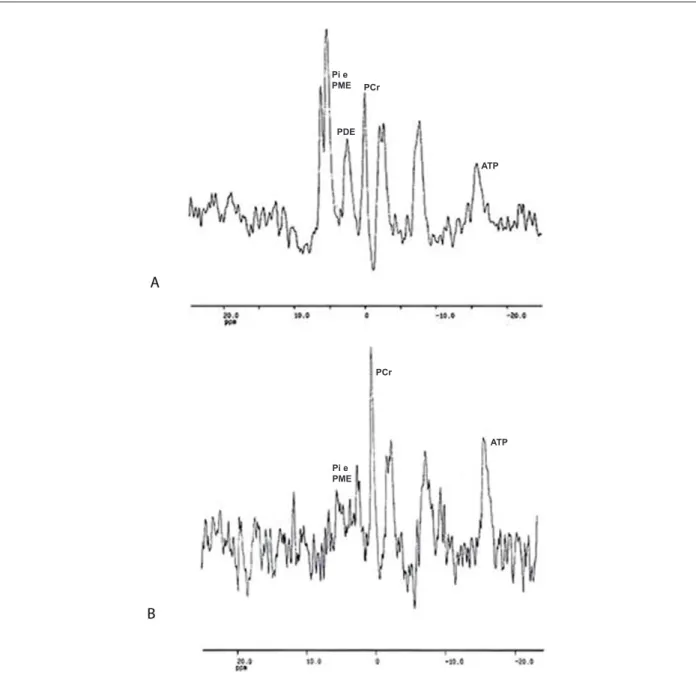
In C Group, the PCr/b-ATP ratio was 1.88 ± 0.08; in VD Group, 0.89 ± 0.31; in EKGalt Group, 1.52 ± 0.30, and in Ind Group, 1.40 ± 0.37, all of which were found to be smaller comparing to C Group (p < 0.001). Figure 1 shows examples of phosphorus-31 spectra.
We performed a comparative study of the resting energy metabolism in patients with ventricular dysfunction (VD Group) and those with preserved ventricular function (PF Group), by joining the patients from EKGalt groups and Ind Group.
• Clinical analysis - There was no significant difference between the two groups analyzed in relation to age or gender. There was a significant difference between the two groups only in relation to ejection fraction, VD Group presented a decrease value compared to PF Group (p <0.001).
• Comparative analysis of PCr/b-ATP ratios and ejection fraction - Cardiac PCr/b-ATP ratio levels were reduced in VD Group (0.89 ± 0.31) compared with the PF Group (1.47 ± 0.34, Graphic 1), and both had reduced levels compared to C Group (1.88 ± 0.08) (p <0.001).There was a positive correlation between left ventricular ejection fraction PCr/b -ATP ratios in a all 39 patients investigated (r = 0.64, p < 0.001, Graphic 2).
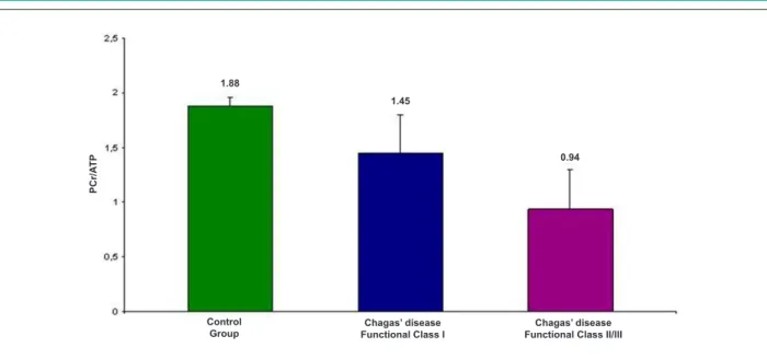
•Comparative analysis of NYHA functional class and PCr/b -ATP - patients in functional class I (22 patients) had PCr/b-ATP of 1.45 ± 0.35, and those in class II and III (17 patients), PCr/b-ATP of 0.94 ± 0.36 (p <0.001, Graphic 3).In this study, there was a good correlation between NYHA functional class and ejection fraction on echocardiography (15 out of the 16 patients studied with ventricular dysfunction were in class II or III of NYHA, 21 out of the 23 patients with preserved ventricular function were in NYHA class I).
Discussion
Figure 1 -(A) Phosphorus-31 spectrum of a patient from the ventricular dysfunction group. Observe the peak of the zero point phosphocreatine and ATP at position -16. The PCr/ATP ratio of this patient is 0.80. The peak of inorganic phosphorus (Pi) and phosphomonoester (PME) is high. (B) Phosphorus-31 spectrum of a patient from the group with preserved ventricular function and electrocardiogram with alterations. Observe the peak of zero point phosphocreatine and ATP in position - 16. The PCr/ ATP ratio of this patient is 1.60. The peak of inorganic phosphorus (Pi) and phosphomonester (PME) is not high. PCr - phosphocreatine, ATP - adenosine triphosphate, Pi - inorganic phosphorus; PME - phosphomonoester, PDE - phosphodiester; ppm - parts per million.
Pi e PME PCr
PDE
ATP
Pi e PME
PCr
ATP
myocardium. It does not require exposure to ionizing radiation or intravenous contrast14,15,19.
The initial in vivo application of MRS was in 197620, and
the first clinical application occurred in 198521. Since then,
it has become an important tool for noninvasive evaluation of multiple metabolic ways in the heart. The most important
Graphic 1 - Reduced cardiac levels of PCr/ATP in the group with ventricular dysfunction in relation to the group with preserved function and from these in relation to the control group (p <0.001). PCr - phosphocreatine; ATP - adenosine triphosphate.
PC
r/
A
T
P
1.88
0.89
1.47
Control
Ventricular dysfunction Preserved ventricular function
leads to a reduction in the PCr/ATP ratio. ATP levels fall only when PCr levels are very low13,14.
The phosphorus-31 spectrum in the human heart is shown as amplitudes or peaks in MRS (Figure 1), and the PCr peak, by convention, is considered the reference peak at 0.0 ppm. The values are negative on the right and represent ATP peaks (g, α, b), and are positive on the left, and represent phosphomonoester, inorganic phosphate and phosphodiester19-23 (fig 1).
In our study, we used the b-ATP peak, which is
considered the most useful of the three because it is not contaminated by other signals19. Thus, we evaluated the
relationship between PCr and ATP, rather than the absolute concentration of each component. Thus, the value of the first is approximately 10 mmol/kg dry weight, while the second is 5 to 6 mmol/kg dry weight.
The technique used in our study enables the analysis of the myocardial energy metabolism, as was previously done in the study of the pathogenesis of other heart diseases24. For
instance, in patients with heart failure, there is a reduction of
Graphic 2 - Positive correlation between the left ventricular ejection fraction on echocardiography and PCr/ATP in 39 patients with Chagas’ disease. PCr - phosphocreatine;
ATP - adenosine triphosphate; EF - left ventricular ejection fraction.
Graphic 3 - Reduction of PCr/ATP ratio in patients in NYHA functional class I in relation to the control group and patients in function class II and III in relation to other groups (p < 0.001). PCr - phosphocreatine, ATP - adenosine triphosphate; NYHA - New York Heart Association.
PC
r/
A
T
P
Control Group
Chagas’ disease Functional Class I
Chagas’ disease Functional Class II/III 1.88
1.45
0.94
the PCr/ATP ratio and after the use of beta blockers there is a reversal of this ratio25,26. In our study, we observed that the
resting energy metabolism of high-energy phosphates is altered in patients with Chagas’ disease, as demonstrated by the reduced levels of PCr/b-ATP, and this reduction is proportional to the degree of ventricular dysfunction and NYHA functional classes. Similar data for the relationship between the NYHA class and PCr/b-ATP have previously been documented in 19 patients with dilated cardiomyopathy, showing a relation between the severity of heart failure and myocardial energy metabolism25. The PCr/b-ATP values of Group C, found in our
study, were similar to those found in literature, which range from 1.6 to 2.027,28.
In literature, the relationship between left ventricular ejection fraction and myocardial energy metabolism in patients with dilated cardiomyopathy is controversial25,26,29,30. However,
our study found a clear positive correlation between left ventricular ejection fraction and PCr/b-ATP levels in Chagas’ disease, demonstrating a greater impairment of myocardial energy reserves at rest in these patients.
In our study, patients with CD without ventricular dysfunction showed, unlike the work done in patients with mild idiopathic dilated cardiomyopathy25,30,31, alterations in
energy metabolism at rest. Such alterations were less severe than those presented by patients with myocardial dysfunction, but still statistically significant. A possible explanation for this finding is the presence of myocardial degenerative impairment in the early stages of Chagas’ disease6,32 demonstrated by the
alterations found within the myocyte nucleus, in the T-tubule system and in mitochondrias, as well as by the alterations in the functions of ATPase myosin, monoamine oxidase and succinate dehydrogenase enzymes, involved in the maintenance of cellular energy homeostasis32.The inflammatory process in
Chagas’ cardiomyopathy is also more intense and persistent
than in idiopathic dilated cardiomyopathy, accounting for the highest degree of fibrosis and microcirculatory impairment detected by comparative studies performed by biopsy in these two pathologies33.
The impairment of the integrity of the myocardial cellular energy has been considered as a contributing factor to the deterioration of global myocardial function in various heart diseases23. One of the first clinical studies with phosphorus-31
MRS revealed small alterations in the cardiac energy metabolism of patients with dilated cardiomyopathy31. Later, it
was demonstrated in 20 patients with dilated cardiomyopathy (9 of ischemic origin and 11 of idiopathic origin), a significant reduction in PCr/b-ATP levels in relation to the control group (PCr/b-ATP of 1.46 ± 0.07 vs dilated cardiomyopathy vs. PCr/
b-ATP of 1.80 ± 0.06 in the control group, p < 0.001)30. We
also found a tendency for the presence of lower levels of PCr/b -ATP in patients with more severe myocardial dysfunction, which is an evidence of the relation between the impairment of the myocardial energy system and cardiac function. Another study also found reduced levels of PCr/b-ATP in individuals with advanced degrees of heart failure secondary to dilated cardiomyopathy, and no difference was found in individuals with milder degrees of the disease25.
Conclusion
Our study confirms the presence of alterations in the cardiac energy metabolism at rest in patients with Chagas’ disease that are related directly to the severity of cardiac involvement.
Future prospects
References
1. Bern C, Montgomery SP, Herwaldt BL, Rassi A Jr, Marin-Neto JA, Dantas RO, et al. Evaluation and treatment of Chagas’ disease in the United States: a systematic review. JAMA. 2007; 298 (18): 2171-81.
2. Mady C, Ianni BM, Arteaga E, Salemi VM, Frimm CC. Maximal functional capacity in patients with Chagas’ cardiomyopathy without congestive heart failure. J Card Fail. 2000; 6 (3): 220-4.
3. Higuchi ML. Chronic chagasic cardiopathy: the product of a turbulent host-parasite relationship. Rev Inst Med Trop S Paulo. 1997; 39: 53-60.
4. Marin-Neto JA. Cardiac dysautonomia and pathogenesis of Chagas’ heart disease. Int J Cardiol. 1998; 66 (2): 129-31.
5. Rossi MA. Microvascular changes as a cause of chronic cardiomyopathy in Chagas’ disease. Am Heart J. 1990; 120 (1): 233-6.
6. Rossi MA. The pattern of myocardial fibrosis in chronic Chagas’ heart disease. Int J Cardiol. 1991; 30 (1): 335-40.
7. Marin-Neto JA, Marzullo P, Marcassa C, Gallo L, Maciel BC, Bellina R, et al. Myocardial perfusion defects in chronic Chagas’ disease as detected by thallium-201 scintigraphy. Am J Cardiol. 1992; 69 (8): 780-4.
8. Acquatella H, Pérez JE, Condado JA, Sanchez I. Limited myocardial contractile reserve and chronotropic incompetence in patients with chronic Chagas’ disease. assessment by dobutamine stress echocardiography. J Am Coll Cardiol. 1999; 33 (2): 522-9.
9. Tanowitz HB, Kaul DK, Chen B, Morris SA, Factor SM, Weiss LM, et al. Compromised microcirculation in acute murine Trypanosoma cruzi infection. J Parasitol. 1996; 82 (1): 124-30.
10. Tanowitz HB, Burns ER, Sinha AK, Kahn NN, Morris SA, Factor SM, et al. Enhanced platelet adherence and aggregation in Chagas’ disease: a potential pathogenic mechanism for cardiomyopathy. Am J Trop Med Hyg. 1990; 43 (3): 274-81.
11. Ingwall JS. Is cardiac failure a consequence of decreased energy reserve? Circulation. 1993; 87: VII-58 – VII-62.
12. Kalil Fillho R, De Albuquerque CP, Weiss RG, Mocelim A, Bellotti G, Cerri G, Pileggi F. Normal high energy phosphate ratios in “stunned” human myocardium. J Am Coll Cardiol. 1997; 30 (5): 1228-32.
13. Ten Hove M, Neubauer S. MR spectroscopy in heart failure--clinical and experimental findings. Heart Fail Rev. 2007; 12 (1): 48-57.
14. Hudsmith LE, Neubauer S. Detection of myocardial disorders by magnetic resonance spectroscopy. Nat Clin Pract Cardiovasc Med. 2008; 5 (Suppl 2): S49-56.
15. Lamb HJ, van der Meer RW, de Roos A, Bax JJ. Cardiovascular molecular MR imaging. Eur J Nucl Med Mol Imaging. 2007; 34 (Suppl 1): S99-104.
16. Camargo ME, Oshino-Shimizu S, Macedo V, Peres BA, Castro C. Diagnóstico sorológico da infecção humana pelo Trypanosoma cruzi. Estudo comparativo de testes de fixação do complemento, imunofluorescência, hemaglutinação e floculação em 3.624 soros. Rev Inst Med Trop São Paulo. 1977; 19: 254-60.
17. Moffa PJ, Sanches PCR. Eletrocardiograma: normal e patológico. São Paulo: Editora Roca; 2001.
18. Lang RM, Bierig M, Devereux RB, Flachskampf FA, Foster E, Pellikka PA, et al. Recommendations for chamber quantification: a report from the American
Society of Echocardiography’s Guidelines and Standards Committee and the Chamber Quantification Writing Group, developed in conjunction with the European Association of Echocardiography, a branch of the European Society of Cardiology. J Am Soc Echocardiogr. 2005;18(12):1440-63.
19. Sardanelli F, Quarenghi M. MR spectroscopy of the heart. Radiol Med. 2006; 111 (8): 1025-34.
20. Dawson J, Gadian DG, Wilkie DR. Proceedings: living muscle studied by 31P nuclear magnetic resonance. J Physiol. 1976; 258 (2): 82P-83P.
21. Ordidge RJ, Connelly A, Lohman JAB. Image-selected in vivo spectroscopy (ISIS): a new technique for spatially selective NMR spectroscopy. J Magn Reson. 1985; 66: 283-94.
22. Bottomley PA. Noninvasive study of high-energy phosphate metabolism in human heart by depth-resolved 31P NMR spectroscopy. Science. 1985; 229 (4715): 769-72.
23. Beer M. Cardiac spectroscopy: techniques, indications and clinical results. Eur Radiol. 2004; 14 (6): 1034-47.
24. Tyler DJ, Hudsmith LE, Clarke K, Neubauer S, Robson MD. A comparison of cardiac (31) P MRS at 1.5 and 3 T. NMR Biomed. 2008; 21 (8): 793-8.
25. Neubauer S, Krahe T, Schindler R. 31P magnetic resonance spectroscopy in dilated cardiomyopathy and coronary artery disease: altered cardiac high-energy phosphate metabolism in heart failure. Circulation. 1992; 86 (6): 1810-8.
26. Neubauer S, Horn M, Pabst T, Gödde M, Lübke D, Jilling B, et al. Contributions of 31P-magnetic resonance spectroscopy to the understanding of dilated heart muscle disease. Eur Heart J. 1995; 16 (Suppl O): 115-8.
27. Bottomley PA. MR spectroscopy of the human heart: the status and the challenges. Radiology. 1994; 191 (3): 593-612.
28. Klug G, Wolf C, Trieb T, Frick M, Koehler A, Schocke MF, et al. Evaluation of transient apical ballooning with cardiac magnetic resonance imaging and 31-phosphorous magnetic resonance spectroscopy. Int J Cardiol. 2007; 118 (2): 249-52.
29. Hansch A, Rzanny R, Heyne JP, Leder U, Reichenbach JR, Kaiser WA. Noninvasive measurements of cardiac high-energy phosphate metabolites in dilated cardiomyopathy by using 31P spectroscopic chemical shift imaging. Eur Radiol. 2005; 15 (2): 319-23.
30. Hardy CJ, Weiss RG, Bottomley PA, Gerstenblith G. Altered myocardial high-energy phosphate metabolites in patients with dilated cardiomyopathy. Am Heart J. 1991; 122 (3 Pt 1): 795-801.
31. Schaefer S, Gober JR, Schwartz GG, Twieg DB, Weiner MW, Massie B. In vivo phosphorus-31 spectroscopy imaging in patients with global myocardial disease. Am J Cardiol. 1990; 65 (16): 1154-61.
32. Carrasco-Guerra HA, Palacios-Pru E, Dagert de Scorza C, Molina C, Inglessis G, Mendoza RVB. Clinical, histochemical and ultrastructural correlation in septal endomyocardial biopsies from chronic chagasic patients: detection of early myocardial damage. Am. Heart J. 1987; 113 (3): 716-24.
33. Higuchi ML, Fukasawa S, De Brito T, Parzianello LC, Bellotti G, Ramires JA. Different microcirculatory and interstitial matrix patterns in idiopathic dilated cardiomyopathy and Chagas’ disease: a three dimensional confocal microscopy study. Heart. 1999; 82 (3): 279-85.
making it possible to evaluate dynamic alterations in metabolite concentrations across the heart.
Potential Conflict of Interest
No potential conflict of interest relevant to this article was reported.
Sources of Funding
There were no external funding sources for this study.
Study Association