Accuracy of sonographic findings in breast
cancer: correlation between BI-RADS® categories
and histological findings*
Acurácia dos achados ultrassonográficos do câncer de mama: correlação da classificação BI-RADS® e achados histológicos
José Hermes Ribas do Nascimento1, Vinicius Duval da Silva2, Antonio Carlos Maciel3
OBJECTIVE: The main purpose of the present study is to evaluate the accuracy of ultrasonography (BI-RADS) in the diagnosis of breast cancer whereas the additional specific objectives are to describe the frequency of different sonographic findings and evaluating interobserver agreement. MATERIALS AND METHODS: Images of 110 patients who had been referred for biopsy with previous diagnosis of breast nodules were independently reviewed by two specialists according to the BI-RADS classification. Histological findings were utilized as a gold-standard. The accuracy of findings was determined. The chi-squared test for categorical variables was utilized in the analysis of the differences resulting from the groups comparison, and the interobserver agreement was calculated with kappa (κκκκκ) statistics. RESULTS: Among 110 breast masses evaluated by ultrasonography, 76 (69%) were benign and 34 (30.9%) were malignant. According to the radiologists, the sensitivity ranged from 70.5% to 82.3%, negative predictive value, from 81.1% to 87.5%, positive predictive value, from 42.1% to 45.1%, specificity from 56.58% to 55.2%, and accuracy from 60.9% to 63.6%. The global interobserver agreement was considered as moderate (κκκκκ = 0.50). CONCLUSION: The fourth edition of BI-RADS provides radiologists with an accurate clinical decision support system for the diagnosis and management of breast disease.
Keywords: Breast cancer; Ultrasonography; BI-RADS; Anatomopathological; Accuracy.
OBJETIVO: O objetivo geral do estudo é avaliar a acurácia da ultrassonografia (BI-RADS) no diagnóstico do câncer de mama, e os objetivos específicos, descrever a frequência de apresentação dos diferentes achados ultrassonográficos e a avaliação da concordância entre observadores. MATERIAIS E MÉTODOS: Exames de 110 pacientes encaminhados para biópsia, com diagnóstico prévio de nódulos, foram reanalisados indepen-dentemente por dois médicos especialistas utilizando a nomenclatura do BI-RADS. Os achados histológicos foram utilizados como padrão-ouro. A acurácia dos achados foi determinada. As diferenças nos grupos de comparação foram analisadas com teste qui-quadrado para variáveis categóricas e a concordância entre os médicos foi calculada por meio da estatística kappa (κκκκκ). RESULTADOS: Cento e dez massas mamárias foram avaliadas pelo ultrassom, sendo que 76 (69%) foram benignas e 34 (30,9%), malignas. Foram observados, entre os radiologistas, sensibilidade variando entre 70,5% e 82,3%, valor preditivo negativo entre 81,1% e 87,5%, valor preditivo positivo entre 42,1% e 45,1%, especificidade entre 56,58% e 55,2% e acurácia entre 60,9% e 63,6%. Na avaliação entre observadores foi obtida concordância global considerada mode-rada (κκκκκ = 0,50). CONCLUSÃO: O BI-RADS 4ª edição é um acurado sistema para auxiliar os médicos na des-crição das lesões mamárias e na tomada de condutas.
Unitermos: Câncer de mama; Ultrassonografia; BI-RADS; Anatomopatológico; Acurácia.
Abstract
Resumo
* Study developed at Pontifícia Universidade Católica do Rio Grande do Sul (PUCRS), Porto Alegre, RS, Brazil.
1. Master, MD, Radiologist, Pontifícia Universidade Católica do Rio Grande do Sul (PUCRS), Porto Alegre, RS, Brazil.
2. PhD, MD, Pathologist, Associate ProfessorProfessor, Pon-tifícia Universidade Católica do Rio Grande do Sul (PUCRS), Porto Alegre, RS, Brazil.
3. Head for the Unit of Radiology – Santa Casa de Misericór-dia de Porto Alegre, MD, Radiologist – Hospital de Clínicas de Porto Alegre, Porto Alegre, RS, Brazil.
Mailing address: Dr. José Hermes Ribas do Nascimento. Rua Marechal Floriano, 774, Meller Sul. Santo Ângelo, RS, Brazil, 98801-650. E-mail: [email protected]
Received December 27, 2008. Accepted after revision April 22, 2009.
ers provide high-resolution images, depth penetration and a high number of scanning lines(2,3).
Although breast ultrasonography has been historically utilized for differentiating solid from liquid lesions, there is an in-creasing interest in the utilization of this method for differentiating malignant from benign masses. Additionally, ultrasonogra-phy has become a valuable tool in the char-acterization of nodules found at
mammog-Nascimento JHR, Silva VD, Maciel AC. Accuracy of sonographic findings in breast cancer: correlation between BI-RADS® categories and histological findings. Radiol Bras. 2009;42(4):235–240.
INTRODUCTION
transduc-raphy, thus avoiding unnecessary biopsies and eliminating the necessity of follow-up mammography(4–6). The breast ultrasonog-raphy sensitivity has been reported as su-perior to that of mammography(7,8) in pre-menopausal women and, recently, sono-graphic screening has also been recom-mended for evaluation of dense breasts(9). Studies have demonstrated the usefulness of ultrasonography for detection of clini-cally and mammographiclini-cally occult, non-palpable breast carcinomas(10,11).
The Breast Imaging Reporting and Data System (BI-RADS®) lexicon for ultra-sonography was developed by the Ameri-can College of Radiology (ACR) aiming at increasing the clinical efficacy of the method and at standardizing the reports organization and wording. There is a spe-cific vocabulary for describing each lesion, and, at the end of the report, the lesion is classified into categories ranging from 0 to 6 according to the findings suspicion de-gree based on the positive predictive value (PPV) of the study for breast cancer(12–14). The BI-RADS lexicon includes a sono-graphic description of breast nodules or masses considering contours, orientation, margins, lesions limits, internal echoes pattern, characterization of posterior acous-tic shadowing, borders and abnormalities in adjacent tissues. At the end of the de-scription, the lesion is assigned to a BI-RADS category(13,15).
The present study primarily proposes an evaluation of the sonographic BI-RADS classification accuracy for differentiating benign lesions from malignant masses. The secondary objectives were the description of the frequency of different sonographic findings and the evaluation of the interob-server agreement.
MATERIALS AND METHODS
Two physicians specialized in breast im-aging diagnosis independently reviewed studies of 110 patients referred to a clinic in the Northwestern region of the Rio Grande do Sul state (Brazil) for core bi-opsy. Previously, all of them had been sonographically diagnosed with breast nodules or masses classified into BI-RADS categories 3, 4 or 5. Each specialist, with more than ten years of professional
expe-rience, course of residency in radiology, specialist title and/or course of specializa-tion in mammography by Colégio Brasi-leiro de Radiologia e Diagnóstico por Ima-gem (CBR), blindly reviewed the sono-graphic studies, utilizing the BI-RADS ter-minology, evaluation and recommenda-tions included in the most recent lexicon for echography. Later, the reviewed stud-ies were compared with the anatomopa-thological results.
The ultrasonography studies were per-formed with a high-resolution Sonoline G50 (Siemens Medical Solutions; Berlin, Germany) equipment with 7.5 MHz and 10 MHz linear array transducers.
The accuracy of the BI-RADS classifi-cation in ultrasonography was evaluated by calculating sensitivity, specificity, PPV and negative predictive value (NPV) for each of the described characteristics, and in the differentiation between benign and malig-nant lesions. Histological findings were utilized as standard criteria.
The interobserver agreement for the fi-nal categories and separately for each cat-egory was analyzed by the kappa test (κ) and the differences between groups were analyzed through the chi-square test for categorical variables.
The BI-RADS lexicon for ultrasonog-raphy considers the following terms for de-scribing breast nodules: shape, margins, orientation of the nodule in relation to the skin axis, lesion borders, internal echoes pattern, posterior acoustic characteristics and alterations in adjacent tissues.
After their description in compliance with the BI-RADS criteria, all the lesions were classified into categories 3, 4 or 5 (Chart 1).
Category 3 included the well-defined le-sions, ovoid or rounded in shape (contour),
with a parallel orientation in relation to the skin axis, circumscribed margins, absent posterior acoustic shadowing or presence of posterior acoustic enhancement, and ab-sence of alterations in adjacent tissues. Le-sions associating at least three signs of ma-lignancy were classified into BI-RADS cat-egory 5 including irregular contour, non-parallel orientation in relation to the skin axis, noncircumscribed margins, presence of hyperechogenic halo, posterior acoustic shadowing and alteration in the adjacent tissues.
BI-RADS category 4 included the le-sions that did not met the benignity crite-ria neither combined three signs of malig-nancy, so being classified as indeterminate. The patient’s age, the site and size of the lesion were also taken into consideration. Histological findings were compared with sonographic characteristics.
BI-RADS lexicon diagnostic accuracy, sensitivity, specificity, PPV and NPV for ultrasonography were calculated, including category 3 in the benign group, and unify-ing categories 4 (probably benign) and 5 in the malignant lesions group. VPPs and NPVs for each category and description were obtained.
RESULTS
The present study included 110 breast nodules, 108 in female patients and 2 in male patients. All the lesions were assessed by ultrasonography and later submitted to histological study. The patients’ mean age was 49.67 ± 12.09 years.
Based on the sonographic BI-RADS classification, the lesions were distributed as follows: observer A – 53 (48.18%) cat-egory 3, 39 (35.46%) catcat-egory 4, and 18 (16.4%) category 5; observer B – 48
Chart 1 Final clinical conduct according to BI-RADS classification(12).
Incomplete evaluation
Category 0 (zero): requires additional imaging evaluation
Complete evaluation
Category 1: negative
Category 2: negative findings
Category 3: probably benign findings – suggesting short interval follow-up
Category 4: suggestive of abnormality – biopsy should be considered (indeterminate)
Category 5: highly suggestive of malignancy – an appropriate conduct should be adopted
(43.64%) category 3, 44 (40%) category 4 and 18 (16.4%) category 5. No lesion was classified as categories 0, 1, 2 and 6.
Amongst all the cases included in the present study, 76 (69%) were benign, and 34 were malignant at the anatomopatho-logical study.
According to the observer A, NPV was 81.1%, PPV, 42.10%, sensitivity, 70.0%, specificity, 56.5%, and accuracy, 60.9%.
On the other hand, according to the ob-server B, NPV was 87.5%, PPV, 46.6%, sensitivity, 82%, specificity, 55.2% and accuracy, 63.6% (Tables 1 and 2).
Sonographic nodules characteristics
Sonographically, the nodules demon-strated the following morphological char-acteristics: lesions contour, margins, inter-nal echoes pattern, orientation in relation to the skin axis, posterior acoustic charac-teristics, borders and alterations in adjacent tissues(14).
a) Evaluation of the lesions shape
According to the observer A, the lesions were round-shaped in 71 cases, ovoid in 13 cases, and irregular in 25 cases. Among the 71 round-shaped lesions, 83.1% were be-nign, and 16.9% malignant. Among the ir-regular nodules, 21 were malignant, and four were benign for a PPV de 84%. The NPV for ovoid lesions was 42.9%,and for round-shaped lesions, 83.1%.
According to the observer B, round-shaped was described in 67 masses, ovoid in 22, and irregular in 21. Among the 67 rounded-shaped lesions, 76.1% were be-nign and 23.9%, malignant. The NPV for rounded-shaped lesions was 76.1%, and 77.3% for ovoid lesions. Among the irregu-lar lesions, 14 were malignant for a PPV of 65%.
b) Evaluation of the lesions margins
According to the observer A, the mar-gins were circumscribed in 68 cases, and
Table 1 Distribution of false- and true-positive results and false- and true-negative results based on pathological and discriminate diagnoses – observer A.
Positive disease Negative disease Total
Test result
T+ (category 4, 5)
T – (category3)
Total
Parameters
Sensitivity
Specificity
Positive predictive value
Negative predictive value
Accuracy
n
24
10
34
(%)
(42.11) TP
(18.87) FN
(30.9)
n
33
43
76
(%)
(57.89) FP
(81.13) TN
(69.1)
n
57
53
110 (%)
(100)
(100)
(100)
Formula
TP/(TP + FN)
TN/(TN + FP)
TP/(TP + FP)
TN/(TN + FN)
(TP + TN)/Total
%
70.59 (with disease and positive test)
56.58 (without disease and negative test)
42.1
81.1
60.9
T+, positive test (lesion rated as category 4 or 5); T –, negative test (lesion rated as category 3); TP, true-positive; FP, false-positive; TN, true-negative; FN, false-negative.
Table 2 Distribution of false- and true-positive results and false- and true-negative results based on pathological and discriminate diagnoses – observer B.
Positive disease Negative disease Total
Test result
T+ (category 4, 5)
T – (category 3)
Total
Parameters
Sensitivity
Specificity
Positive predictive value
Negative predictive value
Accuracy
n
28
6
34
(%)
(45.16) TP
(12.5) FN
(30.9)
n
34
42
76
(%)
(54.84) FP
(87.5) TN
(69.1)
n
62
48
110 (%)
(100)
(100)
(100)
Formula
TP/(TP + FN)
TN/(TN + FP)
TP/(TP + FP)
TN/(TN + FN)
(TP + TN)/Total
%
82.3 (with disease and positive test)
55.2 (without disease and negative test)
45.1
87.5
63.6
T+, positive test (lesion rated as category 4 or 5); T –, negative test (lesion rated as category 3); TP, true-positive; FP, false-positive; TN, true-negative; FN, false-negative.
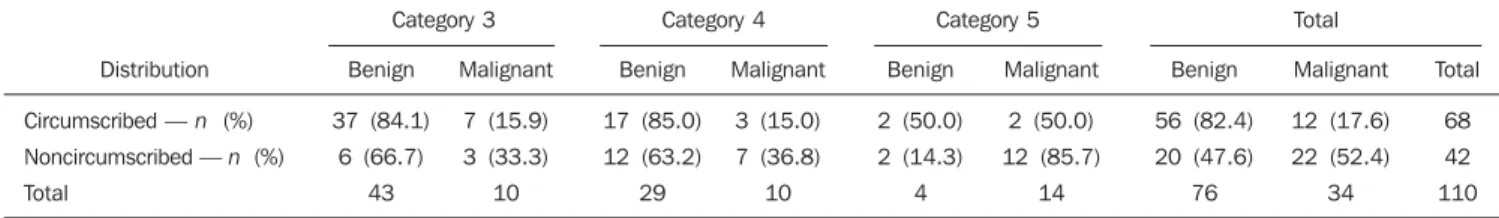
Table 3 Distribution of margins and relationship with the BI-RADS classification for ultrasonography – observer A.
Distribution
Circumscribed — n (%)
Noncircumscribed — n (%)
Total
Category 3 Category 4 Category 5 Total
Benign
37 (84.1)
6 (66.7)
43
Malignant
7 (15.9)
3 (33.3)
10
Benign
17 (85.0)
12 (63.2)
29
Malignant
3 (15.0)
7 (36.8)
10
Benign
2 (50.0)
2 (14.3)
4
Malignant
2 (50.0)
12 (85.7)
14
Benign
56 (82.4)
20 (47.6)
76
Malignant
12 (17.6)
22 (52.4)
34
Total
68
42
110 noncircumscribed in 42. Only 12 (17.6%)
of the 68 lesions with circumscribed mar-gins were malignant. Only four (6%) among the cases with circumscribed mar-gins were classified in category 5 (Table 3). Out of 42 masses, 22 with noncircum-scribed margins were malignant. The PPV for noncircumscribed margins was of 52.4%, and the NPV for circumscribed margins was of 82.4%. Sensitivity was 64.7%, and specificity, 73.7%.
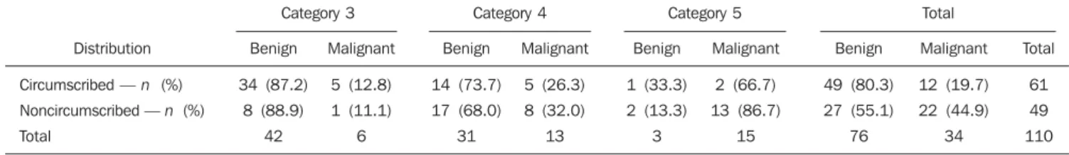
According to the observer B, margins were circumscribed in 61 cases and noncir-cumscribed in 49. Only 12 (19.7%) of the 61 lesions with circumscribed were malig-nant. Only three cases (4.9%) with circum-scribed margins were classified as category 5 (Table 4).
NPV for circumscribed margins was of 80.3%. Sensitivity was 64.7%, and speci-ficity, 64.5%.
c) Evaluation of internal echoes patterns
Internal echoes patterns were observed as follows: hypoechoic in 85 cases, iso-echoic in two, hyperiso-echoic in two, and complex in 14 cases. All the hyperechoic lesions were benign, and 71.4% of lesions with complex pattern were benign. Among the 85 (77.3%) hypoechoic nodules, 26 were malignant, with PPV of 30.6%.
According to the observer B, the inter-nal echoes pattern were hypoechoic in 79 cases, isoechoic in two, hyperechoic in five, and complex in 17 cases. Particularly, the complex pattern represented 15 (92.8%) of the nodules classified as catego-ries 4 and 5. All the hyperechoic lesions were benign, and 58.8% of lesions with complex pattern were benign. Among the 79 (71.8%) hypoechoic nodules, 22 were malignant, with PPV of 27.8%.
d) Evaluation of nodule orientation
in relation to the skin axis
For both observers, the mean size of the lesions parallel to the skin axis was 14.2 ± 9.9 mm, and 9.4 ± 4.5 mm in the lesions with vertical orientation, with prevalence in the upper breast quadrants.
Parallel orientation in relation to the skin axis was present in 101 cases (28 ma-lignant, and 73 benign lesions) for a NPV of 72.3%. Antiparallel orientation was present in seven cases (four malignant and three benign lesions) for a PPV of 72.3%.
e) Description of the posterior acoustic
characteristic
According to the observer A, the ab-sence of posterior acoustic characteristic presented a NPV of 46.7%. Out of the 110 lesions, 49 presented posterior acoustic enhancement and, among them, 40 were benign, with NPV of 81.6%; and posterior
acoustic shadowing was described in 13 cases, of which two were malignant, with PPV of 15%.
According the observer B, the absence of posterior acoustic characteristic pre-sented a NPV of 58.6%. Out of the 110 lesions, 34 presented posterior acoustic en-hancement and, among them, 29 were be-nign, with NPV of 85.3%; and posterior acoustic shadowing was described in 11 cases, of which four were malignant, with PPV of 36.4%.
f) Description of lesions borders
According to the observer A, 38 (34.5%) of 110 cases presented abrupt interface and 65 (59.0%) demonstrated echogenic halo. Echogenic halo was described in 27 cases classified as category 3, with NPV of 72.3%. Abrupt interface presented NPV of 68.4%.
According to the observer B, 31 (28.1%) of cases presented abrupt interface, and 66 (60%) of the 110 cases demonstrated echo-genic halo. Echoecho-genic halo was described in 24 cases classified as category 3, with NPV of 72.1%. Abrupt interface presented NPV of 61.2%.
g) Adjacent tissues appearance
According the observer A, 96 of the 110 described masses did not present alteration in adjacent tissues and, among them 52 were classified as category 3, with NPV of 76.6%. Out of 24 malignant lesions, 11 (45%) presented alteration in adjacent tissues, with PPV of 45%. Skin thickening was not observed in any case.
According the observer B, 96 of the 110 described masses did not present alteration in adjacent tissues and, among them 45 were classified as category 3, with NPV of 72.6%. Out of 34 malignant lesions, 26 (76%) presented alteration in adjacent tissues, with PPV of 76%. Skin thickening was not observed in any case.
h) Interobserver agreement (Table 5)
As far as the sonographic description is concerned, a moderate interobserver agree-ment was observed in the evaluation of nodules orientation (κ = 0.52), that was described as parallel or antiparallel in re-lation to the skin axis.
A moderate agreement was observed in the evaluation of the lesions contour (κ = 0.50). Low interobserver agreement (κ = 0.29) was observed in the evaluation of the lesion borders.
Moderate agreement was also observed in the evaluation of the lesion margins (κ = 0.53) and in the description of internal echoes pattern (κ = 0.56).
The different terms utilized for describ-ing posterior acoustic characteristic has also determined a moderate interobserver agreement (κ = 0.51).
A moderate interobserver agreement (κ = 0.51) was also observed in the evaluation of adjacent tissues, especially in cases where no alteration was found.
The κ value, for unified categories 4 and 5 was 0.36. The prevalence of breast can-cer in the present study was of 34 (30.9%).
DISCUSSION
The BI-RADS classification for mam-mography was the first attempt to standard-Table 4 Distribution of margins and relationship with the BI-RADS classification for ultrasonography – observer B.
Distribution
Circumscribed — n (%)
Noncircumscribed — n (%)
Total
Category 3 Category 4 Category 5 Total
Benign
34 (87.2)
8 (88.9)
42
Malignant
5 (12.8)
1 (11.1)
6
Benign
14 (73.7)
17 (68.0)
31
Malignant
5 (26.3)
8 (32.0)
13
Benign
1 (33.3)
2 (13.3)
3
Malignant
2 (66.7)
13 (86.7)
15
Benign
49 (80.3)
27 (55.1)
76
Malignant
12 (19.7)
22 (44.9)
34
Total
61
49
110
Masses description
Lesions contour
Lesions margins
Internal echoes pattern
Lesions borders
Orientation in relation to the skin axis
Posterior acoustic pattern
Adjacent tissues appearance
BI-RADS
κ
values
0.50
0.53
0.56
0.29
0.52
0.51
0.51
ize imaging findings in descriptive terms, constituting an important tool for aiding physicians both in the suspicion of malig-nancy and in the decision making about the strategy to be adopted(16–18). In 2003, the BI-RADS lexicon was updated, with a re-finement in the description of microcal-cifications and the inclusion of topics re-garding breast ultrasonography and mag-netic resonance imaging.
Because of the frequency of overlap-ping between radiological and echographic findings and the great PPV variability among BI-RADS categories 3, 4 and 5 in mammography(18), breast lesions indicative of malignancy detected at mammography and ultrasonography have been evaluated by biopsy to prove their malignancy or be-nignity(4,8,19). A high number of biopsies is performed for benign lesions because of several factors; among them the patients’ dread; physicians’ uncertainty or even the standard protocols utilized(1,20).
Ultrasonography should not be utilized only for differentiating cystic from solid nodules and in the evaluation of dense breasts. This method must be exploited with an accurate interpretation of the char-acteristics of each suspicious lesion in or-der to reduce the number of biopsies in benign lesions(21).
Improvements in the sonographic diag-nosis have been achieved with the intro-duction by the American College of Radi-ology, of the BI-RADS classification to aid radiologists in the description of sono-graphic findings and that define the final classification into categories associated with a better clinical management of the cases(12).
In the present study, both observers found a sensitivity ranging between 70% and 80% (identification of malignant le-sions patients with breast cancer) and high NPV, between 81% and 87% (identifica-tion of negative findings in cancer-free patients), in relation to characteristics de-scribed in the BI-RADS with 18% FN. However, BI-RADS presented a low speci-ficity, between 55% and 56% (cancer-free patients with negative studies) because of the high number of false-positive findings. The PPV (number of cancers for sono-graphic characteristics) ranged between 45.1% and 42.1%.
In the present study, the sonographic ac-curacy ranged from 60.9% to 63.6% in the differentiation between benign and malig-nant lesions with the utilization of the BI-RADS. The NPV for the category 3 ranged from 81.1% to 87.5% between the observ-ers, with a PPV ranging between 42.1% and 45.1%, similarly to the studies devel-oped by Costantini et al.(15) and Roveda Jr et al.(20), who have demonstrated a NPV ranging between, respectively, 92.3% and 70.58% for category 3.
Thus, the utilization of the category 3, as probably benign, is a tool utilized by ra-diologists to avoid unnecessary biopsies, considering that the risk for malignancy of lesions described in this category corre-sponds to less than 2%(12). If an increase in the lesions dimensions were observed in the follow-up, there would be a trend to-wards changing to BI-RADS category 4, so that the biopsy could be appropriate.
The classification of breast nodules into category 4 presents the same clinical im-pact and meaning as those described for category 5, since in both cases biopsy would be indicated. In the present study, the PPV for categories 4 and 5 was o, respec-tively, 45.2% and 42.2%, similarly to the study developed by Roveda Jr et al.(20), with a 50% PPV in category 4.
The analysis of the sonographic charac-teristics associated with the classification into categories 4 and 5 demonstrated that lesions with proved malignancy were fre-quently associated with hypoechogenicity, irregular contours, noncircumscribed mar-gins, and antiparallel orientation in relation to the skin axis, although many of the be-nign nodules classified as BI-RADS cat-egories 4 and 5 were hypoechoic, even being associated with circumscribed mar-gins and parallel orientation in relation to the skin axis.
It could be observed that with the pres-ence of three of the following findings, such as posterior acoustic shadowing, ir-regular contours, noncircumscribed mar-gins, hypoechoic halo and antiparallel ori-entation in relation to the skin axis, the le-sions were normally classified into catego-ries 4 and 5, in accordance with the find-ings described by Chen et al.(22).
Masses demonstrating more than three characteristics suggestive of malignancy
were classified into category 5 by both observers.
Nodule margins represented a relevant criterion in the differentiation between be-nign and malignant lesions, with a NPV ranging between 82.4% and 80.3% for cir-cumscribed margins, not very different from the findings reported by Calas et al.(23), who had observed a NPV of 97% for circumscribed lesions. In the present study, the PPV for noncircumscribed margins ranged between 52.4% and 44.9%, differ-ently from the NPV described by Calas et al.(23), corresponding to 70.4%.
Rounded contour (shape) was associ-ated with high NPV that ranged between 83.3% and 76.1%; and irregular contour, with high PPV, that ranged between 84% and 65% for both observers.
Hypoechoic halo demonstrated a lower PPV than irregular contour and noncircum-scribed margins. Echogenic halo presented a NPV ranging between 72.3% and 72.1%, and abrupt interface presented a NPV rang-ing from 68.4% to 61.2% between the ob-servers.
Antiparallel nodule orientation in rela-tion to the skin axis presented a high PPV (72.3%). In the study developed by Calas et al.(23) a PPV of 57.6% has been observed. The posterior acoustic characteristic is a result of the sound attenuation. The pos-terior acoustic enhancement presents a NPV between 81% and 85%, for both ob-servers. Posterior acoustic shadowing pre-sented a low PPV, ranging between 15% and 35% in the BI-RADS categories 4 and 5. Although posterior acoustic shadowing is a sonographic characteristic of malignant lesions(15), this finding was not confirmed in the present study, being also observed in benign lesions. Fine bilateral shadow-ing was considered as a sign of benign le-sion.
CONCLUSION
The sonographic evaluation of breasts utilizing the BI-RADS classification is an accurate method, with the interobserver variability ranging between 60.9% and 63.3% in the differentiation of malignant from benign lesions. The most frequent sonographic findings of neoplasms were irregular nodules with noncircumscribed margins and antiparallel orientation. In the present study, complex or hypoechoic in-ternal echoes pattern, the hypoechoic bor-ders of the lesions, and posterior acoustic shadowing presented low PPV. The over-all interobserver variability was moderate. It is believed that the practice, the sys-tematic follow-up periods, the double-read-ing technique, and BI-RADS traindouble-read-ing courses for physicians should be imple-mented to improve even more the accuracy in the diagnosis of breast diseases, thus reducing the number of unnecessary and expensive invasive procedures.
REFERENCES
1. Chala LF, Barros N. Avaliação das mamas com métodos de imagem [editorial]. Radiol Bras. 2007;40(1):iv–vi.
2. Guiseppetti GM, Giuliani F, Baldassarre S, et al. Metodologia e semiologia. In: Veronesi U, editor. Mastologia oncológica. Rio de Janeiro: Medsi; 2002. p. 95–106.
3. Baker JA, Soo MS. Breast US: assessment of tech-nical quality and image interpretation. Radiology. 2002;223:229–38.
4. Parker SH, Stavros AT, Dennis MA. Needle bi-opsy techniques. Radiol Clin North Am. 1995;33: 1171–86.
5. Stavros AT, Thickman D, Rapp CL, et al. Solid breast nodules: use of sonography to distinguish between benign and malignant lesions. Radiol-ogy. 1995;196:123–34.
6. Bassett LW, Kim CH. Breast imaging: mammog-raphy and ultrasonogmammog-raphy. Magn Reson Imaging Clin N Am. 2001;9:251–71.
7. Schroeder RJ, Bostanjoglo M, Rademaker J, et al. Role of power Doppler techniques and ultrasound contrast enhancement in the differential diagno-sis of focal breast lesions. Eur Radiol. 2003;13: 68–79.
8. Leconte I, Feger C, Galant C, et al. Mammogra-phy and subsequent whole-breast sonograMammogra-phy of nonpalpable breast cancers: the importance of radiologic breast density. AJR Am J Roentgenol. 2003;180:1675–9.
9. Kolb TM, Lichy J, Newhouse JH. Occult cancer in women with dense breasts: detection with screening US – diagnostic yield and tumor char-acteristics. Radiology. 1998;207:191–9.
10. Kolb TM, Lichy J, Newhouse JH. Comparison of the performance of screening mammography, physical examination, and breast US and evalu-ation of factors that influence them: an analysis of 27,825 patient evaluations. Radiology. 2002; 225:165–75.
11. Saarenmaa I, Salminen T, Geiger U, et al. The ef-fect of age and density of the breast on the sensi-tivity of breast cancer diagnostic by mammogra-phy and ultrasonogramammogra-phy. Breast Cancer Res Treat. 2001;67:117–23.
12. American College of Radiology. BI-RADS: ultra-sound, 1st ed. In: Breast Imaging Reporting and Data System: BI-RADS atlas, 4th ed. Reston: American College of Radiology; 2003. 13. Colégio Brasileiro de Radiologia. BI-RADS –
Sistema de laudos e registro de dados de imagem da mama. São Paulo: Colégio Brasileiro de Ra-diologia; 2005.
14. Miller AB, To T, Baines CJ, et al. Canadian Na-tional Breast Screening Study-2: 13-year results of a randomized trial in women aged 50-59 years. J Natl Cancer Inst. 2000;92:1490–9.
15. Costantini M, Belli P, Lombardi R, et al.
Charac-terization of solid breast masses: use of the sonographic breast imaging reporting and data system lexicon. J Ultrasound Med. 2006;25:649– 59.
16. Melhado VC, Alvares BR, Almeida OJ. Correla-ção radiológica e histológica de lesões mamárias não-palpáveis em pacientes submetidas a marca-ção pré-cirúrgica, utilizando-se o sistema BI-RADS. Radiol Bras. 2007;40:9–11.
17. Liberman L, Abramson A, Squires FB, et al. The breast imaging report and data system: positive predictive value of mammographic features and final assessment categories. AJR Am J Roentgenol. 1998;171:35–40.
18. Kestelman FP, Souza GA, Thuler LC, et al. Breast Imaging Reporting and Data System – BI-RADS®: valor preditivo positivo das categorias
3, 4 e 5. Revisão sistemática da literatura. Radiol Bras. 2007;40:173–7.
19. Fleury EFC, Rinaldi JF, Piato S, et al. Apresenta-ção das lesões mamárias císticas à ultra-sonogra-fia utilizando a elastograultra-sonogra-fia. Radiol Bras. 2008; 41:167–72.
20. Roveda Jr D, Piato S, Oliveira VM, et al. Valores preditivos das categorias 3, 4 e 5 do sistema BI-RADS em lesões mamárias nodulares não-palpá-veis avaliadas por mamografia, ultra-sonografia e ressonância magnética. Radiol Bras. 2007;40: 93–8.
21. Ciatto S, Houssami N, Apruzzese A, et al. Reader variability in reporting breast imaging according to BI-RADS assessment categories (the Florence experience). Breast. 2006;15:44–51.
22. Chen SC, Cheung YC, Su CH, et al. Analysis of sonographic features for the differentiation of benign and malignant breast tumors of different sizes. Ultrasound Obstet Gynecol. 2004;23:188– 93.