A preliminary experiment utilizing “aneurysm neck
remodeling technique” for management of complex
wide-necked renal artery aneurysms*
Experiência preliminar com o uso da técnica de “remodelagem de colo” para tratamento endovascular de aneurismas complexos da artéria renal
Eduardo Wajnberg1, Diogo Aquino2, Gabriela Spilberg2
OBJECTIVE: To report preliminary results of aneurysm neck remodeling in the management of wide-necked renal artery aneurysms. MATERIALS AND METHODS: Five patients (three women and two men between 49–72 years; mean age, 62 years) with wide-necked renal artery aneurysms measuring from 10 to 25 mm in diameter were submitted to balloon-assisted coil embolization along a three-year period. The micro-balloon was placed along the aneurysm neck and temporarily inflated for introduction of detachable microcoils into the aneurysmal sac. RESULTS: Balloon positioning and microcoil embolization were successfully completed in all of the cases with achievement of complete aneurysm occlusion without coil protrusion or parent vessel obstruction. CONCLUSION: The present preliminary experiment indicates that the “aneurysm neck remodeling” is technically feasible and effective in the management of complex renal artery aneurysms without sacrificing any arterial branch.
Keywords: Aneurysms; Renal; Endovascular treatment; Remodeling technique.
OBJETIVO: Relatar os resultados preliminares da aplicação da técnica de “remodelagem do colo” no trata-mento dos aneurismas de colo largo da artéria renal. MATERIAIS E MÉTODOS: Cinco pacientes (três mulhe-res e dois homens, com idade média de 62 anos, intervalo de 49–72 anos) com aneurismas da artéria renal variando de 10 a 25 mm de diâmetro, de colo largo, foram tratados com técnicas de embolização assistidas por “remodelagem do colo” com balão durante o período de três anos. O microbalão era posicionado diante do colo do aneurisma e insuflado, temporariamente, durante a colocação das micromolas destacáveis no interior do aneurisma. RESULTADOS: O posicionamento do balão e a colocação das micromolas foram rea-lizados com êxito em todos os casos. Oclusão completa do aneurisma, sem protrusão de micromolas ou obstrução do vaso parental, foi alcançada em todos os pacientes. CONCLUSÃO: Nossa experiência prelimi-nar indica que a aplicação da técnica de “remodelagem do colo” no tratamento dos aneurismas de colo largo da artéria renal é tecnicamente viável e eficaz para o tratamento endovascular de aneurismas complexos da artéria renal, sem o sacrifício de qualquer ramo arterial.
Unitermos: Aneurismas; Renal; Tratamento endovascular; Remodelagem.
Abstract
Resumo
* Study developed at Unit of Interventional Radiology, Hospi-tal Universitário Clementino Fraga Filho – Universidade Federal do Rio de Janeiro (HUCFF-UFRJ), Rio de Janeiro, RJ, Brazil.
1. Specialist in Interventional Neuroradiology and Diagnostic Imaging, MD, Unit of Interventional Radiology, Hospital Univer-sitário Clementino Fraga Filho – Universidade Federal do Rio de Janeiro (HUCFF-UFRJ), Rio de Janeiro, RJ, Brazil.
2. MDs, Residents at Department of Radiology and Diagnos-tic Imaging, Universidade Federal do Rio de Janeiro (UFRJ), Rio de Janeiro, RJ, Brazil.
Mailing address: Dr. Eduardo Wajnberg. Rua Lopes Quintas, 100, Bloco I/602, Jardim Botânico. Rio de Janeiro, RJ, Brazil, 22460-010. E-mail: [email protected]
management of RAAs may be performed by means of balloon-catheters, liquid em-bolic agents, covered stents, traditional platinum microcoils or controlled-release microcoils, such as the Guglielmi detach-able microcoils (GDC)(7–9). This type of microcoils allows a precise release once its correct positioning is confirmed by arteri-ography.
Endovascular occlusion of lateral wall or narrow neck aneurysms with microcoils generally is not very laborious. The utili-zation of endoprostheses (or covered stents) also is feasible for the management of such aneurysms. However, if the aneu-rysm involves an arterial bifurcation, or
Wajnberg E, Aquino D, Spilberg G. A preliminary experiment utilizing “aneurysm neck remodeling technique” for manage-ment of complex wide-necked renal artery aneurysms. Radiol Bras. 2010;43(1):29–33.
been diagnosed with higher frequency be-cause of the increasing utilization of non-invasive diagnostic imaging methods. Re-nal artery aneurysms may be treated with conventional surgery(3–5) and with endovas-cular therapy, which is less invasive so that recently it has become preferable to sur-gery. The development of microcatheters, guide-wires and novel embolic devices that were firstly utilized in interventional neuroradiology, allows a selective embo-lization even in cases of complex wide-necked lesions(6). Endovascular therapy for INTRODUCTION
Renal artery aneurysms (RAAs) repre-sent 22% of visceral aneurysms(1) and 1% of all aneurysms(2,3). However, despite their low incidence, renal artery aneurysms have
even is located only few millimeters from a vascular bifurcation, such endovascular treatment modalities may place the perme-ability of these adjacent vascular branches at risk(10).
Currently, the aneurysm neck remodel-ing technique (Moret technique) or bal-loon-assisted embolization is a technique widely utilized in the management of in-tracranial wide-necked aneurysms. Such technique has considerably increased the feasibility and usefulness of the endovas-cular therapy in cases of more complex aneurysms.
The authors present preliminary out-comes in patients with complex wide-necked renal artery aneurysms submitted to this technique originally adopted for neu-rovascular applications.
MATERIALS AND METHODS
The authors retrospectively analyze five cases of wide-necked renal artery aneu-rysms endovascularly treated with the neck remodeling technique by the team of Hos-pital Universitário Clementino Fraga Filho – Universidade Federal do Rio de Janeiro, RJ, Brazil, in the period from March/2006 to March/2009. The patients were in the age range between 49 and 72 year, and the aneurysms sizes ranged from 10 to 25 mm. During this three-year period, five wide-necked renal artery aneurysms were treated by means of balloon-assisted embolization with the neck remodeling technique. Ex-clusion criteria were the presence of uncorrectable coagulopathy and narrow neck aneurysm (body:neck ratio > 2).
Three of the aneurysms were initially di-agnosed at computed tomography angiog-raphy and two at color Doppler ultrasonog-raphy. Detailed study of the aneurysmal neck and analysis of the origin and course of arterial branches at risk for occlusion or
microcoil protrusion during embolization were reserved for digital catheter angiog-raphy. The mentioned aneurysms were con-sidered as complex because of their loca-tion in the renal artery bi- or trifurcaloca-tion, or involving the origin of segmental artery branches and, for this reason indicating a balloon- or stent-assisted procedure to pro-tect the parent artery. In the author’s insti-tution, the balloon-assisted neck remodel-ing technique is generally adopted to avoid the necessity of extended platelet antiag-gregation therapy.
Patients’ clinical data and aneurysms’ characteristics are shown on Table 1. The contralateral kidney was healthy in all of the five cases. The main indication for the treatment was the presence of difficult-to-manage hypertension in two of the patients, increase in the aneurysm size observed at follow-up in one, flank pain associated with hematuria in one, and size > 2 cm in one patient. A term of free and informed consent was signed by all the patients.
A 60 cm-long 6Fr sheath (Cook, Inc.; Bloomington, USA) was introduced by means of puncture of the right femoral ar-tery and positioned at the origin of the re-nal artery of interest for digital angiogra-phy. An intravenous 5,000 U heparin bo-lus was performed at the beginning of the process to keep the ACT (activated coagu-lation time) at two-threefold higher than the basal time. . The aneurysms and parent vessels were measured with the aid of a standard software for anatomic measure-ments. Through the 6Fr sheath, a Hyper-form 4 × 20 mm compliant balloon cath-eter (Figure 1) with a Silverspeed 10 micro-guidewire (MTI Microtherapeutics; Irvine, USA) was introduced into the main seg-mental artery branch and positioned along the aneurysmal neck. Through the same 6Fr sheath, a microcatheter with two radio-paque marks (SL 1018) with Transend 14
guidewire (Boston Scientific; Natick, USA) was advanced within the aneurysm, successively releasing platinum microcoils (GDC-Boston Scientific; Natick, USA) with different diameters and lengths, the first microcoil presenting a 3D shape, with a maximum size approximately similar to the aneurysm size. A road map was utilized in this phase of the procedure. Before each microcoil detachment, the balloon was deinflated to check whether there was a microcoil protrusion through the aneurys-mal neck. Once each microcoil was suc-cessful positioned, the balloon was dein-flated and an angiogram was performed to confirm the arterial patency. Then, control arteriography demonstrated complete an-eurysm obliteration and a dense microcoils agglomerate within the aneurysm, with main renal artery and respective segmen-tal branches permeability and normal nephrogram (Figure 2). The 6Fr sheath was drawn back and the femoral hemostasis was achieved with 6F Angio-Seal (St Jude Medical; Minnetonka, USA).
RESULTS
Embolization with neck remodeling technique was technically successful in all of the five patients. Angiography demon-strated complete aneurysmal occlusion without microcoil protrusion into the par-ent vessel after the treatmpar-ent completion in all of the cases, with no arterial flow com-promise. None of the patients presented significant post-procedural increase in lev-els of nitrogen waste. Three of the patients were submitted to follow-up with com-puted tomography angiography within 6 to 14 months after the embolization (mean follow-up period, 10 months). The other two patients could not be reached or re-fused to undergo follow-up studies. At such follow-up studies, the aneurysms remained
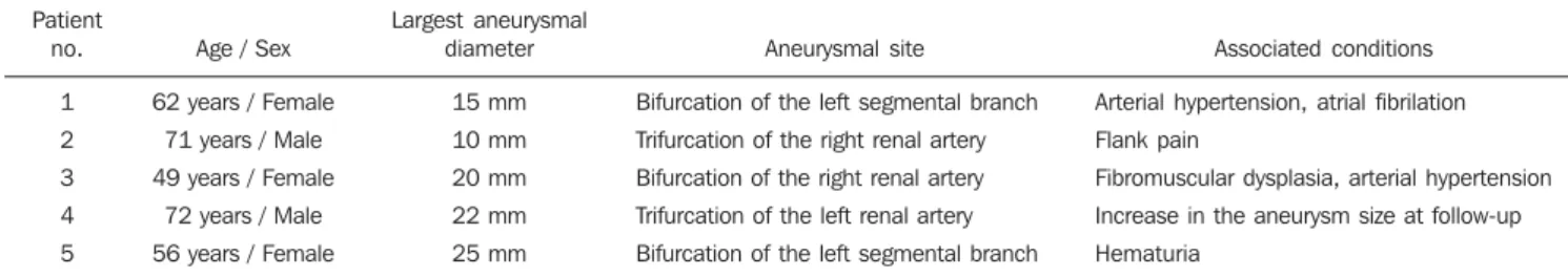
Table 1 Clinical and angiographic characteristics of renal artery aneurysms.
Patient no. 1 2 3 4 5
Age / Sex
62 years / Female
71 years / Male
49 years / Female
72 years / Male
56 years / Female
Largest aneurysmal diameter 15 mm 10 mm 20 mm 22 mm 25 mm Aneurysmal site
Bifurcation of the left segmental branch
Trifurcation of the right renal artery
Bifurcation of the right renal artery
Trifurcation of the left renal artery
Bifurcation of the left segmental branch
Associated conditions
Arterial hypertension, atrial fibrilation
Flank pain
Fibromuscular dysplasia, arterial hypertension
Increase in the aneurysm size at follow-up
totally occluded, with no aneurysmal reca-nalization or microcoils compaction. At follow-up, two of the patients presented improvement in their previously observed arterial hypertension.
DISCUSSION
Renal artery aneurysms represent one of the most common visceral aneurysms (15% to 22% of cases)(8), being found in 0.3% to
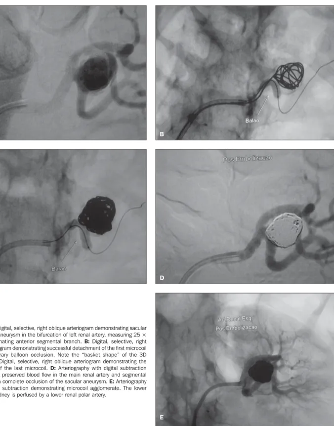
0.7% of autopsies and in up to 1% of renal arteriograms(4). Such aneurysms are most frequently found in women. Most of them present as a non-calcified, sacular dilata-tion, tending to occur in the bifurcation of Figure 1. A: Digital, selective, right oblique arteriogram demonstrating sacular
wide-necked aneurysm in the bifurcation of left renal artery, measuring 25 × 12 mm, originating anterior segmental branch. B: Digital, selective, right oblique arteriogram demonstrating successful detachment of the first microcoil during temporary balloon occlusion. Note the “basket shape” of the 3D microcoil. C: Digital, selective, right oblique arteriogram demonstrating the detachment of the last microcoil. D: Arteriography with digital subtraction demonstrating preserved blood flow in the main renal artery and segmental branches, with complete occlusion of the sacular aneurysm. E: Arteriography without digital subtraction demonstrating microcoil agglomerate. The lower pole of this kidney is perfused by a lower renal polar artery.
A B
C D
the main renal artery. The primary etiology is degenerative, with atherosclerosis and fibromuscular dysplasia being the most fre-quent causes. Vasculitis (for example, nodous polyarteritis)(11), trauma(12), neo-plasias (for example, angiomyolipoma), mycotic and iatrogenic aneurysms (for ex-ample, post-biopsy aneurysms), besides the idiopathic ones, constitute other less fre-quent causes(13,14).
In most of cases, RAAs are asymptom-atic, although rupture may occur with ret-roperitoneal hemorrhage, peripheral ves-sels embolization or even arterial thrombo-sis. The RAA is associated with renovas-cular hypertension in up to 73% of cases(15). Other complications include dissection, renal infarction and arteriovenous fistula.
Improvement in the arterial hypertension is a consensus in the literature(11), and hema-turia resolution is described in 30% of RAAs(16,17). The therapeutic decisions should be based, or not, on symptoms, pa-tient’s sex, hypertension severity, antici-pated pregnancy, childbearing age, ana-tomic characteristics of the aneurysm, rup-ture, size, expansion and distal emboliza-tion. Fusiform type and arterial wall calci-fication suggest protection against rupture. While size > 2 cm is considered as the start-ing point for vascular treatment, rupture of aneurysms < 2 cm has been reported(18–20). Young women, particularly those with anticipated pregnancy, are considered as patients at a higher risk for aneurysm rup-ture. Generally, such patients are
asymp-tomatic, but complications such as expan-sion, rupture or thrombi embolism and re-nal infarction may occur. In some cases, these microembolisms may cause renal is-chemia and consequential renovascular hy-pertension, although such relation still re-mains controversial.
Renal artery rupture occurs in less than 3% of cases, and is most frequently ob-served in cases of intrarenal aneurysms.
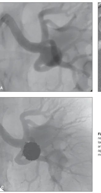
The mortality rate among pregnant women with RAA rupture is around 80%(4). Despite the establishment of size > 2 cm as the starting point for interventional treat-ment, studies in the literature advocate a conservative approach(5,19). For aneurysms with < 2 cm in size, follow-up with com-puted tomography or MRI is appropriate. Figure 2. A: Digital, selective, right oblique arteriogram demonstrating saccular, wide-necked aneurysm in the trifurcation of the left renal artery, measuring 22 mm in the largest diameters. B: Digital, selective, right oblique arteriogram demonstrating the final angiographic result. C: Arteriography without digital subtraction demonstrating microcoils agglomerate determining complete occlusion of sacular aneurysm, with preserved seg-mental branches.
A B
The management of intracranial aneu-rysms with detachable microcoils was firstly described in 1991 by Guglielmi et al.(21,22). Guglielmi detachable coils (GDC) comprise a proximal teflon guidewire dis-tally connected with a platinum microcoil with several circular memory sizes. The microcoils detachment from their guide-wires occurs by means of electrolysis, as an electric current is applied, with the positive pole connected to the distal portion of the guidewire, and the negative pole (earth wire) connected to the patient.
The balloon-assisted embolization (neck remodeling technique) was firstly de-scribed by Moret et al.(18), consisting in the utilization of a microcatheter with a com-pliant balloon that temporarily occludes de intracranial aneurysmal neck during the placement of the microcoils, preventing their possible migration into the parent ar-tery(16). Klein et al.(8) have described the utilization of platinum microcoils for renal artery aneurysms, in the first published study on the dilemma in the management of a sacular, wide-necked renal artery an-eurysm. By utilizing temporary balloon-assisted occlusion with the neck remodel-ing technique(18), the microcoils were suc-cessfully and safely positioned, function-ing as a barrier against migration and as a support for their positioning, besides pro-viding stability to the microcatheter, allow-ing the microcoils to most easily assume the 3D shape of the aneurysm.
Besides describing the neck remodeling technique, Abath et al.(6) have also classi-fied the RAAs and described the best alter-natives endovascular management for each aneurysm type, as follows: type I (saccular aneurysms of the main renal artery) would be best treated either with covered stent
implantation or microcoils embolization; type II (aneurysms in the renal artery bifur-cation), with detachable microcoils by means of the neck remodeling technique, supplemented, or not, with liquid embolic agents (Onix® or Histoacril®); and type III
(aneurysms of small segmental intraparen-chymatous branches), by occlusion of the parent artery with microcoils or liquid embolic agents(6).
The controlled and precise nature of the remodeling technique is already estab-lished and with proved efficacy, as reported by the medical literature approaching in-tracranial aneurysms. Such technique can be easily adapted for endovascular manage-ment of complex RAAs, reducing the risks for coil migration and consequential, unde-sirable vascular occlusion, adding safety to the endovascular treatment and yielding better clinical and angiographic results.
REFERENCES
1. Deterling RA. Aneurysms of the visceral arteries. J Cardiovasc Surg. 1981;12:309–22.
2. Post K, Hupp T, Roeren T, et al. Nierenarterien-aneurysmen. Radiologe. 1991;31:56–61. 3. Hageman JH, Smith RF, Szilagyi S, et al.
Aneu-rysms of the renal artery: problems of prognosis and surgical management. Surgery. 1978;84:563– 72.
4. Tham G, Ekelund L, Herrlin K, et al. Renal artery aneurysms: natural history and prognosis. Ann Surg. 1983;197:348–52.
5. Bulbul MA, Farrow GA. Renal artery aneurysms. Urology. 1992;40:124–6.
6. Abath C, Andrade G, Cavalcanti D, et al. Com-plex renal artery aneurysms: liquids or coils? Tech Vasc Interv Radiol. 2007;10:299–307. 7. Klein GE, Szolar DH, Karaic R, et al.
Extracra-nial aneurysm and arteriovenous fistula: embo-lization with the Guglielmi detachable coil. Ra-diology. 1996;201:489–94.
8. Klein GE, Szolar DH, Breinl JR, et al. Endovas-cular treatment of renal artery aneurysms with conventional non-detachable microcoils and Guglielmi detachable coils. Br J Urol. 1997;79: 852–60.
9. Bui BT, Oliva VL, Leclerc G, et al. Renal artery aneurysm: treatment with percutaneous place-ment of a stent-graft. Radiology. 1995;195:181–2.
10. Beaujeux R, Saussine C, al-Fakir A, et al. Super-selective endo-vascular treatment of renal vascu-lar lesions. J Urol. 1995;153:14–7.
11. Routh WD, Keller FS, Gross GM. Transcatheter thrombosis of a leaking saccular aneurysm of the main renal artery with preservation of renal blood flow. AJR Am J Roentgenol. 1990;154:1097–9.
12. Morse SS, Clark RA, Puffenbarger A. Platinum microcoils for therapeutic embolization: nonneu-roradiologic applications. AJR Am J Roentgenol. 1990;155:401–3.
13. Uflacker R. Transcatheter embolisation of arterial aneurysms. Br J Radio1. 1986;59:317–24.
14. Dib M, Sedat J, Raffaelli C, et al. Endovascular treatment of a wide-neck renal artery bifurcation aneurysm. J Vasc Interv Radiol. 2003;14:1461–4. 15. Manninen HI, Berg M, Vanninen RL. Stent-as-sisted coil embolization of wide-necked renal artery bifurcation aneurysms. J Vasc Interv Radiol. 2008;19:487–92.
16. Malacrida G, Dalainas I, Medda M, et al. Endo-vascular treatment of a renal artery branch aneu-rysm. Cardiovasc Intervent Radiol. 2007;30:118– 20.
17.Ôahin S, Okbay M, Çinar B, et al. Wide-necked renal artery aneurysm: endovascular treatment with stent-graft. Diagn Interv Radiol. 2007;13: 42–5.
18. Moret J, Pierot L, Boulin A, et al. Endovascular treatment of anterior communicating artery aneu-rysms using Guglielmi detachable coils. Neuro-radiology. 1996;38:800–5.
19. Henke PK, Cardneau JD, Welling TH 3rd, et al. Renal artery aneurysms: a 35-year clinical expe-rience with 252 aneurysms in 168 patients. Ann Surg. 2001;234:454–63.
20. English WP, Pearce JD, Craven TE, et al. Surgi-cal management of renal artery aneurysms. J Vasc Surg. 2004;40:53–60.
21. Guglielmi G, Viñuela F, Sepetka I, et al. Electro-thrombosis of saccular aneurysms via endovascu-lar approach. Part 1: electrochemical basis, tech-nique, and experimental results. J Neurosurg. 1991;75:l–7.