ABSTRACT
http://dx.doi.org/10.1590/1678-775720140311
Differential expression of immunologic proteins
in gingiva after socket preservation in mini pigs
Seunggon JUNG1*, Hee-Young YANG2*, Tae-Hoon LEE2,3
1- Department of Oral and Maxillofacial Surgery, School of Dentistry, Chonnam National University, Gwangju, Republic of Korea.
2- Department of Oral Biochemistry, Dental Science Research Institute, Medical Research Center for Biomineralization Disorders, School of Dentistry, Chonnam National University, Gwangju, Republic of Korea.
3- Department of Molecular Medicine, Graduate School, Chonnam National University, Gwangju, Republic of Korea.
*These authors contributed equally to this work.
Corresponding address: Tae-Hoon Lee - Department of Oral Biochemistry - School of Dentistry - Chonnam National University - 77 Yongbong-ro - Buk-gu - Gwangju 500-757 - Republic of Korea - Phone: +82-62-530-4842 - Fax: +82-62-530-4848 - e-mail: [email protected]
Submitted: August 14, 2014 - Modiication: January 11, 2015 - Accepted: January 27, 2015
D
uring healing following tooth extraction, inlammation and the immune response within the extraction socket are related to bone resorption. Objective: We sought to identify how the alloplastic material used for socket preservation affects the immune responses and osteoclastic activity within extraction sockets. Material and Methods: Using a porcine model, we extracted teeth and grafted biphasic calcium phosphate into the extraction sockets. We then performed a peptide analysis with samples of gingival tissue from adjacent to the sockets and compared the extraction only (EO) and extraction with socket preservation (SP) groups. We also used real-time polymerase chain reaction (PCR) to evaluate the expression level of immunoglobulins, chemokines and other factors related to osteoclastogenesis. Differences between the groups were analyzed for statistical signiicance using paired t tests. Results: Levels of IgM, IgG and IGL expression were higher in the EO group than in the SP group 1 week post-extraction, as were the levels of CCL3, CCL5, CXCL2, IFN-γ and TNF-α expression (p<0.05). In addition, receptor activator of nuclear factor kappa-B ligand (RANKL) was also signiicantly upregulated in the EO group (p<0.05), as were IL-1β, IL-6 and IL-8 (p<0.05). Conclusions: These results suggest that the beneicial effect of socket preservation can be explained by suppression of immune responses and inlammation.Keywords: Tooth socket. Tooth extraction. Alveolar bone loss. Cytokines. Preprosthetic
oral surgical procedures.
INTRODUCTION
Healing after tooth extraction and the subsequent dimensional changes related to alveolar bone resorption are well documented2,24,25. To minimize alveolar bone resorption after tooth extraction and to obtain better outcomes with dental implants, various techniques for socket preservation have been developed. Autogenous bone is the gold standard for bone grafts16. In practice, however, alloplastic materials are used more often24. Moreover, numerous studies have shown that there is less bone resorption when socket preservation is performed after extraction than when there is additional treatment, and a beneicial effect is
obtained irrespective of the type of graft material used24,28,31. On the other hand, there have been no reports suggesting the mechanism by which socket preservation reduces bone resorption. Furthermore, previous studies are mainly focused on the healing process in the alveolar socket and/ or alveolar bone24,28,31. Therefore, it is necessary to study healing process in gingiva adjacent to alveolar bone, especially the crestal area showing major post-extraction resorption.
arthritis21. Immunoglobulins produced by B cells are present at sites of acute inlammation23. In addition, the inlammatory cytokine interleukin (IL)-1β and chemokines CXCL2 and CXCL5 are immediately up-regulated after tooth extraction, whereas CXCL12 levels rise gradually22. Finally, tumor necrosis factor-alpha (TNF-α) plays a key role in lipopolysaccharide (LPS)-induced inhibition
of osteogenesis in a murine tooth extraction
model29. Taken together, these indings suggest that inlammation and immune response are related to the alveolar bone resorption seen after tooth extraction.
Both osteoblastic and osteoclastic activities are observed during bone healing5. Osteoclastogenesis is activated by receptor activator of nuclear factor kappa-B ligand (RANKL) and macrophage colony-stimulating factor (M-CSF), as well as by various immune cell products19. It therefore seems plausible
that an immune response in extraction socket
could increase osteoclastic activity, leading to bone resorption. We hypothesized that alloplastic bone graft material suppresses osteoclastogenesis by suppressing immune responses. To test that idea, we investigated the immune response that occurs during wound healing after dental extraction, focusing on the bone resorption process, which might be altered by socket preservation.
MATERIAL AND METHODS
Animal experimental procedures
Nine miniature pigs (Sus scrofa; PWG Genetics Korea, Ltd., Pyeongtaek, Republic of Korea) were maintained under speciic-pathogen free conditions. All animal-related procedures were reviewed and approved under the Animal Care Regulations (ACR) of Chonnam National University (No. CNU
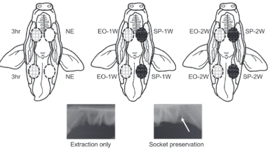
IACUC-YB-2011-3). Nine pigs were divided into three groups (n=3 in each group), depending on the time point of their sacriice, as depicted in Figure 1. In three animals, the left premolars were used as controls, and the right premolars were extracted without socket preservation. These animals were sacrificed 3 h after the extraction (right: 3 h after the extraction; left: no extraction/control, NE). In the remaining six animals, maxillary and mandibular premolars (PM1, PM2, and PM3) were extracted bilaterally, and the left extraction sockets were illed with graft material (right: extraction only, EO; left: extraction and socket preservation, SP). Three of these animals were sacriiced 1 week after the procedure, and the inal three, 2 weeks after the procedure (1 week after the procedure, 1W; 2 weeks after the procedure, 2W). All extraction sockets were closed primarily. Biphasic calcium phosphate (BCP; Bone Plus™ BCP Eagle eye, MegaGen Implant Co., Ltd., Seoul, Republic of Korea) composed of 60% hydroxyapatite (HA) and 40% beta-tricalcium phosphate (β-TCP) was used for socket preservation. After surgical procedures, the animals were monitored carefully, and no sign of postoperative infection was noted around the extraction site. The animals were euthanized humanely just before the collection of specimens. After the mini pigs were sacriiced, the alveolar process of the maxilla and mandibular body were harvested immediately. The specimens were stored in liquid nitrogen until analyzed. About 0.3 g of the gingiva of crestal area was carved out of the frozen specimens for analysis.
LC-MS/MS peptide analysis
For peptide analysis, gingival tissue was homogenized in buffer containing 1% Triton X-100, 20 mM Tris-HCl (pH 7.5), 150 mM NaCl, 1 mM EDTA,
Figure 1- Overview of the animal experiment presented with a schematic drawing of swine oral cavity. Circles of dotted outline represent the following; 3hr: 3 hours after the extraction, right premolar extraction sockets; NE: no extraction/ control, left premolars; EO-1W: extraction only, 1 week post-extraction; SP-1W: extraction and socket preservation, 1 week after the procedures; EO-2W: extraction only, 2 weeks post-extraction; SP-2W: extraction and socket preservation, 2
1 mM EGTA, 2.5 mM sodium pyrophosphate, 1 mM β-glycerolphosphate, 1 mM sodium orthovanadate, 25 mM sodium luoride, 1 µg/ml leupeptin and 1 mM PMSF. Proteins extracted from gingiva after tooth extraction were analyzed using LC-MS/MS as described previously32. To search via ProteinLynx GlobalServer search version 2.3.3 (Waters Corporation, Milford, MA, USA), the parent ion tolerance was set at 100 ppm, and the fragment ion tolerance was set at 0.2 Da. Carbamidomethylation (+57 Da) of cysteine and methionine oxidation (+16 Da) were chosen as the ixed and variable modiications, respectively.
Analysis of quantitative changes in protein abundance, based on measurements of peptide ion peak intensities observed in the low collision energy mode (MS) in a triplicate set, was carried out using the Expression™ software (version 2). To normalize comparative proteomic data, the “auto normalization” function of ProteinLynx GlobalServer was used. Protein identiication was allowed only if the conidence was greater than 95% on the basis of the IDENTITYE algorithm.
Quantitative real-time PCR
Total RNA was isolated from samples using
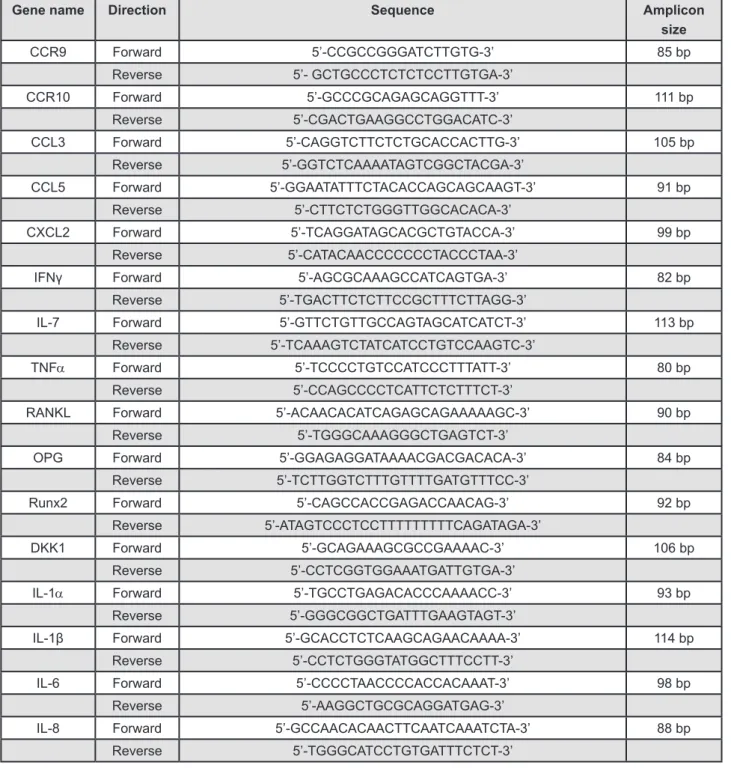
Gene name Direction Sequence Amplicon
size
CCR9 Forward 5’-CCGCCGGGATCTTGTG-3’ 85 bp
Reverse 5’- GCTGCCCTCTCTCCTTGTGA-3’
CCR10 Forward 5’-GCCCGCAGAGCAGGTTT-3’ 111 bp
Reverse 5’-CGACTGAAGGCCTGGACATC-3’
CCL3 Forward 5’-CAGGTCTTCTCTGCACCACTTG-3’ 105 bp
Reverse 5’-GGTCTCAAAATAGTCGGCTACGA-3’
CCL5 Forward 5’-GGAATATTTCTACACCAGCAGCAAGT-3’ 91 bp
Reverse 5’-CTTCTCTGGGTTGGCACACA-3’
CXCL2 Forward 5’-TCAGGATAGCACGCTGTACCA-3’ 99 bp
Reverse 5’-CATACAACCCCCCCTACCCTAA-3’
IFNγ Forward 5’-AGCGCAAAGCCATCAGTGA-3’ 82 bp
Reverse 5’-TGACTTCTCTTCCGCTTTCTTAGG-3’
IL-7 Forward 5’-GTTCTGTTGCCAGTAGCATCATCT-3’ 113 bp
Reverse 5’-TCAAAGTCTATCATCCTGTCCAAGTC-3’
TNFα Forward 5’-TCCCCTGTCCATCCCTTTATT-3’ 80 bp
Reverse 5’-CCAGCCCCTCATTCTCTTTCT-3’
RANKL Forward 5’-ACAACACATCAGAGCAGAAAAAGC-3’ 90 bp
Reverse 5’-TGGGCAAAGGGCTGAGTCT-3’
OPG Forward 5’-GGAGAGGATAAAACGACGACACA-3’ 84 bp
Reverse 5’-TCTTGGTCTTTGTTTTGATGTTTCC-3’
Runx2 Forward 5’-CAGCCACCGAGACCAACAG-3’ 92 bp
Reverse 5’-ATAGTCCCTCCTTTTTTTTTCAGATAGA-3’
DKK1 Forward 5’-GCAGAAAGCGCCGAAAAC-3’ 106 bp
Reverse 5’-CCTCGGTGGAAATGATTGTGA-3’
IL-1α Forward 5’-TGCCTGAGACACCCAAAACC-3’ 93 bp
Reverse 5’-GGGCGGCTGATTTGAAGTAGT-3’
IL-1β Forward 5’-GCACCTCTCAAGCAGAACAAAA-3’ 114 bp
Reverse 5’-CCTCTGGGTATGGCTTTCCTT-3’
IL-6 Forward 5’-CCCCTAACCCCACCACAAAT-3’ 98 bp
Reverse 5’-AAGGCTGCGCAGGATGAG-3’
IL-8 Forward 5’-GCCAACACAACTTCAATCAAATCTA-3’ 88 bp
Reverse 5’-TGGGCATCCTGTGATTTCTCT-3’
QIAzol® RNA Lysis reagent (QIAGEN, Hilden,
Germany), after which cDNAs were synthesized using a PrimeScript™ RT reagent Kit for RT-PCR (Takara Bio Inc., Otsu, Japan) according to the manufacturer’s instructions. Quantitative PCR was performed using an ABI 7300 Prism SDS real-time PCR detection system (Applied Biosystems, Foster City, CA, USA) with a SYBR® Premix Ex Tag kit (Takara Bio Inc., Otsu, Japan) and a standard temperature protocol. The results obtained using CT (cycle threshold) were expressed as relative quantities and were calculated using the 2-ΔΔCT method (expressed as relative fold ratio). Hypoxanthine phosphoribosyltransferase 1 (HPRT1) was used as a control gene for normalization, and three separate experiments were performed. Detailed primer information is shown in Figure 2.
Statistical analysis
The results of real-time PCR were expressed as mean values with standard deviations (±SD). For statistical signiicance, changes of mRNA expression level in control and experimental groups were analyzed using ANOVA. Differences between the control and 3 h and those between the EO and SP groups at same time point were analyzed using paired t-tests. Values of p<0.05 were considered signiicant. All statistical tests were performed using SigmaPlot software version 10.0 (Systat Software Inc., San Jose, CA, USA).
RESULTS
Peptide analysis of immunoglobulins expressed in gingiva
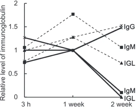
The relative expression levels of immunoglobulin peptides are summarized in Figure 3. In the EO group, levels of immunoglobulin M (IgM) and immunoglobulin light chain (IGL) were respectively 1.7-fold and 1.3-fold higher 1 week after tooth extraction than they were 3 h after tooth extraction. By 2 weeks, however, the expression levels had fallen to the same levels seen after 3 h. By contrast, immunoglobulin G (IgG) expression increased throughout the experimental period. In the SP group, IgM and IGL declined throughout the experimental period and were dramatically reduced after 2 weeks. Levels of IgG, on the other hand, were lower after 1 week than after 3 h, but the level was somewhat higher after 2 weeks. Overall, the expression levels of all three immunoglobulins (IgG, IgM and IGL) were higher in the EO group than in the SP group. In addition, the pattern of immunoglobulin expression differed between the EO and SP groups. In particular, whereas IgM and IGL levels peaked after 1 week in the EO group, they declined over the entire course of the experiment in the SP group.
Assessing the immune response: cytokines and chemokines in the EO and SP groups
To evaluate the effect of socket preservation on the immune response following tooth extraction,
we examined the mRNA expression of several cytokines and chemokines (Figure 4). Three hours after extraction, levels of most of the inlammatory cytokines and chemokines tested (CCR10, CCR9, CCL3, CXCL2, IFNγ, and TNF-α) increased slightly, as compared to control (NE), while levels of CCL5 and IL-7 were lower than in the NE group.
Differences between the groups were signiicant in CCR9, CCL5, CXCL2, IL7, and TNF-α. The cytokine and chemokine responses seen at 3 h are known as the early immune response, which is triggered by bleeding following tooth extraction32. At 1 week post-extraction, levels of the inlammatory chemokines CCL3, CCL5, and CXCL2 and the chemokine receptors CCR9 and CCR10 had declined in the SP group, as compared to the EO group (however, CCR9 showed no statistical signiicance.). Similarly, expression of the pro-inlammatory cytokines TNF-α
and interferon-gamma (IFN-γ) were signiicantly lower in the SP group than in the EO group, whereas expression of the anti-inlammatory cytokine IL-7 was signiicantly higher in the SP group than in the EO group. Within 2 weeks after tooth extraction, however, there were no differences in the levels of cytokine and chemokine expression between the EO and SP groups.
C y t o k i n e s r e l a t e d t o c a n o n i c a l osteoclastogenesis
To assess osteoblastic activity, we examined the mRNA levels of RANKL, osteoprotegerin (OPG), runt-related transcription factor (2Runx2) and Dickkopf-related protein 1 (DKK1). OPG and Runx2 presented signiicant differences. Overall, the expression levels of all of the
osteoblast-Figure 5- (A) mRNA expression related to osteoclastic activation. Levels of receptor activator of nuclear factor kappa-B
ligand (RANKL) expression were signiicantly higher in the EO group 1 week after tooth extraction (1W). Osteoprotegerin
(OPG) levels did not differ between groups; (B) mRNA expression related to osteoblastic activation. Levels of DKK1
expression were signiicantly higher in the EO group 1 week after tooth extraction. Levels of Runx2 mRNA were signiicantly
higher in the EO than in the SP group 2 weeks post-extraction (2W). NE: no extraction; EO: extraction only; SP: extraction
and socket preservation; NS: non-signiicant; *: p<0.05 in paired t-test; **: p<0.05 in ANOVA
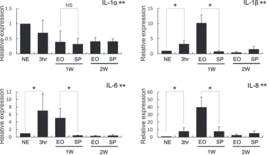
Figure 6- Expression of interleukins related to non-canonical osteoclastogenesis. Expression levels of IL-1β, IL-6 and IL-8 were signiicantly higher in the EO group than in the SP group 1 week after tooth extraction (1W). While IL-1α expression level decreased after the extraction, the levels of EO and SP were similar at 1 week and 2 weeks (1W, 2W). NE: no
related genes were higher in the EO group than the SP group 1 week post-extraction (Figure 5). In particular, RANKL expression was increased to a dramatically greater degree in the EO group (34.1-fold) than in the SP group (5.6-(34.1-fold) at 1 week. By 2 weeks, however, the expression was similarly increased in the two groups (6.4-fold). The level of OPG expression gradually increased over time, and did not differ between the EO (2.3-fold at 1 week; 3.1-fold at 2 weeks) and SP (2.3-fold at 1 week; 3-fold at 2 weeks) groups. The level of Runx2 expression was higher in the EO than the SP group 1 week post-extraction (3.4-fold in EO group vs. 2.6-fold in SP group), and the difference reached signiicance by 2 weeks. Finally, the level of DKK1 expression was signiicantly higher in the EO group (14.2-fold) than in the SP group (3.2-fold) after 1 week, but the expression levels in the two groups were the same as the expression levels at 2 weeks (3.7-fold).
Interleukins related to non-canonical osteoclastogenesis
We also investigated the post-extraction
expression of interleukins known to activate
osteoclasts, and changes in IL-1α, IL-1β, IL6, and IL8 were signiicant (Figure 6). One week post-extraction, gingival expression of IL-1β was dramatically higher in the EO group than in the SP group. In addition, the levels of IL-6 and IL-8 expression were also signiicantly higher in the EO group. However, IL-6 expression was also dramatically increased 3 h after tooth extraction, suggesting it may be involved in the acute immune response induced by bleeding following the extraction.
DISCUSSION
Osteoclasts have been observed in the crestal region of alveolar bone during the healing process
after tooth extraction2. This suggests osteoclasts are likely responsible for the bone resorption seen after extraction, and the factors related to bone resorption present in the adjacent gingiva should be evaluated.
Bleeding after extraction may induce nonspeciic gene expression related to the early immune
response9. We compared the ongoing expression 3 h after extraction in the NE, EO and SP groups to avoid misinterpreting the results due to the inluence of bleeding. Both B cells and T cells are abundant
within the extraction socket30, and the presence of immunoglobulin would reflect lymphocyte iniltration into the adjacent gingiva, activating an inlammatory response around the extraction socket. Levels of all the identiied immunoglobulins, IgM, IgG and IGL, were higher in the EO group than
in the SP group (Figure 3). Higher levels of IGL suggest greater B cell activation and inlammation in the EO group15. The importance of inlammation to alveolar bone resorption is illustrated by the inding that oral Porphyromonas gingivalis infection induces less alveolar bone resorption in immunodeicient mice than in immunocompetent
mice3. Similarly, the stronger immune response in the EO group may lead to greater bone resorption.
A variety of cytokines and chemokines contribute to the post-extraction immune response13, and several are involved in osteoclastogenesis. For example, CCL3 and CCL5 are related to osteoclast
activation or formation18,27, while CXCL2 plays a key role in neutrophil chemotaxis. Moreover, CXCL2 is reportedly induced by RANKL, which in
turn enhances osteoclastogenesis11. IFN-γ is a pro-inlammatory cytokine that activates plasma cells, B cells and macrophages, and is also reported to stimulate bone resorption through stimulation of RANKL and TNF-α production in T cells10. Conversely, IL-7, which is required for normal adult progenitor B cell development, reportedly inhibits osteoclastogenesis in a murine model1. As shown in Figure 3, the stronger expression of these cytokines and chemokines in the EO group compared with the SP group is consistent with the higher immunoglobulin levels revealed by the peptide analysis. This suggests that B cells are activated to a greater degree in the EO group, and that B cells are a likely source of RANKL in compromised gingiva17.
Runx2 is a well-known mediator of osteoblastic differentiation, while RANKL and OPG respectively induce and inhibit osteoclastogenesis4. Levels of Runx2, RANKL and OPG expression were all increased 1 and 2 weeks after tooth extraction, suggesting activation of osteoblasts. Interestingly, Runx2 expression was signiicantly higher in the EO group 2 weeks post-extraction. This might be related to repressed bone healing in socket, as in a previous study which reported that biomaterials grafted to fresh extraction sockets may interfere
with the normal healing processes of the alveolar
bone and present poor bone quality7. While the level of OPG and Runx2 expression did not differ signiicantly between the EO and SP groups, RANKL expression was signiicantly higher in the EO group than in the SP group 1 week post-extraction. RANKL is secreted by osteoblasts as well as immune
model and in human rheumatoid arthritis8. It also appears TNF-α may play a major role in LPS-induced inhibition of osteogenesis in areas of inlammation29, and upregulation of DKK1 is thought to be related to inlammation-induced upregulation of TNF-α (Figure 4 and 5B).
IL-1, which is produced by polymorphonuclear neutrophils, monocytes and macrophages in the periodontium, stimulates bone resorption via effects
on osteoclasts19 and plays a signiicant role in the pathogenesis of periodontal tissue destruction14. IL-6, which is initially upregulated by stimulation of IL-1, is reportedly capable of inducing osteoclast
formation from circulating cells20. Consistent with that inding, the alveolar bone resorption in an oral infection model was diminished in the absence of IL-63. In addition, IL-8, which is stimulated by RANKL, enhances RANKL-induced osteoclastogenesis18. Interestingly, IL-1, IL-6 and IL-8 together coordinate osteoclastogenesis in a RANKL-independent
manner19. Collectively, these indings indicate that the upregulation of IL-1, IL-6 and IL-8 may account for the greater osteoclastogenesis seen in the EO group compared with the SP group 1 week after tooth extraction.
In this study, we observed that levels of cytokines associated with inflammation and osteoclastogenesis were lower in the SP group than in the EO group. This suggests that grafting
alloplastic material into the extraction socket after
tooth extraction may suppress the inlammatory cytokines and osteoclastogenic mediators in the gingiva adjacent to the socket. It might be related to ibrin, which is a major component of blood clot. Since graft material ills the alveolar socket, the amount of blood clot is smaller in the SP group. It was recently suggested that ibrin may be an exogenous activator of the bone remodeling unit, favoring bone resorption6. Since, however, the forementioned study described a systemic effect of ibrin accumulation, it is considered that further study about the local effect of ibrin on bone healing should be followed.
ACKNOWLEDGEMENTS
This work was supported by the Basic Science Research Program through the National Research Foundation of Korea (NRF) funded by the Ministry of Science, ICT and future Planning (MSIP) (2014R1A2A2A01005448), and by the National Research Foundation of Korea (NRF) grant funded by the Korean government (2011-0030121).
REFERENCES
1- Aguila HL, Mun SH, Kalinowski J, Adams DJ, Lorenzo JA, Lee SK. Osteoblast-speciic overexpression of human interleukin-7 rescues the bone mass phenotype of interleukin-7-deicient female mice. J Bone Miner Res. 2012;27(5):1030-42.
2- Araújo MG, Lindhe J. Dimensional ridge alterations following tooth extraction. An experimental study in the dog. J Clin Periodontol. 2005;32(2):212-8.
3- Baker PJ, Dixon M, Evans RT, Dufour L, Johnson E, Roopenian DC. Cd4(+) t cells and the proinlammatory cytokines gamma interferon and interleukin-6 contribute to alveolar bone loss in mice. Infect Immun. 1999;67(6):2804-9.
4- Boyce BF, Xing L. Functions of RANKL/RANK/OPG in bone modeling and remodeling. Arch Biochem Biophys. 2008;473(2):139-46.
5- Cardaropoli G, Araújo M, Lindhe J. Dynamics of bone tissue formation in tooth extraction sites. An experimental study in dogs. J Clin Periodontol. 2003;30(9):809-18.
6- Cole HA, Ohba T, Nyman JS, Hirotaka H, Cates JM, Flick MJ, et al. Fibrin accumulation secondary to loss of plasmin-mediated ibrinolysis drives inlammatory osteoporosis in mice. Arthritis Rheumatol. 2014;66(8):2222-33.
7- De Coster P, Browaeys H, De Bruyn H. Healing of extraction sockets illed with BoneCeramic® prior to implant placement:
preliminary histological indings. Clin Implant Dent Relat Res. 2011;13(1):34-45.
8- Diarra D, Stolina M, Polzer K, Zwerina J, Ominsky MS, Dwyer D, et al. Dickkopf-1 is a master regulator of joint remodeling. Nat Med. 2007;13(2):156-63.
9- Esmon CT. The interactions between inflammation and coagulation. Br J Haematol. 2005;131(4):417-30.
10- Gao Y, Grassi F, Ryan MR, Terauchi M, Page K, Yang X, et al. Ifn-gamma stimulates osteoclast formation and bone
loss in vivo via antigen-driven T cell activation. J Clin Invest.
2007;117(1):122-32.
11- Ha J, Choi HS, Lee Y, Kwon HJ, Song YW, Kim HH. CXC chemokine ligand 2 induced by receptor activator of NF-kappa b ligand enhances osteoclastogenesis. J Immunol. 2010;184(9):4717-24.
12- Horowitz MC, Fretz JA, Lorenzo JA. How b cells inluence bone biology in health and disease. Bone. 2010;47(3):472-9. 13- Horuk R. Chemokine receptors. Cytokine Growth Factor Rev. 2001;12(4):313-35.
14- Hou LT, Liu CM, Liu BY, Lin SJ, Liao CS, Rossomando EF. Interleukin-1beta, clinical parameters and matched cellular-histopathologic changes of biopsied gingival tissue from periodontitis patients. J Periodontal Res. 2003;38(3):247-54. 15- Hutchison CA, Landgren O. Polyclonal immunoglobulin free light chains as a potential biomarker of immune stimulation and inlammation. Clin Chem. 2011;57(10):1387-9.
16- Kao ST, Scott DD. A review of bone substitutes. Oral Maxillofac Surg Clin North Am. 2007;19(4):513-21.
17- Kawai T, Matsuyama T, Hosokawa Y, Makihira S, Seki M, Karimbux NY, et al. B and t lymphocytes are the primary sources of RANKL in the bone resorptive lesion of periodontal disease. Am J Pathol. 2006;169(3):987-98.
18- Kim MS, Day CJ, Morrison NA. MCP-1 is induced by receptor activator of nuclear factor-{kappa}B ligand, promotes human osteoclast fusion, and rescues granulocyte macrophage colony-stimulating factor suppression of osteoclast formation. J Biol Chem. 2005;280(16):16163-9.
19- Knowles HJ, Athanasou NA. Canonical and non-canonical pathways of osteoclast formation. Histol Histopathol. 2009;24(3):337-46.
21- Lerner UH. Inflammation-induced bone remodeling in periodontal disease and the influence of post-menopausal osteoporosis. J Dent Res. 2006;85(7):596-607.
22- Lin Z, Rios HF, Volk SL, Sugai JV, Jin Q, Giannobile WV. Gene expression dynamics during bone healing and osseointegration. J Periodontol. 2011;82(7):1007-17.
23- Male D. Mechanisms of innate immunity. In: Male D, Brostoff J, Roth D, Roitt I, ed. Immunology. London: Elsevier Health Sciences; 2012. p. 109-16.
24- Morjaria KR, Wilson R, Palmer RM. Bone healing after tooth extraction with or without an intervention: a systematic review of randomized controlled trials. Clin Implant Dent Relat Res. 2014;16(1):1-20.
25- Scala A, Lang NP, Schweikert MT, Oliveira JA, Rangel-Garcia I Jr, Botticelli D. Sequential healing of open extraction sockets. An experimental study in monkeys. Clin Oral Implants Res. 2014;25(3):288-95.
26- Steen BM, Gerstenfeld LC, Einhorn TA. The role of the immune system in fracture healing. In: Lorenzo J, Choi Y, Horowitz M, Takayanagi H, ed. Osteoimmunology. London: Academic Press; 2010. p. 343-67.
27- Terpos E, Politou M, Viniou N, Rahemtulla A. Signiicance of macrophage inlammatory protein-1 alpha (MIP-1alpha) in multiple myeloma. Leuk Lymphoma. 2005;46(12):1699-707. 28- Tomlin EM, Nelson SJ, Rossmann JA. Ridge preservation for implant therapy: a review of the literature. Open Dent J. 2014;8:66-76.
29- Tomomatsu N, Aoki K, Alles N, Soysa NS, Hussain A, Nakachi H, et al. LPS-induced inhibition of osteogenesis is TNF-alpha dependent in a murine tooth extraction model. J Bone Miner Res. 2009;24(10):1770-81.
30- Trombelli L, Farina R, Marzola A, Bozzi L, Liljenberg B, Lindhe J. Modeling and remodeling of human extraction sockets. J Clin Periodontol. 2008;35(7):630-9.
31- Wang RE, Lang NP. Ridge preservation after tooth extraction. Clin Oral Implants Res. 2012;23 Suppl 6(147-56.