Comparison of the changes of alveolar bone thickness
in maxillary incisor area in extraction and non-extraction
cases: Computerized tomography evaluation
Paulo Roberto Barroso Picanço1, Fabricio Pinelli Valarelli2, Rodrigo Hermont Cançado2, Karina Maria Salvatore de Freitas3, Gracemia Vasconcelos Picanço1
How to cite this article: Picanço PRB, Valarelli FP, Cançado RH, Freitas KMS, Picanço GV. Comparison of the changes of alveolar bone thickness in maxillary incisor area in extraction and non-extraction cases: Computerized tomography evaluation. Dental Press J Orthod. 2013 Sept-Oct;18(5):91-8.
Submitted: October 21, 2010 - Revised and accepted: October 22, 2011
Contact address: Fabrício Valarelli
Rua Manoel Pereira Rolla, 12-75, apto 503 – Bauru/SP – CEP: 17012-190 E-mail: [email protected]
1 MSc in Orthodontics, Uningá. Professor, Paulo Picanço Center of
Orthodontics.
2 Adjunct Professor, Uningá.
3 Post-Doc in Orthodontics, University of Toronto. Professora, Uningá.
» The authors report no commercial, proprietary or inancial interest in the prod-ucts or companies described in this article.
Objective:To compare, through computed tomography, alveolar bone thickness changes at the maxillary incisors area during orthodontic treatment with and without tooth extraction. Methods: Twelve patients were evaluated. They were divided into 2 groups: G1 - 6 patients treated with extraction of right and left maxillary first premolars, with mean initial age of 15.83 years and mean treatment length of 2.53 years; G2 - 6 patients treated without extraction, with mean initial age of 18.26 years and mean treatment length of 2.39 years. Computed tomographies, lateral cephalograms and periapical radiographs were used at the beginning of the treatment (T1) and 18 months after the treatment had started (T2). Extrac-tion space closure occurred in the extracExtrac-tion cases. Intragroup and intergroup comparisons were performed by depen-dent and independepen-dent t test, respectively. Results: In G1, the central incisor was retracted and uprighted, while in G2 this tooth showed vestibularization. Additionally, G1 presented a higher increase of labial alveolar bone thickness at the cervical third in comparison with G2. The incidence of root resorption did not present significant differences between groups. Conclusion: There were no changes in alveolar bone thickness when extraction and nonextraction cases were compared, except for the labial alveolar bone thickness at the cervical third of maxillary incisors.
Keywords:Alveolar ridge. Tooth movement. Tooth extraction. Tomography.
Objetivo:comparar, por meio de tomografia computadorizada, a alteração da espessura óssea alveolar na região de incisivos superiores durante o tratamento ortodôntico, com e sem extração dentária. Métodos: foram avaliados 12 pa-cientes, divididos em dois grupos: G1, seis pacientes tratados com extrações de dois primeiros pré-molares superiores, com idade média inicial de 15,83 anos, e tratados por um tempo médio de 2,53 anos; G2, seis pacientes tratados sem extrações, com idade média inicial de 18,26 anos e tratados por um período de 2,39 anos. Foram utilizadas tomografias computado-rizadas, telerradiografias em norma lateral e radiografias periapicais ao início (T1) e após 18 meses de tratamento (T2), desde que o espaço da extração já estivesse fechado nos casos tratados com extrações. A comparação intragrupo foi realizada por meio do teste t dependente, e a comparação intergrupos por meio do com o teste t independente. Resultados: o grupo 1
apresentou uma retração e verticalização do incisivo central, enquanto o grupo 2 apresentou uma vestibularização des-se dente. Além disso, o grupo 1 apredes-sentou maior aumento da espessura ósdes-sea cervical vestibular durante o tratamento, quando comparado ao grupo 2. A incidência de reabsorção radicular não apresentou diferenças significativas entre os grupos. Conclusões: não houve alteração nas espessuras ósseas alveolares quando comparados casos tratados com e sem extrações, com exceção da espessura óssea vestibular na região cervical dos incisivos superiores.
INTRODUCTION
Orthodontic movement can be quick or slow, depend-ing on the physical characteristics of the applied force, the
size and the biological response of the periodontal ligament.1
According to Vardimon, Oren and Ben-Bassat,2 there is an
axiom in orthodontics that says: “tooth movement leaves marks on the bone”, however, this fact is not always fa-vorable. In vertical direction, during tooth extrusion, the changes in the underlying bone tissue may not follow tooth displacement, leading to an increase in clinical length of the tooth crown, otentimes undesirable. In transverse and an-teroposterior directions, bone dehiscence and fenestration have been reported when the incisors are either protruded
or retracted.3,4 According to Handelman,5 labial and lingual/
palatal bone cortical plates at incisors’ apexes may represent anatomical limits to orthodontic tooth movement. The lit-erature has speculated that protrusion and vestibularization of the maxillary incisors may produce labial bone cortical dehiscence while teeth retraction afects the palatal bone plate, although this would be reversible only if the teeth
re-turned to their original position.6
Designing the limits of tooth movement before be-ginning the orthodontic treatment may be extremely beneicial, especially in situations in which skeletal dis-crepancy is severe or the maxilla and/or mandible can accommodate, in a limited way, the repositioning of the
teeth ater orthodontic movement.5,7
To detect bone levels, the following methods may be used: periapical radiographs, lateral cephalograms and panoramic radiographs. Notwithstanding, bidimensional radiographs reveal some limitations, such as image super-imposition and distortions as well as the inability of mea-suring bone thickness in panoramic and periapical
radio-graphs.7,8 Taking these factors into account, computed
tomography (CT) has been considered of paramount im-portance for diagnosing initial bone levels as well as bone
level changes during orthodontic treatment.9,10
The aim of this study was to compare, through com-puted tomography, the alveolar bone thickness changes at the maxillary incisors area during orthodontic treat-ment with or without maxillary premolar extractions.
MATERIAL AND METHODS
Sample
Twelve patients of both genders were selected in the orthodontic clinic Paulo Picanço. Inclusion crite-ria were: 1) permanent dentition; 2) absence of systemic
disease that may alter bone metabolism; 3) nonsmoker; 4) patients who are not using steroid-based drugs; 5) ab-sence of chronic kidney disease; 6) if female, patients who do not present low level of estrogen; 7) presence of all six maxillary anterior teeth; 8) patients who have not undergone tooth trauma, alveolar bone fracture or luxation in maxillary incisors; 9) patients who do not present a prosthetic crown on maxillary incisors; 10) ab-sence of alveolar clet in the maxillary anterior area.
The sample was divided into two groups: G1 – 6 patients (5 male; 1 female) with mean initial age of 15.83 ± 4.87 years, presenting Class II malocclusion, treated with 2 maxillary premolar extractions during a mean period of 2.53 ± 0.49 years; G2 – 6 patients (5 male; 1 female), with mean initial age of 18.26 ± 6.42 years, 3 showing Class I and 3 Class II malocclusion, treated without extractions during a mean treatment period of 2.39 ± 0.66 years.
Patients were treated by post-graduation students oriented by the same professor, following the same diag-nosis pattern, treatment planning and orthodontic me-chanics (Edgewise, 0.018 x 0.025-in brackets – Morelli – Sorocaba, SP, Brazil).
Methods
Computed tomographies were performed at the
beginning of the orthodontic treatment (T1) and 18
months after the treatment had started, (T2). The
ex-ams confirmed extraction space closure.
Addition-ally, periapical radiographs were performed at T1 and
T2 using the parallelism technique in order to
evalu-ate external root resorption of the maxillary incisors. Lateral cephalograms were performed to evaluate an-teroposterior and vertical changes as well as the incli-nation of these teeth.
For cephalograms and tomographs evaluation, image digitalization and measurement processing, Dolphin Imaging Premium 10.5 software (Dolphin Imaging & Management Solutions, Chatsworth, CA, USA) was used.
On the lateral cephalograms, the following variables were analyzed: 1-PTV incisal, 1-PTV apical, FMA, PFH/AFH, Wits, 1.NA, H-11 and UL+UP.
The degree on initial and inal root resorption was an-alysed through periapical radiographs, based on the scores
of Levander and Malmgren’s score system:11 0 – lack
of root resorption; 1 – presence of apical irregularities; 2 – presence of root resorption up to 2 mm; 3 – presence of root resorption from 2 mm to one third of the root original length; 4 – presence of root resorption greater than one third of the original length of the root. Root length was obtained from measuring the distance from root apex to ECJ, following the incisor long axis (Fig 2).
Method error
To determine intraexaminer error, both lateral cepha-lograms and tomographies were reevaluated in 6 ran-domly selected patients ater a month interval. Systematic error was determined by dependent t test while casual error was determined by the Dahlberg’s formula. Kappa test was used to establish root resorption score error.
Statistical analysis
The following statistical tests were employed: difer-ence between two means for carrying out the sample calculation, chi-square test for intergroup comparison
concerning the malocclusion type; independent t test for intergroup age and treatment period comparison; dependent t test for intragroup initial and inal stages comparison; independent t test for intergroup initial and inal stages as well as treatment changes comparison. All tests were performed with Statistica sotware (Statistica for Windows, version 7.0, Statsot, 2005). The results were considered signiicant at p < 0.05.
Figure 1 - Alveolar bone thickness assessment evaluated through computed tomography.
Figure 2 - Crown-root ratio and maxillary central incisor length assessment.
Crown-root ratio Example: Degree 4
Crown = 11 mm
11 --- 100% 10 --- X
X = 1000 ÷ 11/100
X= 0.9
Ratio = 1:09
Apical
Middle
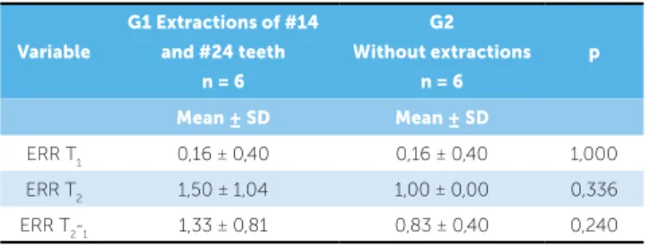
With regard to root resorption degree, there were no significant differences between groups at any of the evaluated periods (Table 8).
DISCUSSION
Handelman5 claims that a thin tooth alveolus or an
inappropriate alveolar cavity for the amount of desir-able tooth movement must be considered as a risk for RESULTS
The 1-PTV apical variable showed the greatest casual error (1.57 mm). Systematic error occurred only for the following variables: 1-PTV incisal and UP middle. Kappa coefficient demonstrated a con-cordance percentage of 90%.
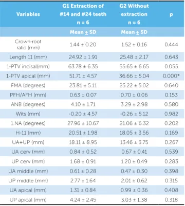
There were differences in the distribution of the malocclusion type between groups (Table 1). The groups were compatible regarding gender, initial and final ages as well as treatment period (Table 2).
At the initial stage (T1), only 1-PTV apical
showed statistically significant difference between groups, indicating that in G1 the maxillary incisor was more protruded than in G2 (Table 3).
The comparison between G1 (treated with two
maxillary premolar extractions) stages (T1 and T2)
dem-onstrated that there was a decrease in the crown-root ratio and in the central incisor length, a retraction of these teeth both in apical (1-PTV apical) and incisal (1-PTV incisal) measurements, a decrease in anteropos-terior discrepancy (ANB), an increase of the UL cervi-cal (labial cervicervi-cal third) measurement and decrease of the UP cervical (palatal cervical third) and UP middle (palatal middle third) measurements (Table 4).
In G2 (treated without extractions), the
compari-son between T1 and T2 demonstrated that there was
a decrease in the crown-root ratio, in the central in-cisor length, in FMA angle, in the relation between posterior and anterior face height, vestibularization of the maxillary incisors (1.NA) and a decrease in the UP middle measurement as well (Table 5).
At T2, there was statistically significant difference
between the groups in two variables: 1-PTV incisal and UL cervical. The difference in 1-PTV incisal indicated that, at the end of the treatment, central incisors in G1 were more retruded than in G2 while the difference in UL cervical showed higher bone thickness at this area in G1 than in G2 (Table 6).
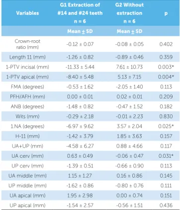
With regard to treatment changes (T2-T1), the
1-PTV incisal, 1-PTV apical and 1.NA measurements were statistically signiicant diferent between groups, revealing that G1 showed maxillary incisor’s retraction and uprighting while G2 exhibited this tooth protru-sion and vestibularization (Table 7). Additionally, UL cervical measurement was also signiicantly diferent between groups indicating an increase of labial bone thickness in G1 when compared to G2 (Table 7).
Group Class I Class II Total
1 0 6 6
2 3 3 6
Total 3 9 12
χ2 = 4.00; GL= 1; p = 0.045*
Table 1 - Intergroup comparison of malocclusion type (chi-square test).
*Statistically significant difference (P<0.05).
Table 2 - Intergroup comparison of initial and final ages as well as treatment period (independent t tests).
Variables (years)
G1 Extraction of #14 and #24 teeth (n = 6)
G2 Without
extractions (n = 6) p
Mean ± SD Mean ± SD
Initial age 15.83 ± 4.87 18.26 ± 6.42 0.477 Final age 18.36 ± 4.84 20.65 ± 6.45 0.502 Treatment
period 2.53 ± 0.49 2.39 ± 0.66 0.682
Table 3 - Intergroup comparison of variables studied during the initial stage (T1) (independent t tests).
Variables
G1 Extraction of #14 and #24 teeth
n = 6
G2 Without extraction
n = 6
p
Mean ± SD Mean ± SD
Crown-root
ratio (mm) 1.44 ± 0.20 1.52 ± 0.16 0.444
Length 11 (mm) 24.92 ± 1.91 25.48 ± 2.17 0.643 1-PTV incisal(mm) 63.78 ± 6.35 55.65 ± 6.65 0.055 1-PTV apical (mm) 51.71 ± 4.57 36.66 ± 5.04 0.000*
Table 4 - Comparison between initial (T1) and final (T2) stages of group 1, with premolar extractions (dependent t tests).
Table 5 - Comparison between initial (T1) and final (T2) stages of group 2, without extractions (dependent t tests).
*Statistically significant difference (P<0.05). *Statistically significant difference (P<0.05).
Variables Initial stage
(T1) n = 6
Final stage (T2) n = 6
Changes
T2-T1 p
Mean ± SD Mean ± SD
Crown-root
ratio (mm) 1:1.44 ± 0.20 1:1.32 ± 0.25 -0.12 0.009* Length. 11 (mm) 24.92 ± 1.91 23.65 ± 2.12 -1.27 0.013* 1-PTV incisal (mm) 63.78 ± 6.35 52.45 ± 4.18 -11.33 0.003* 1-PTV apical (mm) 51.71 ± 4.57 43.30 ± 5.17 -8.41 0.013* FMA (degrees) 23.81 ± 5.11 23.28 ± 5.13 -0.53 0.458 PFH/AFH (mm) 0.63 ± 0.07 0.64 ± 0.08 0.01 0.258 ANB (degrees) 4.10 ± 1.71 2.61 ± 1.38 -1.49 0.006*
Wits (mm) -0.20 ± 4.57 -0.50 ± 5.05 -0.30 0.752 1.NA (degrees) 27.96 ± 10.67 20.99 ± 4.08 -6.97 0.136 H-11 (mm) 20.51 ± 1.98 19.09 ± 2.09 -1.42 0.399 UA+UP (mm) 18.11 ± 8.95 13.53 ± 3.18 -4.58 0.133 UA cerv (mm) 0.84 ± 0.52 1.48 ± 0.40 0.64 0.025* UP cerv (mm) 1.68 ± 0.91 0.28 ± 0.69 -1.40 0.001* UA middle (mm) 0.61 ± 0.28 1.77 ± 1.43 1.16 0.077 UP middle (mm) 2.77 ± 1.64 1.15 ± 0.96 -1.62 0.005*
UA apical (mm) 1.31 ± 0.84 3.27 ± 3.44 1.96 0.170 UP apical (mm) 4.24 ± 2.45 2.69 ± 2.09 -1.55 0.200
Variables Initial stage
(T1) n = 6
Final stage (T2) n = 6
Changes
T2-T1 p
Mean ± SD Mean ± SD
Crown-root
ratio (mm) 1.52 ± 0.16 1.43 ± 0.14 -0.09 0.013* Length 11 (mm) 25.48 ± 2.17 24.59 ± 1.86 -0.89 0.005* 1-PTV incisal (mm) 55.65 ± 6.65 63.26 ± 6.00 7.61 0.142 1-PTV apical (mm) 36.66 ± 5.04 41.80 ± 4.04 5.14 0.138 FMA (degrees) 25.22 ± 5.02 23.16 ± 4.60 -2.06 0.015* PFH/AFH (mm) 0.70 ± 0.06 0.72 ± 0.07 0.02 0.034* ANB (degrees) 3.29 ± 2.98 2.82 ± 2.34 -0.47 0.482
Wits (mm) -0.26 ± 5.12 -0.28 ± 5.51 0.02 0.986 1.NA (degrees) 21.06 ± 6.32 24.63 ± 6.61 3.57 0.007* H-11 (mm) 18.05 ± 3.56 19.90 ± 3.27 1.85 0.267 UA+UP (mm) 13.46 ± 3.75 14.35 ± 4.89 0.89 0.662 UA cerv (mm) 0.67 ± 0.41 0.61 ± 0.57 -0.06 0.762 UP cerv (mm) 1.20 ± 0.49 0.53 ± 0.59 -0.67 0.132 UA middle (mm) 0.47 ± 0.30 0.63 ± 0.61 0.16 0.658 UP middle (mm) 2.01 ± 0.62 1.21 ± 0.81 -0.80 0.049*
UA apical (mm) 0.99 ± 0.36 0.99 ± 0.78 0.00 0.995 UP apical (mm) 3.03 ± 1.38 2.47 ± 1.28 -0.56 0.406
Variables
G1 Extraction of
#14 and #24 teeth n = 6
G2 Without
extraction n = 6
p
Mean ± SD Mean ± SD
Crown-root
ratio (mm) 1.32 ± 0.25 1.43 ± 0.14 0.343 Length 11 (mm) 23.65 ± 2.12 24.59 ± 1.86 0.434 1-PTV incisal (mm) 52.45 ± 4.18 63.26 ± 6.00 0.004* 1-PTV apical (mm) 43.30 ± 5.17 41.80 ± 4.04 0.586
FMA (degrees) 23.28 ± 5.13 23.16 ± 4.60 0.968 PFH/AFH (mm) 0.64 ± 0.08 0.72 ± 0.07 0.133 ANB (degrees) 2.61 ± 1.38 2.82 ± 2.34 0.855 Wits (mm) -0.50 ± 5.05 -0.28 ± 5.51 0.944 1.NA (degrees) 20.99 ± 4.08 24.63 ± 6.61 0.277 H-11 (mm) 19.09 ± 2.09 19.90 ± 3.27 0.621 UA+UP (mm) 13.53 ± 3.18 14.35 ± 4.89 0.738 UA cerv (mm) 1.48 ± 0.40 0.61 ± 0.57 0.012* UP cerv (mm) 0.28 ± 0.69 0.53 ± 0.59 0.515 UA middle (mm) 1.77 ± 1.43 0.63 ± 0.61 0.103 UP middle (mm) 1.15 ± 0.96 1.21 ± 0.81 0.907 UA apical (mm) 3.27 ± 3.44 0.99 ± 0.78 0.146 UP apical (mm) 2.69 ± 2.09 2.47 ± 1.28 0.828
Table 6 - Intergroup comparison of the studied variables at the final stage (T2)
(independent t tests).
Table 7 - Intergroup comparison of the studied variables concerning treat-ment changes (T2-T1) (independent t tests).
*Statistically significant difference (P < 0.05). *Statistically significant difference (P < 0.05).
Variables
G1 Extraction of
#14 and #24 teeth n = 6
G2 Without
extraction n = 6
p
Mean ± SD Mean ± SD
Crown-root
ratio (mm) -0.12 ± 0.07 -0.08 ± 0.05 0.402 Length 11 (mm) -1.26 ± 0.82 -0.89 ± 0.46 0.359 1-PTV incisal (mm) -11.33 ± 5.44 7.61 ± 10.73 0.003* 1-PTV apical (mm) -8.40 ± 5.48 5.13 ± 7.15 0.004* FMA (degrees) -0.53 ± 1.62 -2.05 ± 1.40 0.113 PFH/AFH (mm) 0.00 ± 0.01 0.02 ± 0.01 0.209 ANB (degrees) -1.48 ± 0.82 -0.47 ± 1.52 0.182 Wits (mm) -0.29 ± 2.18 -0.01 ± 2.23 0.830 1.NA (degrees) -6.97 ± 9.62 3.57 ± 2.04 0.025*
the occurrence of unfavorable sequelae to orthodontic movement, especially fenestration, bone dehiscence and root resorption. This information can inluence the patient’s treatment planning which, prior to orthodon-tic treatment, can be diagnosed as unfavorable to great teeth movement. The tridimensional analysis provided by computed tomography is of great importance for an accurate assessment of craniofacial morphology because through this examination, it is possible to obtain more reliable information on the dimensions and levels of fa-cial bone tissues when compared to traditional bidimen-sional examinations. Moreover, CT is considered as a
noninvasive, fast, high-accurate diagnosis method.7,12,13
It is important to underline the diiculty and the merit of obtaining a sample comprising 12 patients not only examined with lateral cephalograms, computed tomographies and periapical radiographs at the begin-ning of the treatment and ater 18 months, but also who meet the aforementioned inclusion criteria of this study methodology. As the study was accomplished using CT, which is diicult to be obtained due to the cost and the ethical question concerning radiation exposure, the sample of 12 patients,, is considered acceptable.
Methods
Measurements were performed on the maxillary central incisor because this is the tooth that shows more resorption
during orthodontic movement.14,15 Periapical radiograph
was the examination chosen to evaluate root resorption be-cause it presents less distortion and more details when
com-pared to panoramic radiograph and lateral cephalograms.11
Lateral cephalograms were used to obtain standard cephalometric measurements as well as to measure the alveolar thickness at the apical area of the right maxillary central incisor from a linear distance traced parallel to the
palatal plane extending from labial to palatal cortical plate.5
Table 8 - Intergroup comparison of external root resorption (ERR) variable at initial and final stages as well as treatment changes (Mann-Whitney).
Variable
G1 Extractions of #14 and #24 teeth
n = 6
G2 Without extractions
n = 6
p
Mean±SD Mean±SD
ERR T1 0,16 ± 0,40 0,16 ± 0,40 1,000 ERR T2 1,50 ± 1,04 1,00 ± 0,00 0,336 ERR T2-1 1,33 ± 0,81 0,83 ± 0,40 0,240
Computed tomography was performed by two dif-ferent radiology centers, by the same examiner in each
one of them. CTs were obtained during T1 (the
begin-ning of the orthodontic treatment) and T2 (ater tooth
extraction space closure). CT scans were used to eval-uate bone thickness at the cervical, middle and apical thirds of the root of the right maxillary central incisor, tracing three lines parallel to the ECJ plane at a 3 mm interval. These measurements aimed to identify lack of bone tissue which may indicate fenestration or bone de-hiscence. Fenestration is the lack of bone tissue in a
re-stricted area of the tooth root16 while dehiscence occurs
when the lack of bone involves the alveolar bone ridge.16
Results
The results demonstrate that there was no difer-ence between genders, i.e., the intergroup comparison showed compatibility regarding the number of males and females in each group. Additionally, there was no diference concerning the variables “age” and “treat-ment period”. Considering the variable “malocclusion type”, the samples were not compatible at the begin-ning of the treatment. G1 exhibited 3 patients with Class II malocclusion which did not inluence the re-sults of this study because the objective was to evaluate the changes in bone thickness at incisors area during the retraction of the anterior teeth, i.e., the importance was in performing or not the retraction of the anterior teeth regardless of the malocclusion type.
Regarding the variables studied during T1, only
1-PTV demonstrated statistically signiicant diference. This occurred because G2 presented less protrusion of maxillary anterior teeth when compared to G1. This result was already expected, since great dentoalveolar protrusion of G1 patients probably inluenced the deci-sion to perform teeth extraction in this group. Premolar extractions have been frequently employed aiming to
reduce dentoalveolar protrusion.17
During treatment, G1 (with extractions) underwent changes in the crown-root ratio, maxillary incisor length, 1-PTV incisal, 1-PTV apical and in the maxilloman-dibular relationship. These changes were expected due to premolar extractions and space closure caused by
re-traction of the maxillary anterior teeth.18 Alveolar bone
As Handelman5 reported, tooth movement can alter the
distance between alveolar cortical plates in relation to the roots of the orthodontically moved teeth, i.e., the antero-posterior movement of the incisors can lead to bone loss
in the direction of the movement.19
The changes occurring in G2 (without extractions), during the treatment phase, were signiicant in the following variables: crown-root ratio, incisor length, FMA, PFH-AFH, 1.NA, and UP middle. Similarly to G1, there were signiicant root resorptions in the studied
teeth due to tooth movement during treatment.20,21 The
maxillary incisors presented signiicant vestibulariza-tion. This occurred for two main reasons: the incisors alignment that were slightly crowded and the Curve of Spee latting during treatment.
At the inal stage (T2), the results of the studied
vari-ables indicated a statistically signiicant diference in two variables: 1-PTV incisal and UL cervical. At this stage, 1-PTV incisal of G2 was greater than 1-PTV incisal of G1, indicating that in G2, the maxillary central incisors were more protruded at the end of the treatment than those of G1. This occurred due to the sum of the statis-tically signiicant retraction (1-PTV incisal and 1-PTV apical) sufered by the incisors of G1 and the vestibu-larization (1.NA) sufered by the incisors of G2 during the treatment period (Tables 4 and 5). UL cervical also presented a statistically signiicant diference between
Groups at T2. The maxillary incisors of G1 presented
a statistically signiicant decrease of the labial alveolar bone thickness at the cervical third in relation to the maxillary incisors of G2. This efect occurred because G1 (with extractions) underwent maxillary incisors re-traction during treatment and G2 (without exre-tractions) presented only vestibularization of these teeth.
The intergroup comparison concerning the variables changes occurring as a result of the treatment (Table 7) demonstrates that 1-PTV incisal and 1-PTV apical ex-hibited statistically signiicant diferences. With regard to the 1-PTV incisal, the group with extractions pre-sented a maxillary incisor retraction of 11.33 mm while the group without extractions presented a protraction of 7.61 mm. In respect to 1-PTV apical, the group with extractions presented a maxillary incisor retrac-tion of 8.40 mm while the group without extracretrac-tions presented a protraction of 5.13 mm. The sum of these changes resulted in the statistically diferences showed by these variables in relation to these groups of study.
Such diferences have already been proved by several previous studies comparing dentoskeletal changes
be-tween extraction and nonextraction cases.22,23,24
The inclination of the incisors evaluated by the variable 1.NA also underwent a statistically signii-cant diference between groups. While in G1 the inci-sors showed a palatal change of 6.97°, in G2, without extractions, the incisors were vestibularized in 3.57°. This result was expected, since G1 presented retraction of maxillary incisors during the extraction space closure and G2 had these teeth vestibularized by the alignment and leveling of the teeth that presented mild crowding and overbite, as described above.
According to Lupi, Handelman and Sadowsky,25 both
the treatment carried out with extractions and the amount of force used for orthodontic movement may inluence alveolar bone loss. These authors also claim that bone de-hiscence and fenestration have been reported when the in-cisors are protracted or retracted; maxillary incisor protrac-tion produces a dehiscence in labial alveolar bone while its
retraction afects the palatal alveolar ridge.25
The change in UL cervical also showed a signiicant diference between groups. In G1, labial bone thickness at the cervical area presented an increase of 0.67 mm while in G2 bone thickness decreased 0.06 mm. These changes were also expected due to the same aforemen-tioned reasons. The other variables analyzed on CT scans, aiming to assess the alveolar bone thickness at other root areas, did not undergo any signiicant chang-es between groups, therefore demonstrating strong evi-dence that the alveolar cortical plates could be submit-ted to re-anatomization, modifying their shape and
po-sition.2,26 These results do not agree with the hypothesis
of Hadelman regarding the limitation of tooth
move-ment by alveolar cortical plates,5 showing that alveolar
bone remodeling is possible during tooth movement
induced by biological forces.2,26
External root resorption
External root resorption is one of the consequences caused by orthodontic movement. This study evalu-ated the degree of external root resorption through the
scores proposed by Levander and Malmgren in 1988.11
According to Cheng et al27 one of the biological factors
factors which the clinician cannot control, also inlu-ence inlammatory root resorption during orthodon-tic movement. Factors related to the treatment are: amount of movement, treatment time and the
magni-tude of the applied force.28,29
This study did not ind statistically signiicant dif-ferences in root resorption between the groups, how-ever, it is not advisable to airm that these resorptions did not occur or were not clinically important. Accord-ing to some authors, patients submitted to retraction of
anterior teeth through lingual root torque presenting low bone thickness, i.e., small alveolar width, clinically demonstrate a decrease in alveolar bone thickness and
a greater tendency towards external root resorption.2,30
CONCLUSIONS
There were no changes in alveolar bone thickness when extractions and nonextraction cases were com-pared, except for labial alveolar bone thickness at the cervical third of the maxillary incisors.
1. Krishnan V, Davidovitch Z. Cellular, molecular, and tissue-level reactions to
orthodontic force. Am J Orthod Dentofacial Orthop. 2006;129(4):469-e1-32.
2. Vardimon AD, Oren E, Ben-Bassat Y. Cortical bone remodeling/tooth movement
ratio during maxillary incisor retraction with tip versus torque movements. Am J Orthod Dentofacial Orthop. 1998;114(5):520-9.
3. Artun J, Krogstad O. Periodontal status of mandibular incisors following excessive
proclination. A study in adults with surgically treated mandibular prognathism. Am J Orthod Dentofacial Orthop. 1987;91(3):225-32.
4. Yared KF, Zenobio EG, Pacheco W. Periodontal status of mandibular central
incisors after orthodontic proclination in adults. Am J Orthod Dentofacial Orthop. 2006;130(1):6 e1-8.
5. Handelman CS. The anterior alveolus: its importance in limiting orthodontic
treatment and its inluence on the occurrence of iatrogenic sequelae. Angle Orthod. 1996;66(2):95-109; discussion 109-10.
6. Karring T, Nyman S, Thilander B, Magnusson I. Bone regeneration in
orthodontically produced alveolar bone dehiscences. J Periodontal Res. 1982 May;17(3):309-15.
7. Garcia R, Claro C, Chagas R, Almeida G. Espessura do processo alveolar da região
anterior da maxila e mandíbula em pacientes com discrepância óssea ântero-posterior. Rev Dental Press Ortod Ortop Facial. 2005;10(5):137-48.
8. Kim HJ, Yun HS, Park HD, Kim DH, Park YC. Soft-tissue and cortical-bone
thickness at orthodontic implant sites. Am J Orthod Dentofacial Orthop. 2006;130(2):177-82.
9. Capelozza Filho L, Fattori L, Maltagliati LA. Um novo método para avaliar as
inclinações dentárias utilizando a tomograia computadorizada. Rev Dental Press Ortod Ortop Facial. 2005;10(5):23-9.
10. De Vos W, Casselman J, Swennen GR. Cone-beam computerized tomography (CBCT) imaging of the oral and maxillofacial region: a systematic review of the literature. Int J Oral Maxillofac Surg. 2009;38(6):609-25.
11. Levander E, Malmgren O. Evaluation of the risk of root resorption during
orthodontic treatment: a study of upper incisors. Eur J Orthod. 1988;10(1):30-8. 12. Consolaro A. Tomograia volumétrica (Odontológica) versus helicoidal (Médica)
no planejamento ortodôntico e no diagnóstico das reabsorções dentárias. Rev Clín Ortod Dental Press. 2007;6(4):108-11.
13. Rodrigues AF, Vitral RWF. Aplicações da tomograia computadorizada na Odontologia. Pesq Bras Odontoped Clín Integrada. 2007;7:317-24. 14. Parker RJ, Harris EF. Directions of orthodontic tooth movements associated
with external apical root resorption of the maxillary central incisor. Am J Orthod Dentofacial Orthop. 1998;114(6):677-83.
15. Janson G, De Luca Canto G, Martins D, Henriques J, De Freitas M. A radiographic comparison of apical root resorption after orthodontic treatment with 3 diferent ixed appliance techniques. Am J Orthod Dentofacial Orthop. 2000;118(3):262-73.
REFERENCES
16. Carranza F, Newman M, Takei H. Periodontia clínica. 9a ed. Rio de Janeiro: Guanabara Koogan; 2002.
17. Baumrind S, Korn EL, Boyd RL, Maxwell R. The decision to extract: part II. Analysis
of clinicians stated reasons for extraction. Am J Orthod Dentofacial Orthop. 1996;109(4):393-402.
18. Luppanapornlarp S, Johnston LE Jr. The efects of premolar-extraction: a long-term comparison of outcomes in “clear-cut” extraction and nonextraction Class II patients. Angle Orthod. 1993;63(4):257-72.
19. Wehrbein H, Bauer W, Diedrich P. Mandibular incisors, alveolar bone, and symphysis after orthodontic treatment. A retrospective study. Am J Orthod Dentofacial Orthop. 1996;110(3):239-46.
20. Consolaro A. Reabsorção dentária na movimentação ortodôntica. In: Reabsorções dentárias nas especialidades clínicas. 1a ed. Maringá: Dental Press; 2002. p. 259-89.
21. Marques LS, Ramos-Jorge ML, Rey AC, Armond MC, Ruellas AC. Severe root resorption in orthodontic patients treated with the edgewise method: prevalence and predictive factors. Am J Orthod Dentofacial Orthop. 2010;137(3):384-8. 22. Basciftci FA, Usumez S. Efects of extraction and nonextraction treatment on
Class I and Class II subjects. Angle Orthod. 2003;73(1):36-42.
23. Kocadereli I. Changes in soft tissue proile after orthodontic treatment with and without extractions. Am J Orthod Dentofacial Orthop. 2002;122(1):67-72. 24. Rains MD, Nanda R. Soft-tissue changes associated with maxillary incisor
retraction. Am J Orthod. 1982;81(6):481-8.
25. Lupi JE, Handelman CS, Sadowsky C. Prevalence and severity of apical root resorption and alveolar bone loss in orthodontically treated adults. Am J Orthod Dentofacial Orthop. 1996;109(1):28-37.
26. Meikle MC. The dentomaxillary complex and overjet correction in Class II, division 1 malocclusion: objectives of skeletal and alveolar remodeling. Am J Orthod. 1980;77(2):184-97.
27. Cheng LL, Turk T, Elekdag-Turk S, Jones AS, Petocz P, Darendeliler MA. Physical properties of root cementum: Part 13. Repair of root resorption 4 and 8 weeks after the application of continuous light and heavy forces for 4 weeks: a microcomputed-tomography study. Am J Orthod Dentofacial Orthop. 2009;136(3):320 e1-10; discussion -1.
28. Brezniak N, Wasserstein A. Orthodontically induced inlammatory root resorption. Part I: The basic science aspects. Angle Orthod. 2002;72(2):175-9.
29. Mirabella AD, Artun J. Risk factors for apical root resorption of maxillary anterior teeth in adult orthodontic patients. Am J Orthod Dentofacial Orthop. 1995;108(1):48-55.