Liliana da Silva Rodrigues
MASTER IN APPLIED BIOCHEMISTRYJuly
| 2016
Quantification of Oxidative Stress Biomarkers:
Development of a Method by Ultra Performance
Liquid Chromatography
Liliana da Silva RodriguesQuantification of Oxidative Stress Biomarkers:
Development of a Method by Ultra Performance
Liquid Chromatography
MASTER DISSERTATION
DIMENSÕES:
45 X 29,7 cm
PAPEL:
COUCHÊ MATE 350 GRAMAS
IMPRESSÃO:
4 CORES (CMYK)
ACABAMENTO:
LAMINAÇÃO MATE
NOTA*
Caso a lombada tenha um tamanho inferior a 2 cm de largura, o logótipo institucional da UMa terá de rodar 90º ,
para que não perca a sua legibilidade|identidade.
Caso a lombada tenha menos de 1,5 cm até 0,7 cm de largura o laoyut da mesma passa a ser aquele que consta
no lado direito da folha.
Nome do Projecto/Relatório/Dissertação de Mestrado e/ou T
ese de Doutoramento |
Nome
do
Liliana da Silva Rodrigues
MASTER IN APPLIED BIOCHEMISTRY
MASTER DISSERTATION
ORIENTADORA
Helena Caldeira Araújo
CO-ORIENTADOR
Quantification of oxidative stress biomarkers:
Development of a Method by Ultra Performance Liquid
Chromatography
Dissertation submitted at the University of Madeira in order to
obtain the degree of Master in Applied Biochemistry
Liliana da Silva Rodrigues
Work developed under the orientation of:
Supervisor Prof. Doctor Helena Cardeira Araújo
Co-supervisor Prof. Doctor José de Sousa Câmara
Funchal, Portugal
To my Family
“Sem eles nada disto seria possível”
I
Acknowledgment
I would like to address an acknowledgement to all the people who collaborated in the accomplishment of this work.
First of all, to my dear family for their unconditional love. Thanks to them, I reached and overcame another important step in my academic and professional life. For all their support and all their lifelong sacrifices. They have always been so important… they are always present… they are my pride. With all my heart... Gracias!
I would like to thank the director of the Applied Biochemistry Master course, Prof. Doctor Helena Gaspar Tomás, for all her guidance and counsels, along the entire journey to obtain this master´s degree.
I thank the support of the Fundação para a Ciência e a Tecnologia (FCT), under the
Centro de Química da Madeira (CQM) grant, PEst-UID/QUI/00674/2013 (CQM is a FCT -
National Research Unit).
To my supervisor Prof. Doctor Helena Caldeira Araújo for having accepted me in this project. For all her guidance, support teaching, comprehension, and counsels, her availability to clarify doubts and the encouragement she always transmitted, my sincere thanks.
To my co-supervisor Prof. Doctor José de Sousa Câmara, for his scientific collaboration on the practical part of this project, for the availability of answering the questions related to the equipment and for its attentive concern demonstrated over all the experimental work.
I thank the researcher and Master Catarina Luís, for all her support and assistance throughout the preparation and performance of this project. I thank all her availability to clarify doubts, for her kind words. For all her understanding and motivation, my deep appreciation.
I thank the cooperation of Paula Andrade and Paula Tem-Tem, the laboratory technicians of Chemistry Department, for all their support and by the help given with some of the materials needed through the preparation of the experimental part.
I show my gratitude to my friends and colleagues, for all their support. Always with friendly and encouragement words in moments of doubts.
I also thank all those who contributed directly or indirectly to this dissertation, to make it a reality.
II
Abstract
Thiol and purine compounds are widely distributed in nature. They are involved in physiological processes, such as, homeostasis and redox signalling, and disturbances of these functions are the basis of many human diseases. Therefore, there has been a great effort to better understand the metabolomics of these compounds.
The goal of the present work was to optimize an Ultra-High Performance Liquid Chromatography method for simultaneous detection and quantification of some thiol, and purine compounds in cells lysates and biological fluids.
For this purpose, an UPLC® system, equipped with an HSS T3 column, with fluorescence and photodiode array detection was used. Preliminary experiences to optimize chromatographic separation of standard compounds were accomplished and involved testing of conditions for sample pre-analytical treatments, mobile phase compositions and detection conditions.
A good resolution and separation was obtained for the standards tested. The thiols Glutathione, Cysteine and Homocysteine eluted at 0.655 0.005, 2.189 0.000 and 3.752 0.001 minutes. Intra-day precision of the method was 12.05, 9.87 and 9.06% for areas. Farther, inter-day precision for the retention times were 29.49, 1.63 and 2.24% and for areas were 41.55, 28.28 and 29.46%, respectively. Purines Adenosine and Inosine eluted at 2.249 0.001 and 2.584 0.005 minutes. Inter-day precision for the retention times were 0.06 and 0.01% and for areas were 6.99 and 6.63%, respectively. Linearity was tested in a range of concentrations from 5 to 100 M for thiols and 25 to 500 M for purines, with good results. Additionally, the detection and quantification limits were too high. Unfortunately, our analyses do not shown intra or inter-day precision. Therefore, the validation was not completed because reproducibility was inconsistent due to the mechanical failure of the chromatographic equipment used. However, we concluded that the identification of these analytes is possible with the methods established.
III
Resumo
Os compostos tiólicos e as purinas encontram-se amplamente distribuídos na natureza. Estão envolvidos em vários processos fisiológicos, como a homeostasia e a sinalização redox. Perturbações destas funções estão na base de muitas patologias humanas. Por essa razão, existe um grande esforço da comunidade científica para entender estes compostos, particularmente ao nível da metabolómica.
O objetivo deste trabalho foi otimizar um método de cromatografia líquida de ultra precisão para a deteção e quantificação destes compostos, em lisados celulares e fluidos biológicos.
Com este propósito, foi usado o sistema cromatográfico UPLC®, equipado com uma coluna HSS T3 acoplado a detetores de fluorescência e fotodiodos em série para os tióis e purinas, respetivamente. Foram realizadas diversas experiências preliminares de otimização da separação dos padrões, onde foram testadas várias condições de pré-tratamento das amostras, diversas composições das fases móveis e condições de deteção.
Foi possível obter uma boa resolução com os compostos tiólicos e com as purinas. O Glutatião, a Cisteína e a Homocisteína eluíram aos 0.655 0.005, 2.189 0.000 e 3.752 0.001 minutos. A precisão inter-diária dos tempos de retenção foi de 29.49, 1.63 e 2.24% e das áreas foi de 41.55, 28.28 e 29.46%, respetivamente. A linearidade do método foi testada numa gama de concentrações entre 5 e 100 M, com bons coeficientes de correlação. Os compostos purínicos Adenosina e Inosina eluíram aos 2.249 0.001 e 2.584 0.005 minutos. A precisão inter-diária dos tempos de retenção foi 0.06 e 0.01% e das áreas 6.99 e 6.63%, respetivamente. A linearidade do método foi testada numa gama de concentrações entre 25 e 500 M, com bons coeficientes de correlação. Infelizmente os resultados foram inconsistentes e a validação não foi alcançada, devido a falhas mecânicas do equipamento cromatográfico utilizado. Conclui-se que a identificação dos analitos é possível com os métodos estabelecidos.
Palavras-chave: Tióis; Purinas; Cromatografia em fase líquida de ultra performance; Stress
IV
Index
Acknowledgment ... I
Abstract ... II
Resumo ... III
List of Figures ... VII
List of Tables ... XI
List of Abbreviations ... XII
Aims ... XVIII
Chapter 1 – Introduction ... 1
1. Oxidative Stress and Redox System ... 1
1.1. Oxidative Stress and Endothelial cells ... 3
1.2. Biomarkers of Oxidative Stress ... 5
1.2.2. Thiol Compounds ... 5
1.2.2.1. Glutathione ... 8
I. Glutathione syntesis ... 8
II. Glutathione and the gamma-glutamyl cycle ... 9
III.
Glutathione oxidation pathway ... 11IV. Physiological levels of glutathione ... 12
V. Specific functions of glutathione ... 12
VI. Oxidative stress and glutathionylation ... 15
VII. Glutathione and pathological relationship ... 16
1.2.2.2. Homocysteine ... 18
I. Relevance of cysteine and methionine ... 18
II. Homocysteine metabolism ... 19
III. Homocysteine and the folate cycle ... 22
IV. Relevance of vitamins ... 23
V. Homocysteine levels ... 24
VI. Homocysteine and pathological relationship ... 25
1.2.3. Purines ... 31
1.2.3.1. Purinesmetabolism ... 31
1.2.3.2. Adenosine and Inosine ... 36
I. Adenosine specificpathway ... 36
V
1.3. Metabolic relationship between all de above-mentioned compounds... 41
2. Analytical Determination of ThiolCompounds ... 43
2.1. Methods for compound identification and quantification ... 43
2.1.1. Chromatography ... 44
2.1.1.1. HistoryandEvolution ... 44
2.1.1.2. Ultra Performance Liquid Chromatography System (UPLC®) ... 46
I. UPLC® operation system ... 46
II. Chromatographicanalysis ... 47
II.A. Column ... 47
II.B. Detector ... 48
II.B1. Photodiode array (PDA) ... 49
II.B2. Fluorescence (FLR) ... 50
2.1.2. Pre-analytical considerations ... 51
Chapter 2 – Experimental ... 53
2.1. Reagents ... 53
2.2. Equipment ... 53
2.3. Preparation of solutions ... 53
2.3.1. Standardsolutions ... 53
2.3.2. Reducingagent... 54
2.3.3. Derivatizingagent ... 54
2.4. Pre-analytical treatment performed in standard solutions ... 54
2.4.1. Standard solutions of purines for UHPLC-PDAanalysis ... 54
2.4.2. Standards solutions of thiol compounds for UHPLC-FLRanalysis ... 54
2.5. Development and optimization of analyticalmethods ... 55
2.5.1. Validation ofmethods ... 55
Chapter 3 – Results and Discussion... 57
3.1. Development and optimization of an analytical method for analysis of thiols by UHPLC with FLRdetection... 57
3.1.1. Thiol analysis by UHPLC with PDAdetection ... 57
I. Sample treatment procedure tested for standard solutions ... 59
3.1.2. Thiol analysis by UHPLC with FLRdetection ... 59
I. Mobile phase composition ... 60
VI
III. Temperature ... 63
IV. Analysis and equilibrating times ... 64
V. Sample treatment procedure tested for standard solutions ... 64
3.1.3. Optimal chromatographic conditions for thiol analysis ... 66
3.2. Development and optimization of an analytical method for analysis of purines by UHPLC with PDA detection ... 67
I. Mobile phase composition ... 69
II. Stationary phase composition ... 72
III. Injection volume ... 73
IV. Temperature ... 75
3.2.1. TableOptimal chromatographic conditions for purine analysis ... 76
3.3. Difficulties encountered during the optimization procedures of PDA and UHPLC-FLR analytical methods ... 77
3.4. Validation of the analytical methods ... 81
4. Conclusions and Future Perspectives ... 88
5. References ... 89
VII
List of Figures
Figure 1 - Counterbalancing of endothelial damage with the regenerative capacity ... 4
Figure 2 - Different thiol compounds that are present in biological systems... 6
Figure 3 - Cardiovascular functions of hydrogen sulfide (H2S) ... 7
Figure 4 - Representation of the molecular structure of glutathione ... 8
Figure 5 - Enzymatic synthesis of glutathione ... 9
Figure 6 - Gamma-glutamyl cycle expressing glutathione metabolism associated with the transport of amino acids ... 10
Figure 7 - Representation of the molecular structure of oxidized glutathione ... 11
Figure 8 - Oxidation and reduction reactions of glutathione... 12
Figure 9 - Detoxification pathway mediated by glutathione ... 13
Figure 10 - Synthesis and neutralization of reactive species ... 15
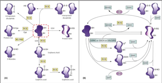
Figure 11 - Oxidative modifications of the thiol containing in proteins (A) and the glutathionylation reaction (B) ... 16
Figure 12 - Representation of the molecular structure of homocysteine, methionine and cysteine ... 18
Figure 13 - Redox reaction of homocysteine ... 18
Figure 14 - Redox reaction of the methionine residue in proteins ... 19
Figure 15 - Homocysteine synthesis, also called demethylation of methionine ... 20
Figure 16 - Remethylation reaction of homocysteine ... 21
Figure 17 - Transsulfuration reaction of homocysteine metabolism ... 21
Figure 18 - Homocysteine metabolism ... 22
Figure 19 - Schematic representation of the folate cycle ... 23
Figure 20 - Reaction of homocysteine thiolactones formation ... 26
Figure 21 - Development and progression of atherosclerosis ... 28
Figure 22 - Development of atherosclerotic plaque, highlighting the endothelial activation pathway and oxidative modifications that precede endothelial injury ... 29
Figure 23 - Representation of the nucleoside and nucleotide structure ... 31
Figure 24 - Synthesis of phosphoribosyl pyrophosphate (PRPP) ... 32
Figure 25 -Schematic representation of purine “de novo” synthesis pathway ... 33
Figure 26 - Inosine monophosphate conversion ... 34
Figure 27 - Regulatory mechanisms in purine metabolism ... 35
VIII
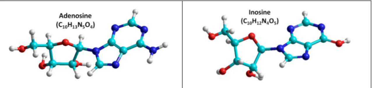
Figure 29 - Representation of the molecular structure of adenosine and inosine ... 36
Figure 30 - Adenosine metabolism ... 37
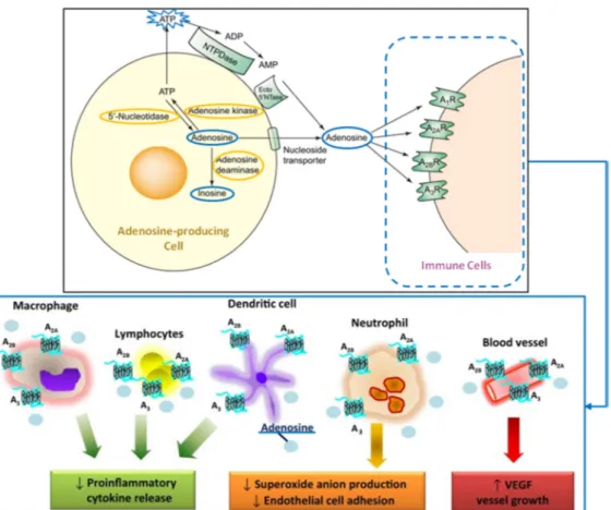
Figure 31 - Interaction of adenosine with immune cells leading to inflammation control response ... 39
Figure 32 - Schematic representation of the metabolic relationship between the studied compounds ... 42
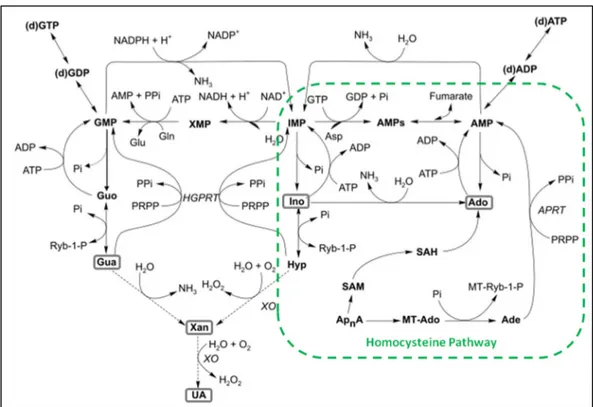
Figure 33 - Purine metabolism emphasizing the link with homocysteine pathway ... 42
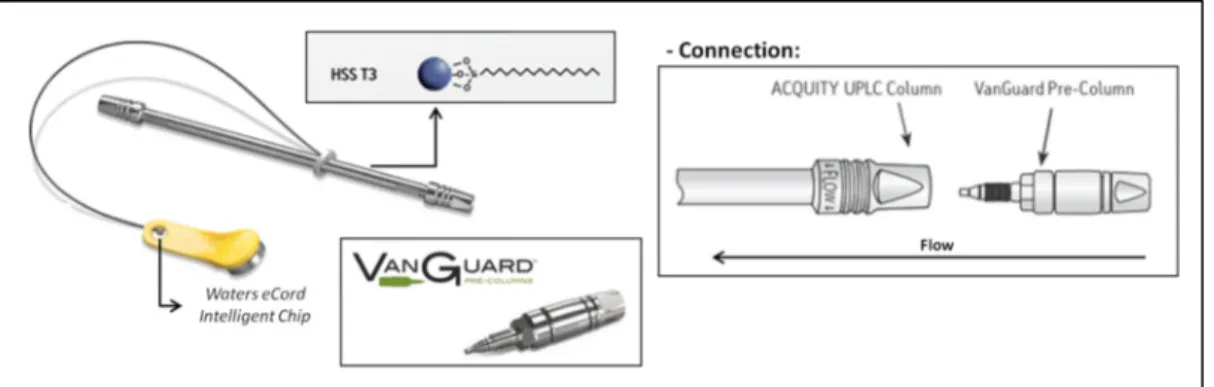
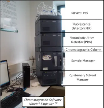
Figure 34 - Characterization of the ultra performance liquid chromatography system ACQUITY UPLC® H-Class of Waters Corporation ... 46
Figure 35 - Schematic representation of the operation system UPLC® ... 47
Figure 36 - ACQUITY UPLC HSS T3 (2.1 x 100 mm, 1.8 µm particle size) column linked to ACQUITY UPLCHSS T3 VanGuardTM (2.1 x 5 mm, 1.8 µm) pre-column and representative scheme of their connection ... 48
Figure 37 - Schematic representation of the ACQUITY UPLC Photodiode Array Detector operating mode in the UPLC© ... 49
Figure 38 - Schematic representation of the ACQUITY UPLC Fluorescence Detector operating mode in the UPLC© ... 50
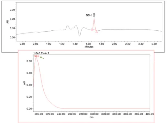
Figure 39 - UHPLC-PDA chromatogram of a mixture of glutathione (100 µM) (A) and homocysteine (7397 µM) (B). ... 57
Figure 40 - Spectra of analyzed glutathione (200 ± 4.7 nm) by the UHPLC-PDA method ... 58
Figure 41 - Spectra of analyzed homocysteine (238 ± 0.0 nm) by the UHPLC-PDA method ... 58
Figure 42 - UHPLC-PDA injection of a mixture of thiol glutathione (100 µM) (A) and homocysteine (7397 µM) (B) ... 59
Figure 43 -UHPLC-FLR injections of cysteine (100 µM) with an isocratic mobile phase at (A) 95%H2O|0.1%FA : 5%ACN, (B) 90%H2O|0.1%FA : 10%ACN and (C) 97.5%H2O|0.1%FA :
2.5%ACN ... 61
Figure 44 - UHPLC-FLR injections of homocysteine (100 µM) with an isocratic mobile phase at (A) 95%H2O|0.1%FA : 5%ACN, (B) 90%H2O|0.1%FA : 10%ACN and (C) 97.5%H2O|0.1%FA :
2.5%ACN. ... 61
Figure 45 – Four UHPLC-FLR chromatograms of glutathione (100 µM) using an isocratic mobile phase 97.2%H2O|0.1%FA : 2.5%ACN ... 62
Figure 46 - Separation of mixture of thiols composed of GSH (1), Cys (2) and Hcy (3), (each 100 µM) in two columns (A) HSS T3 and (B) BEH C18 ... 63
IX
Figure 48 - UHPLC-FLR chromatograms of the thiol standards homocysteíne (A) and homocystine (B) at 10 µM ... 66
Figure 49 - Optimal conditions for the chromatographic analysis of a mixture of thiols glutathione, cysteine and homocysteine (each 100 µM), with FLR detection. ... 67
Figure 50 - Verification of the specific wavelength detection and spectrum of adenosine (254 ± 2.8 nm) ... 68
Figure 51 - Verification of the specific wavelength detection and spectrum of inosine (250 ± 1.8 nm) ... 68
Figure 52 - UHPLC-PDA chromatograms of adenosine (150 µM), using an isocratic mobile phase 95%H2O|0.1%FA:5%ACN at pH of 2.70 (A) and 3.60 (B). ... 71
Figure 53 - Overlap of Inosine (125 µM) chromatograms using an isocratic mobile phase with methanol ... 71
Figure 54 -UHPLC-PDA chromatograms of inosine (250 µM) with (A) HSS T3 and (B) BEH C18 . 72
Figure 55 - UHPLC-PDA chromatograms of adenosine (250 µM) with (A) HSS T3 and (B) BEH C18
... 72
Figure 56 - UHPLC-PDA chromatograms of adenosine and inosine (500 µM) with (A) HSS T3 and (B) BEH C18 both at 254 nm ... 73
Figure 57 - Theoretical equation for the calculation of the empty space of a chromatographic column ... 74
Figure 58 - Overlap of chromatograms of different injected volumes, 2 (red), 4 (blue), 6 (green) and 8 µL (black), of inosine (500 µM) detected at 250 nm ... 74
Figure 59 - Graphical representation of the chromatographic data obtained for inosine when testing injection volume ... 75
Figure 60 - Optimal conditions for the chromatographic analysis of inosine and adenosine (500 µM) with PDA detection at 250 and 254 nm, respectively ... 76
Figure 61 - Chromatographic results of three injections of cysteamine standard (42 µM) showing no intra-day precision ... 77
Figure 62 - Overlap of chromatographic results of three injections of inosine standard (100 µM), showing no intra-day precision ... 78
Figure 63 - Chromatographic results of three injections of inosine standard (100 µM) showing no inter-day precision ... 78
X
Figure 65 - Chromatographic results of inter-day injections of a mixture of standard thiols (100 µM) identifying GSH, Cys, Cyst and Hcy ... 80
Figure 66 - Graphical representation of the inter-day calibration curves of adenosine (n=6). .. 81
Figure 67 - Chromatograms obtained for the calibration curves of the mixture of thiol standards glutathione, cysteine and homocysteine ... 82
Figure 68 - Graphical representation of the calibration curves of glutathione, cysteine and homocysteine (n=3) ... 82
Figure 69 - Chromatograms obtained for the calibration curve of a mixture of standard (A) inosine and (B) adenosine ... 83
Figure 70 – Graphical representation of the calibration curve of standard inosine and adenosine (n=6) ... 84
XI
List of Tables
Table 1 - Different reactive species that are found in organisms, under normal or pathological
conditions ... 2
Table 2 - Analytical methods ... 44
Table 3 - Different mobile phases tested in isocratic conditions with pH adjustments ... 60
Table 4 - Mobile phase conditions tested for chromatographic separation of thiols ... 60
Table 5 - Chromatographic data obtained from separation of a mixture of thiols in the HSS T3 column ... 63
Table 6 - Chromatographic data obtained from homocysteine and cysteine ... 65
Table 7 - Chromatographic data obtained from separation of thiol standard homocysteine and homocystine at 10 µM ... 66
Table 8 - Optimal chromatographic conditions achieved for the determination of thiol compounds ... 66
Table 9 - Chromatographic data obtained from separation of a mixture of thiols glutathione, cysteine and homocysteine (each 100 µM) ... 67
Table 10 - Mobile phase conditions tested for chromatographic separation of purines ... 69
Table 11 - Different mobile phases tested in isocratic conditions with pH adjustments ... 70
Table 12 - Chromatographic data obtained from separation of adenosine in isocratic mobile phase 95%H2O|0.1%FA:5%ACN with pH variation ... 71
Table 13 - Chromatographic data obtained from separation of adenosine and inosine (500 µM) with (A) HSS T3 and (B) BEH C18 ... 73
Table 14 - Variation of injection volume for standard inosine ... 75
Table 15 - Optimal chromatographic conditions achieved for the analysis of purines and related compounds ... 76
Table 16 - Chromatographic data obtained from separation of inosine and adenosine ... 76
Table 17 - Chromatographic data obtained from separation of cysteamine showing no inter-day precision ... 77
Table 18 - Chromatographic data obtained from separation of inosine without precision ... 78
Table 19 - Chromatographic data obtained from separation of inosine showing no inter-day precision ... 79
Table 20 - Chromatographic results of the inter-day precision of a mixture of standards ... 79
Table 21 - Chromatographic result of the inter-day precision of a mixture of standard ... 80
Table 22 - Detection and quantification limits for standards detection with the established methods ... 85
XII
List of abbreviations
Scientific Notation
% Percentage °C Degrees Celsius
µL Microlitres (1 x 10⁻⁶ litres)
µM or µmol/L Micromole per litres (1 x 10⁻⁶ moles) µV MicroVolt units
µV°sec MicroVolt units per second AU Absorbance units
D-amino acid Chirality (-)levorotatory (R-enantiomer) EU Emission units
L-amino acid Chirality (+)dextrorotatory (S-enantiomer) mg Milligrams (1 x 10⁻³ grams)
min Minute
mL Millilitres (1 x 10⁻⁹ litres)
mM or mmol/L Millimole per liter (1 x 10⁻³ moles) nm Nanometer (1 x 10⁻⁹ metres) β Greek letter beta
γ Geek letter gamma
λ Greek letter lambda which expresses the wavelength
A
A Alanine transporter system AC Adenylyl cyclase
ACN Acetonitrile
ADA Adenosine deaminase
ADHD Dimethylarginine dimethylamino hydrolase ADK Adenosine kinase
ADMA Asymmetric dimethylargenine Ado Adenosine
ADP Adenosine 5'diphosphate ADSL Adenylosuccinate lyase ADSS Adenylosuccinate synthetase
AICAR Aminoimidazolecarboxamide ribonucleoside
AICARFT Aminoimidazole carboxamide ribonucleotide formyltransferase AIRC Aminoimidazole ribonucleotide carboxylase
AIRS Aminoimidazole ribonucleotide synthetase ALS Amyotrophic lateral sclerosis
AMP Adenosine 5'monophosphate
APOBEC Apolipoprotein-β mRNA aditing anzyme, catalytic polypeptide-like APRT Adenosine phosphoribosyl transferase
APS Antiphospholipid antibody syndrome Arg Arginine
ASC Alanine-serine-cysteine transporter system Asp Aspartate
ATIC Aminoimidazole carboxamide ribonucleotide transformylase/inosine monophosphate cyclohydrolase
XIII
B
BHMT Betaine-homocysteine methyltransferase B2 | B6 | B12 Cofactors derived from B vitamin complex
C
2Cys Cystine Ca2+ Calcium ion
cAMP Cyclic adenosine monophosphate CBS Cystathionine β-synthase
CEC Circulating endothelial cells
CEPC Circulating endothelial progenitor cells cGMP Cyclic guanosine monophosphate CG-X Conjugate cysteinylglycine CH2 Methylene group
Cl- Chlorine ion
CPK Creatinine phosphokinase CSE Cystathionase γ-liase
CSIF Human cytokine synthesis inhibitory factor CVD Cardiovascular diseases
C-X Cysteine conjugates Cys Cysteine
CysGly Cysteinylglycine Cys-SS-Hcy Cysteinylhomocysteine
D
DBP Diastolic blood pressure DHFR Dihydrofolate reductase DMG Dimethylglycine
DNA Deoxyribonucleic acid
E
e- Free electron EC Endothelial cells
eNOS Endothelial nitric oxide synthase
ENT1 and 2 Equilibrative nucleoside transporter 1 and 2 ERK½ Extracellular signal-regulated kinase ½
ERK1/2 Extracellular signal-regulated protein kinases 1 and 2
F
FA Formic acid
FAD Flavine adenine dinucleotide oxidized (active form of vitamin B2)
FADH2 Flavine adenine dinucleotide reduced
FGAMS Formylglycinamidine ribonucleotide synthase
G
G6PD Glucose-6-phosphate dehydrogenase enzyme GARS Glycinamide ribonucleotide synthetase GART Glycinamide ribonucleotide transformylase GCL Gamma-glutamylcysteine ligase
GGT Gamma-glutamyl transpeptidase Gln Glutamine
XIV GNMT Glycine N-methyltransferase GPx Glutathione peroxidase GR Glutathione reductase Grx Glutaredoxine
GS Glutathione synthetase
GS(O)SG Glutathione disulphide S-monoxide GS● Glutathione radical
GSH Reduced glutathione GSNO S-Nitrosoglutathione GSOH Glutathione sulphenic acid GS-R or GS-X Glutathione conjugates GSSG Oxidized glutathione GST Glutathione S-transferase Gua Guanosine
γ-GGCT Gamma-glutamyl cycle transferase γ-Glu-aa Gamma-glutamylaminoacid γ-GluCys Gamma-glutamylcysteine
H
2Hcy Homocystine H+ Hydrogen cation H2O Water
H2O2 Hydrogen peroxide
H2S Hydrogen sulfide
HCO2
-hydrogen carbonate anion Hcy Homocysteine
HDL High-density lipoprotein
HGPRT Hypoxanthine-guanine phosphoribosyltranferase HHcy Hyperhomocysteine
HMGCR 3-Hydroxy-3-methylglutaryl-CoA reductase HNE 4-Hydroxynonenal
HNO2 Nitrous acid
HO● Hydroxyl radical HOCl Hydrochloric acid
HPLC High Performance Liquid Chromatography HUVEC Human Umbilical Vein Endothelial Cells
I
IL-10 Interleukin
IMP Inosine 5'monophosphate
IMPCH Inosine monophosphate cyclohydrolase
L
L Large branched-chain neutral amino acids transporter system L/T-type Ca2+ L and T-type calcium channels
LDL Low-density lipoprotein LOH Lipid hydroxide
LOO• Lipid
LOOH Lipid hydroperoxides LVCa L-type calcium channels
XV
3MST 3-Mercaptopyruvate sulphur transferase MAPK Mitogen-activated protein kinase MAT Methionine S-adesonyltranferase Met Methionine
meTHF 5,10-Methylenetetrahydrofolate Met-R-O Methionine sulfoxide residues MetRS Methyonyl-tRNA synthetase miRNA Micro RNA
MPx Methionine peroxidase
MRP Protein complex of transporter MS Methionine synthase
MSR Methionine sulfoxide reductase mTHF 5-Methyltetrahydrofolate
MTHFR Methylene tetrahydrofolate reductase
N
N2O3 Dinitrogen trioxide
Na+ Sodium ion NAC-X Mercapturic acid
NAD+ Nicotinamide adenine dinucleotide oxidized NADH Nicotinamide adenine dinucleotide reduced
NADP+ Nicotinamide adenine dinucleotide phosphate oxidized NADPH Nicotinamide adenine dinucleotide phosphate reduced NF-kβ / NrF-2 Nuclear factor
-NH2 Amino group
-NH4 +
Ammonia group NO Nitric oxide NO- Nitroxyl ion
NO● Nitric oxide radical NO2- Nitrite ion
NO2● Nitrogen dioxide radical
NO2Cl Nitryl chloride
NOH Nitroxide
NOOH Nitro hydroperoxides NOOR Nitroperoxide
O
●OH Hydroxyl radical
5-Oxo 5-Oxoprolinase O2 Oxygen molecule
O2●- Superoxide radical anion
OH- Hydroxyl anion ONOO- Peroxynitrite ONOO● Peroxynitrite radical ONOOH Peroxynitrous acid
P
Pept Peptidase PGI2 Prostacyclin
XVI PKA Protein kinase A
PKC Protein kinase C
PLAT Plasminogen tissue activator PLC Phospholipase C
PLP Pyridoxal-5'-phosphate (active form of vitamin B6)
PNP Purine-nucleoside phosphorylase
PPAT Phosphoribosyl pyrophosphate amidotransferase PPP Pentose phosphate pathway
Protein-Met Methionine residue in the protein complex
Protein-Met-O Protein complex with methionine sulfoxide residue PRPP Phosphoribosyl pyrophosphate
PRPPS Phosphoribosyl pyrophosphate synthetase Prx Peroxiredoxins
R
R● Free radicals
R-CH3 Acceptor methylated
RMVEC Rat Microvascular endothelial cells RNA Ribonucleic acid
RNAi Interference RNA RNH Nitroxyl
RNS Reactive nitrogen species ROH Alcohol
R-OH Alcohol group ROOH Hydroperoxide
ROS Reactive oxygen species RS Reactive species
R-S- Thiol thiolate anion R-SH Thiol protein compound R-SO2H Sulfinic acid
R-SO3H Sulfonic acid
R-SOH Sulfenic acid
S
S Sulfur
SAH (or AdoHcy) S-adenosylhomocysteine
SAHH S-adenosyl-homocysteine hydrolase
SAICARS Aminoribosyl-aminoimidazole succinocarboxamide ribonucleotide synthetase
SAM (or AdoMet) S-adenosylmethionine Sar Sarcosine
SBP Systolic blood pressure Ser Serine
SHMT Serine hydroxymethyltransferase SOD Peroxide dismutase
T
XVII tRNA Transfer ribonucleic acid
TRPV Transient receptor potential cation channel TrxOxi Thioredoxin oxidazed form
TrxR Thioredoxin reductase TrxRed Thioredoxin reduced form TX Diffusion or active transport
U
UA Uric acid
UMP Uridine monophosphate
UHPLC Ultra-High Performance Liquid Chromatography
V
VEGF Vascular endothelial growth factor VSMC Vascular smooth muscle cells
X
X Xenobiotic molecule or Toxic compound XAG Aspartate and glutamate system transporter
XVIII
Aims
The project presented in this thesis had as the main purpose to obtain a master's degree in Applied Biochemistry. This work was developed in the Centro de Química (CQM) of the University of Madeira.
Specific objectives
In this dissertation, an extensive bibliographic search and laboratory research were performed. The theoretical introduction highlights all relevant information for the development, analysis and discussion of the experimental work.
The experimental part consisted of two approaches, developed with the following purpose:
First, developed and optimized a pre-analytical procedures for standard treatment of some biomarkers of oxidative stress, for analysis by Ultra-High Performance Liquid Chromatography (UHPLC). The optimization procedure was held for the detection of the standard glutathione in reduced form (GSH), homocysteine (Hcy), cysteine (Cys) and cysteamine (Cyst) with fluorescence (FLR) detection. Additionally, a method of photodiode array (PDA) detection for purine compounds analysis, like adenosine (Ado) and inosine (Ino) was developed.
1
Chapter 1
Introduction
In this chapter, the most relevant aspects of thiol compounds and their derivatives will be reviewed, as biomarkers of oxidative stress. Also, some purine compounds will be mentioned, referring and emphasizing cardiovascular pathologies and some of their factors, like the unbalance of the physiological and homeostatic state of endothelial cells.
Additionally, analytical techniques for their assessment will be focused, including pre-analitical considerations.
1. Oxidative Stress and Redox Systems
The term “stress”, expresses the tension exerted in an organism mediated by a physical or chemical injury, which consequently triggers several mechanisms of defense and adaptation [1]. Cellular stress is a cause of pathology in certain human diseases, such as, hypoxia, immune reactions and infections [2].
The cell is composed of organelles, small structural units bounded by the membrane, which are the basis of their operation and integrity. As such, it can be said that the redox state is also divided into compartments, highlighting the mitochondria, the endoplasmic reticulum, the nucleus and the cytoplasm, as the main areas of redox signaling. Comparatively, it is in the mitochondria, where there is a greater redox activity, since its membrane has five multiprotein complexes, which are responsible for the oxidative phosphorylation. These are the NADH-quinone oxidoreductase, succinate dehydrogenase, coenzyme Q cystochrome reductase, cytochrome oxidase and ATP synthase [3].
In cell metabolism, it is possible to find different redox systems, such as, the pairs flavine adenine dinucleotides (FADH2/FAD), nicotinamide adenine dinucleotides (NADH/NAD+
and NADPH/NADP+), thioredoxins (TrxRed/TrxOxi) and glutathiones (GSH/GSSG) [4, 5]. They
promote a dynamic regulation that maintains the cells in good oxy-reductive conditions, by balancing the concentrations of each coupled form [6].
2
Table 1 - Different reactive species that are found in organisms, under normal or pathological conditions.
Adapted from [8 - 10]:
Reactive Oxygen Species (ROS) Reactive Nitrogem Speies (RNS)
Superoxide radical (O2●-) Nitrogen dioxide radical (NO2●)
Hydrogen peroxide (H2O2) Nitrite ion (NO2-)
Hydroxyl radical (HO●) Nitric oxide radical (NO●)
Hydroxyl ion (HO-) Nitroxyl ion (NO-)
Hydroxyl (ROH) Nitroxyl (RNH) Hydroperoxide (ROOH) Nitroperoxide (NOOR) Lipid peroxyl radical (LOO●) Dinitrogen trioxide (N
2O3)
Lipid hydroxide (LOH) Nitroxide (NOH) Lipid hydroperoxide (LOOH) Nitrous acid (HNO2)
Hydrochloric acid (HOCl) Nitryl chloride (NO2Cl)
Peroxynitrite radical (ONOO●)
Peroxynitrite ion (ONOO-)
Peroxynitrous acid (ONOOH)
Antioxidants are molecules that prevent oxidative reactions caused by ROS and RNS occurring with damage effects [7]. The regulation of antioxidant systems is crucial for a proper cell function, which is achieved by lowering levels of reactive species. Several studies have shown that these systems are important protective agents, because they avoid abrupt and irreversible changes on metabolic molecules and irreparable damage of deoxyribonucleic acid (DNA), which in turn could induce cell death [4].
In some physiological conditions, the regulatory mechanisms may not be effective, for example, when the levels of antioxidants are very low or nonexistent, or when there is absence or inactivation of some of the enzymes involved in those mechanisms [11]. In these cases, excess of reactive species compromises the metabolic balance or even the cell viability. However, the consequences of these possible situations depend on where it happens, the availability of energy and the plasticity of the cells [2]. Therefore, oxidative stress may arise and translate into an imbalance between oxidants and neutralizing compounds, leading to disruption of the redox signalling, loss of homeostatic control and can consequently, cause genetic damage [12]. Therefore, a condition that results from the accumulation of oxidative compounds, in detriment of the reducing ones, reduction of the antioxidant agents, or even, by synergy of these two situations [13].
In aerobic metabolism, the inevitable oxygenated status can be a hostile environment, what is a contradiction of life. The oxygen molecule (O2) is essential in the respiration process
and energy metabolism but, at the same time, is the pathogenic basis of many diseases or degenerative conditions of living beings. The theoretical principle, states that a single inspiration contains O2 capable of producing billions of reactive species that may have signaling
3
oxygenated and stress conditioned environment [12]. Still, there is always a chance of something going wrong, at metabolic level, and oxidative damage may arise.
Accordingly, the main consequences of oxidative stress are the molecular changes in nucleic acids, lipids and proteins that compromise cell stability or even, viability [15]. Thus, in extreme cases, the cell promotes secondary responses to induce death by necrosis or apoptosis. Moreover, in most fortunate cases, the cell is able to restore the redox homeostatic state and the metabolism back to be in equilibrium without negative consequences [11].
1.1. Oxidative Stress and Endothelial Cells
The endothelial cells (EC) line on the entire surface of the vascular system, forming a barrier between the blood and the other organs. It is a versatile and multifunctional monolayer that is essential in the structural support and integrity of the vascular wall throughout all circulation system. These are cells with high metabolic activity, with endocrine, paracrine and autocrine functions. They form a dynamic and semi-permeable barrier, which controls the transport of a large number and variety of metabolites between the different tissues [16]. In its metabolic properties, regulation of the vascular tone that is characterized by the normal state of firmness and elasticity of an organ or tissue, through the production of vasodilator and vasoconstrictor molecules, stands out. At this level, the control of the flow and blood clotting happens by the action of platelet factors and regulation of inflammatory responses [17].
Under physiological conditions, EC express and secrete a number of specific metabolic compounds in response to different stimulations. Among the activating molecules, we present some of those that stand out in the anticoagulant reactions, such as, prostacyclin (PGI2), nitric oxide (NO), thrombomodulin (TM) and plasminogen tissue activator (PLAT); the latter, also presents pro-coagulant action; the vasodilators NO, PGI2 and endothelin, and the vasoconstrictors endothelin, thromboxane and prostaglandins [18, 19]. Furthermore, EC mediate vascular proliferation (angiogenesis) in synergy with platelets, smooth muscle cells and the matrix constituents (fibronectin, laminin, collagen and proteoglycans) [20, 21].
A relevant fact, is that the growth and survival of EC depend on the intracellular production of reactive species, such as, superoxide anion (O2●-) and hydrogen peroxide (H2O2),
4
The syndrome of endothelial dysfunction is a disorder that expresses vascular abnormalities, which are the consequence of damage in the thrombogenic and angiogenic properties. This may result from loss of tone by the vascular smooth muscle or, from blocking of the normal immunologic response [24]. This disturbance is characterized essentially by reduced NO bioavailability and production. Also, by increased adhesion of monocytes and polymorphonucleate cells, cholesterol and oxidized low-density lipoprotein (LDL) accumulation, signaling failure of endothelium derivatives, expression of pro-fibrotic genes and premature cell senescence or apoptosis [24, 25].
Risk factors of oxidative stress and endothelial dysfunction are classified as i) behavioral, such as, smoking, malnutrition, physical inactivity and obesity, and ii) biological determinants, like gender, aging, dyslipidemia, hypertension, diabetes, family history and genetic defects [26, 27]. These factos affect functional integrity of the endothelium, and should always be taken into consideration at the clinical level.
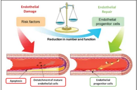
The endothelial health is balanced (figure 1) by damage factors and regenerative capacity [28]. In the cardiovascular system, the regeneration is promoted by a decrease in the inflammatory response, which is achieved by reducing cell interactions, such as, decreasing macrophage phagocytosis of LDL, secreting platelets and inhibiting foam cells formation [29]. That balance is also promoted by the circulating endothelial cells (CEC), which derive from the endothelial layer of vessels, and by the circulating endothelial progenitor cells (CEPC) from bone marrow, in the bloodstream [28]. These cell types are major promoters of the integrity, are responsible for angiogenesis and regeneration of the endothelium. Therefore, they prevent, attenuate or delay the development of atherosclerosis and improve the blood flow [29].
Figure 1 - Counterbalancing of endothelial damage with the regenerative capacity. Image obtained from
5
1.2. Biomarkers of Oxidative Stress
The term biomarker features a measurable compound as an indicator of normal biological metabolism, pathological conditions or pharmacological responses to therapeutic processes [30]. Therefore, a biomarker should help in symptomatic or pre-symptomatic disease diagnosis and provide data results showing the clinical effectiveness of treatments [15]. In theory, to be validated as a biomarker of oxidative stress, a compound must have the following characteristics: i) be stable, not susceptible to oxidative induction or loss due to handling, processing, analysis or storage; ii) be the main product that is directly involved in the initiation or progression of the pathological condition; iii) be accessible on the target tissue and quantitatively translate the metabolic changes [30]; iv) be a significant product present in high concentrations and free of confounding factors, such as from diet; v) be identified and quantified by noninvasive and painless methods; vi) its analysis must be specific, sensitive and reproducible [30]; vii) be easy to detect and quantify between populations; viii) have relatively stable concentrations, which do not vary widely in the same person, in different times and conditions; ix) be measurable with relatively small variation in intra and inter sample tests [30].
Validation requires multiple steps that are often difficult to achieve, because of the complexity of the metabolic pathways.
Measurement of oxidative stress and antioxidant defense biomarkers is a major challenge in research studies. Next, the most relevant topics for each thiol and purine compound analyzed in this dissertation are presented, including properties, functional characteristics, metabolic and regulatory pathways, as well as, some pathological associations. Finally, the relationship and metabolic link between them is showed.
1.2.2. Thiol Compounds
Thiol compounds and their derivatives are widely distributed in nature, either in prokaryotic or eukaryotic cells, in plant and animal tissue [31]. These compounds are synthesized and then sent to the required locations, where they are essential for the proper functioning of organisms. Thiols are involved in crucial physiological processes that include elimination of toxins, redox signalling, transport, metabolic storage and protein functionality, as well as, gene expression, proliferation, differentiation and even in cell death [32]. Several studies have shown that the maladjustment of these roles is the basis of many human diseases, which is why there is a great effort of the scientific community to better understand their metabolic implications.
6
linked to a carbon atom, also known as thiol or mercaptan group. The R-SH group is analogue to alcohol group (R-OH) but form weaker hydrogen bonds. Therefore, these compounds are volatile [35], highly polar, soluble in water and with low boiling points [36]. The sulfur (S) is an abundant non-metal atom, which can be incorporated into molecules, giving rise to sulfate or be reduced to sulfite, the main substrate in cysteine synthesis [37]. Thiol group is considered to be the most reactive group present in cells. Actually, it is involved in many metabolic reactions that depend on the oxidative capacity, a specific biochemical property that distinguishes them from other molecules with metabolic functions [37]. In figure 2 the most relevant organic sulfur compounds in cell metabolism, are highlighted.
The thiolate anions (R-S-) are more reactive than the sulfhydryl group, becouse they
are easily oxidized to different products, such as, sulfenic acid (R-SOH), sulfinic acid (R-SO2H)
and sulfonic acid (R-SO3H) [7]. The sulfite compound is less reactive than the thiol that gives
rise to it, but it is extremely important in the stability of protein complexes, in polypeptide chains with tertiary and quaternary three-dimensional structure, as well as, in the functionality of the active site of some metabolic enzymes. The biosyntheses of organosulfur compounds occur mainly in bacteria and plants, while the oxidation commonly occurs in animals [37].
Figure 2 - Different thiol compounds that are present in biological systems. Abbreviations: H2O2,
hydrogen peroxide; ●NO, nitric oxide; Srx, sulfiredoxin. Image obtained from Ma [7].
Endogenoushydrogen sulfide (H2S) is considered an important signaling molecule for
EC and cardiovascular system (Figure 3) [38]. It is produced by enzymatic reactions of cystathionase γ-liase (CGL), cystathionine β-synthase (CBS) and 3-mercaptopyruvate sulphur transferase (3MST). Subsequently, it suffers different conversions. It may be oxidized to hydrosulfide ion (HS-) or thiolsulfate forms, may produce sulfhemoglobin reacting with
7
Figure 3 - Cardiovascular functions of hydrogen sulfide (H2S). Abbreviations: H2O2, hydrogen peroxide;
3MST, 3-mercaptopyruvate sulphur transferase; SH-, hydrosulfide ion; H+, free hydrogen cation; CAT,
cysteine amino transferase; CGL, cystathionase γ-liase. Image obtained from Calvert et al. [38].
According to its concentration, H2S (ranging from 10 to 100 µM [38]) can produce
cytoprotection actions, through i) protein modifications (S-sulfhydration); ii) modulation of ion channels, such as, vascular KATP channels, transient receptor potential cation channel (TRPV)
and calcium channel (L/T-type Ca2+) iii) stimulation of the production of antioxidant molecules,
like nuclear factors (NrF-2) and glutathione [39]; and iv) decrease of mitochondrial functions [40]. The purpose is to prevent oxidative stress, apoptoctic and necrotic mechanisms. In some conditions, like in heart failure, administration of H2S has shown to be a promoter of
remodelling processes [41]. Similarly, in atherosclerosis, this molecule modulates vasodilatation or vasoconstriction reactions, inhibits the expression of leukocyte adhesion molecules and formation of foam cells. In addition, it activates pro-angiogenic mechanisms, prevents edema formation and it is an anti-apoptotic mediator [39]. This is a compound related to the thiol homocysteine, and its metabolic importance will be specified further on.
Also important in this group of thiol compounds, are some amino acids, which have been implicated in the pathophysiology of a wide range of chronic conditions [42]. As a matter of fact, sulphur amino acids like methionine (Met), homocysteine (Hcy) and cysteine (Cys) are being investigated as potential indicators of health status and disease risk. Glutathione, the most abundant mammalian antioxidant, a tripeptide that is linked to Hcy metabolism via the transsulphuration pathway, which will be specified later on.
8
Alzheimer disease, dementia, sleep apnea, renal failure, diabetes, erectile dysfunction, among others [43].
1.2.2.1. Glutathione
In 1888, Rey-Pailhade identified a compound that he called Philothione, a Greek
expression, which means “sulfur friend”, in yeasts. Later in 1927, he changed the name to glutathione (GSH), when Hopkins identified it as a dipeptide of glutamic acid (Glu) and Cys [44]. However, in the same year, Hunter and Eagles conducted more studies and realized that there was an error [45]. As such, in 1929 [46] the compound was reanalyzed in yeast and erythrocytes, and GSH was recharacterized as a tripeptide also composed by glycine (Gly) (figure 4). Hopkins [45, 46] noted that, biochemically, this molecule exhibited an exceptional behavior, that it was a very unstable compound due to the sulfur atom in the Cys residue. He even stated that this instability resided in all molecules formed by Cys or its derivatives, which could lead to GSH misidentification [45, 46].
Figure 4 - Representation of the molecular structure of glutathione.
I.
Glutathione syntesis
The thiol compound GSH results from enzymatic reactions, which occur in the
9
Figure 5 – Enzymatic synthesis of glutathione. Abbreviations: ADP, adenosine 5'diphosphate; ATP,
adenosine 5'triphosphate; GCL, γ-glutamylcysteine ligase; GS, glutathione synthetase; Cys, cysteine; L-Glu, glutamic acid; L-Gly, glycine; Pi, inorganic phosphate; γ-GluCys, gamma-glutamylcysteine; 1.
Chemical bond between the γ-carbonyl group of glutamic acid and the amine group of cysteine; 2.
Chemical bond between the α-carbonyl group of cysteine and the amine group of glycine. Adapted from Lu [47].
The biosynthesis of GSH is initially regulated by gene expression and metabolism is further controlled by feedback signaling mechanisms, following homeostatic conditions [49]. GSH synthesis is limited by ATP bioavailability and by the catalytic action of GCL and MS enzymes. Besides, GCL is inhibited by negative feedback when GSH concentrations reach high levels [4].
In GSH molecule, each amino acid has unique features that are critical in its general skills and properties. GSH presents a unique structure due to the binding between Glu and Cys, which uses the γ-carbonyl group rather than the typical α-carboxyl bond. This rare union is resistant to the endogenous action of peptidases and is only degraded by the gamma-glutamyl transpeptidase (GGT) [50]. GGT is an ecto-enzyme, it has an active centre facing the outside and a higher affinity for Met, Glu, Cys and arginine (Arg) [51]. Orlowski and Meister [51] studied the activity of this enzyme in mice kidney and found that the reactions of GSH synthesis and degradation are coupled to major transport ways.
II.
Glutathione and the gamma-glutamyl cycle
10
and, at the same time, the latest mediates the uptake of amino acids by other specific carriers, from the extracellular space up to the cytoplasm [51, 52]. So, GGT converts GSH into γ-glutamylaminoacid (γ-Glu-aa) releasing cysteinylglycine (CysGly), in the extracellular space. This dipeptide, suffers degradation by some exopeptidases and their amino acids are provided for new metabolic reactions. The γ-Glu-aa is then imported and, in the intracellular space, it is catalysed by γ-glutamyl cycle transferase (γ-GGCT), which releases the transported amino acid and 5-oxoproline complex. It is then converted to Gluthrough the action of 5-oxoprolinase (5-Oxo) [47, 51]. Following the cycle, GSH is regenerated by the subsequent reactions, which have already been described and schematized in figure 5.
GGT is a canalicular enzyme [1], which has been identified through histological studies, in the apical surfaces of several epithelial tissues, such as, the cells covering the villi in the jejunum, external portion of pancreas, liver, bile, seminal vesicles, epididymis, fallopian tubes, ovaries, endometrium, prostatic ducts, submaxillary and mammary glands, ependymal cells of the brain and in the endothelium of blood capillaries [51], among others.
Figure 6 - Gamma-glutamyl cycle expressing glutathione metabolism associated with the transport of
amino acids. Abbreviatons: 5-Oxo, 5-oxoprolinase; aa, amino acid; ADP, adenosine 5'diphosphate; ATP, adenosine 5'triphosphate; GCL, gamma-glutamylcysteine ligase; GGT, gamma-glutamyl transpeptidase; GS, glutathione synthetase; L-Cys, cysteine; L-Glu, glutamic acid; L-Glu-Cys, glutamylcysteine; L-Gly, glycine; Pi, inorganic phosphate; T-GSH, glutathione transporter protein complex; T-X, transporter X; γ-GGCT, gamma-glutamyl cyclotransferase; γ-Glu-aa, gamma-glutamyl amino acid. Adapted from [47, 51].
11
In 1970, Meister [51] speculated that the gamma-glutamyl cycle could be crucial for metabolic transport and/or secretion and, that the presence of GSH was indispensable. Fifteen years later [52], he confirmed that the transport of γ-Glu-aa is one step of the cycle, which mediates the uptake of amino acids. He also found that this transport system is inhibited by high GSH concentrations in the extracellular space, as well as, low GSH concentrations inside the cells [52, 53].
A limiting factor of GSH synthesis is the bioavailability of Cys [53]. This amino acid is in higher concentration in the cytoplasm and, externally, it is more abundant in the oxidized form, as cystine (2Cys). Therefore, it seems that cells perform the gamma-glutamyl cycle also as a Cys source [54]. However, numerous studies have tried to clarify the uptake of Cys and other amino acids by cells, but until now, there is no consensus, because in the several reports, different cells were used (for example: astrocytes [55], red blood cells, endothelium or epithelium [56]). The uptake of Cys and 2Cys is surely mediated by different carriers, which can be coupled or in antiport. It also depends on sodium (Na+) or chlorine (Cl-) ions. However, all
studies confirm that the transport of amino acids is an important feature in antioxidant defence mechanisms, as induced by GSH [57, 58].
III.
Glutathione oxidation pathway
GSH is a molecule with a high redox potential [4]. In certain physiological conditions, it may be in the reduced (GSH) or in the oxidized (GSSG) tripeptide form (Figure 7). The latter, results from the intermolecular binding of two reduced forms by a bisulfite bond (GH-S-S-GH), a reaction that is mediated by direct oxidation of ROS or by enzymatic action of glutathione peroxidase (GPx) a key selenoprotein [4, 59].
Figure 7 - Representation of the molecular structure of oxidized glutathione.
12
Figure 8 -Oxidation and reduction reactions of glutathione. Abbreviations: H+, free hydrogen cation; H2O,
water; H2O2, hydrogen peroxide; GPx, glutathione peroxidase; GR, glutathione reductase; GSH, reduced
glutathione; GSSG, oxidized glutathione; NADPH, nicotinamide adenine dinucleotide phosphate reduced; NADP+, nicotinamide adenine dinucleotide phosphate oxidized. Adapted from Lushcak [4].
IV.
Physiological levels of glutathione
GSH is present in all types of cells. GSH total concentration, quantified on plasma samples, ranges from 2 to 20 µM [62] and inside cells varies between 0.1 and 10 mM [63]. These values depend on the type of tissue cell and the overall homeostatic conditions displayed. For instance, in neurons it is approximately 2.5 mM and in astrocytes about 3.8 mM [53]. Intracellular GSH is in the millimolar (mM or mmol/L) range and extracellularly it is in the micromolar (µM or µmol/L) range. GSH levels decrease with increasing transpeptidation [64].
Quantification is not accurate so far. However, it is estimated that cells have three major GSH reservoirs, which are 80 to 85% in the cytoplasm, about 10% in the mitochondria and a small percentage in the endoplasmic reticulum [47, 48]. It is considered that 99.5% of the circulating GSH is linked to the red blood cells [65] and, in the extracellular space, the concentration is very low, because is rapidly metabolized. In 1980, Wendel and Cikryt [66] performed an in vivo experiment and determined that, in human plasma, the halt-life time of
GSH is around 1.6 minutes. It is important to note that GSH concentrations are different between tissues due to its functions. In accordance, there is a higher concentration in the liver. However, it also depends on the prevalence of some pathological conditions.
V.
Specific functions of glutathione
13
elimination of ROS and ERN; ii) detoxification of endogenous and exogenous toxins [68]; iii) maintenance of normal levels of essential thiols, for proteins synthesis and other molecules, such as vitamin C and E; iv) control of Cys storage; v) mediation of copper and iron ions transfer [69]; vi) intervention in hormone metabolism, like prostaglandins, estrogen and leukotriene [4]; vii) mediation of ribonucleotides reduction both in DNA synthesis and signal transduction, by enzymatic regulation; viii) regulation of mitochondrial function and integrity [70, 71]; ix) control of cell proliferation and apoptosis [4].
Some of the above functions are here specified:
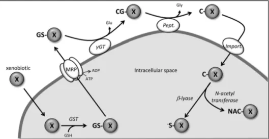
Detoxification - The detoxifying action mediated by GSH is translated in the
mercapturic acid pathway [72], shown in figure 9. It consists of a sequence of reactions that neutralize and eliminate toxic compounds and their metabolic derivatives, harmful to cells. This protection mechanism begins with the conjugation of GSH with xenobiotic (X) molecules, by a spontaneous reaction or, it may be mediated by glutathione S-transferase (GST) enzyme, which produces glutathione conjugates (GS-X) [47]. Subsequently, the conjugate compounds are transported across the membrane by the human multidrug resistant-associated protein (MRP), an ATP-dependent complex. Outside the cell, they are degraded by the GGT enzyme followed by the action of peptidases, giving rise to a conjugate of cysteine (C-X). This comes later into the cell, where it is acetylated to mercapturic acid by N-acetyl transferase enzyme, or is transformed into a very reactive thiol compound, by the action of β-liase enzyme [47, 72]. Therefore, the resulting compounds are polar and highly hydrophilic, which facilitates their cell transportation and elimination. In this way, GGT is an enzyme that protects cells from potentially mutagenic and carcinogenic compounds.
Figure 9 - Detoxification pathway mediated by glutathione. Abbreviatons: ADP, adenosine
5'diphosphate; ATP, adenosine 5'triphosphate; CG-X, conjugate cysteinylglycine; C-X, cysteine conjugate; Glu, glutamic acid; Gly, glycine; GSH, reduced glutathione; GST, glutathione S-transferase; GS-X, conjugate glutathione; MRP, protein complex of transporter; NAC-X, mercapturic acid; Pept, peptidase;
14
Elimination of reactive species - In living organisms, over 90% of the O2 consumed is
directly reduced to H2O by the electron transport chain, at the mitochondrial membrane of
cells, with the purpose of producing energy in ATP form [3]. The remaining O2 is reduced, as
shown in figure 10,by elimination of an electron, giving rise toO2●-. After this, an electron is
removed and two hydrogen cations (H+) are accepted, forming H
2O2. The latter, accepts an
electron, dividing in a hydroxyl radical (HO●) and a hydroxyl anion (HO-), which subsequently,
produce water [11, 49]. Therefore, this is one of the means responsible for the emergence of ROS and free radical (R●) compounds.
The amount and types of species that are generated depends on the physiological state of the organism [3]. It is important to note, that ROS are important signaling molecules, whose action can have negative or positive consequences. It depends on the time and intensity of the products, which can mediate an adaptive or a pathological condition [73]. Therefore, low levels of ROS favour cell proliferation, intermediate levels can lead to senescence and high concentrations may induce cell death by apoptosis or necrosis.
The mechanisms that regulate ROS levels arise not only by direct elimination, but also by the anti-oxidation systems, where low molecular weight compounds like GSH, intervene. Proteins with enzymatic activity, such as, glutathione peroxidase (GPx), glutaredoxin (Grx), peroxiredoxins (Prx) and thioredoxin reductase (TrxR) are associated to those systems [3, 5]. For example, GSH and Trx are involved in the electron transport chain, in an antioxidant pathway of mitochondria [74]. Moreover, the GSH:GSSG pair relies on the availability of each molecule, since glutathione is synthesized in the cytoplasm and is then targeted to the organelles. So, transport is a crucial process [47]. In figure 10 the reaction mediated by peroxide dismutase (SOD), which catalyses the reduction of O2●- originating H2O2 and catalase,
is represented. The scheme shows the reduction of H2O2 by GPx coupled to GSH, originating
H2O and GSSG [4, 11]. In addition, the oxidation products from fatty acids and phospholipids,
like lipid hydroperoxides (LOOH) and nitro hydroperoxides (NOOH), which are neutralized by GSH associated to GPx or GST, are also shown. Similar protective actions can be mediated by GSH over the RNS compounds, like the peroxynitrites (ONOO-) [11].
15
Figure 10 - Synthesis and neutralization of reactive species. Abbreviatons: e-, free electron; GPx,
glutathione peroxidase; GR, glutathione reductase; GS●, glutathione radical; GSH, reduced glutathione;
GSSG, oxidized glutathione; GST, glutathione S-transferase; H+, hydrogen cation; H2, free hydrogen; H2O,
water; H2O2, hydrogen peroxide; HNE, 4-hydroxynonenal; HO●, hydroxyl radical; LOH, lipid hydroxide;
LOOH, lipid hydroperoxide; NADP+, nicotinamide adenine dinucleotide phosphate oxidazed; NADPH,
nicotinamide adenine dinucleotide phosphate reduced; NOH, nitroxide; NOOR, nitroperoxide; O2,
oxygen; O2●-, superoxide radical; ROH, alcohol; ROOH, hydroperoxide; SOD, dismutase peroxide.
Adapted from [4, 11].
VI.
Oxidative stress and glutathionylation
GSH is seen as the main cellular redox buffer. Therefore, the detection and quantification of the GSH:GSSG ratio has been considered an important indicator of the redox state of cells [75]. In case of oxidative stress, protein modifications mediated by GSH molecules emerge, being called glutathionylation. These are considered important functional changes, which are responsible for metabolic, transcriptional and signalling processes [76]. That reaction is also a way to prevent the loss of GSH, by cellular exportation of the GSSG form (in case of oxidative stress, where GSSG is accumulated).
16
have positive (activation) or negative (inhibition) consequences in signaling, regulation and protein functionality of cells, particularly in proteins with enzymatic action [76]. At this level, the presence of GSH and the reaction of glutathionylation (Figure 11B) are decisive in redox regulation [77], and we emphasize the reversibility and specificity of this reaction.
Figure 11 - Oxidative modifications of the thiol containing in proteins (A) and the glutathionylation
reaction (B). Abbreviatons: GPx, glutathione peroxidase; GS(O)SG, glutathione disulphide S-monoxide; GS●, glutathione radical; GSH, reduced glutathione; GSNO, S-nitrosoglutathione; GSOH, glutathione
sulphenic acid; GSSG, oxidized glutathione; NRS, reactive nitrogen species; ROS, reactive oxygen species. Adapted from Dalle-Donne et al. [77].
These post-transcriptional modifications, which occur in protein complexes and rely on redox reactions are important in the cell-based mechanisms, such as, gene expression, protein signaling, energy metabolism, ionic transport, protein folding, degradation and even in cell death and survival [78, 79]. On the one hand, it appears that proteins with thiol groups should be in a reduced state, because it facilitates their transport and mobilization. On the other hand, changes that occur in sulfur residues are crucial in cell signalling or protein activation. Hence, dysregulation of redox balance or protein glutathionylation may give rise to human pathology.
VII.
Glutathione and pathological relationship
GSH deficiency manifests itself mainly through an increase in susceptibility to oxidative stress. Moreover, high levels of GSH increase the antioxidant capacity and the resistance to damage.
17
enzyme deficiency, inhibition or inactivation (truncated/non-functional or destabilized/short-lived enzymes) that leads to metabolic dysregulation [81].
As mentioned previously, on the one hand the presence of ROS is one of the determinant factors in endothelial activation and that leads to the emergence of atherosclerosis. On the other hand, GSH is a crucial antioxidant compound, and disturbance of its metabolism may also promote this disease. Some studies have shown this relationship, as held by Ashfaq et al. [82], where they proved that the GSH:GSSG ratio is a novel biomarker
that helps prevent the onset of atherosclerosis, through early detection. Also, Lapenna et al.
[83] analyzed mammary arteries in a normal state and with atherosclerotic plaques. They observed that in the plaque condition there was no activity of GPx enzyme and that in both cases, GST enzyme had a similar level of function. The first enzyme, promotes the elimination of reactive species and the second acts on toxins. Additionaly, Prasad et al. [84], conducted a
study, which showed that administering GSH to patients with atherosclerosis reduced the risk factors and the endothelial injury and, consequently, increased NO concentration and antioxidant action.
Hypertension is a chronic increase of blood pressure. It may result from genetic trait or acquired condition and presents different degrees of intensity, such as, mild, moderate and severe [85]. Some studies, conducted in animals and humans, suggest that an increased concentration of reactive species and oxidative stress are the cause and the consequence, respectively, of that condition. The vascular system has antioxidant defense mechanisms, such as, SOD, GSH, catalase, Trx and Prx mentioned above, but the failure of these systems in synergy with increase of ROS, causes hypertension [86, 87]. It has been observed that the GSH:GSSG ratio is superior in whole blood and in EC of hypertensive individuals, and that inhibition of GSH pathways induces an increase of blood pressure [87].
18
1.2.2.2. Homocysteine
Hcy is a non-protein amino acid, discovered in 1931 by DuVigneaud [90] when he tried to get 2Cys by making Met react with sulphuric acid. Hcy is an endogenous thiol that, in physiological conditions, is not incorporated into functional or structural proteins [91]. It derives from Met and, when it is further catabolized, results in Cys [92]. So, these three amino acids are structurally similar. Met has one more methylene group (–CH2) than Hcy and Cys has
one less –CH2 (Figure 12). These thiols present a sulphur atom in their molecular structure,
which determines a large reactive capacity. Met has another biochemical feature that stands out, the methyl group (-CH3). It provides a high hydrophobic character and is important for
proteins at structural and fuctional levels [93] (for example in interaction with lipid barriers [94]).
The conversion of Met to Cys is an irreversible reaction, the bioavailability of the latter is directly related to dietary habits and the metabolism of Hcy, which will be discussed ahead [95]. These three amino acids are important molecules for the normal functioning of living organisms, and nutritionally, only Met is considered essential, since it comes from food.
Figure 12 - Representation of the molecular structure of homocysteine, methionine and cysteine.
I.
Relevance of cysteine and methionine
The Cys residue of Hcy is responsible for dimer formation, through disulfide bonds between homocysteines, known as homocystine (2Hcy), or with other Cys, called cystinylhomocysteine (Cys-SS-Hcy). These oxidation and reduction reactions occur spontaneously (Figure 13).
![Figure 35 - Schematic representation of the operation system UPLC®. Image obtained from [203]](https://thumb-eu.123doks.com/thumbv2/123dok_br/15672286.624153/71.892.154.741.87.448/figure-schematic-representation-operation-uplc-image-obtained.webp)