Caries experience, mutans streptococci and total protein
concentrations in children with protein-energy
undernutrition
T Rodrigues Ribeiro,* K Shangela da Silva Alves,* AC de Miranda Mota,* D Pereira Costa,*
C Barreto Mano de Carvalho,* CF Santos,†
AJ Monteiro,* CSR Fonteles*
*Federal University of Ceara, Fortaleza, Brazil.
†State University of Ceara, Fortaleza, Brazil.
ABSTRACT
Background: The highest prevalence of protein-energy undernutrition is observed during early childhood, being also a
time in which the presence of dental caries can be unusually aggressive. The present study aimed to verify if different levels of undernutrition could influence the risk of early childhood caries (ECC), in the presence of other predisposing factors.
Methods: One hundred and twenty undernourished 12–70 month old children, with or without ECC, were selected.
Undernourished children were classified as being mildly, moderately or severely undernourished. All children were exam-ined for determination of decayed, missing and filled surfaces (dmfs). Total protein concentration in saliva was analysed by the Bradford method. For microbiological analysis, mitis salivarius-bacitracin agar medium was used. A binary logis-tic regression model was applied to test the simultaneous influence of different variables over caries experience.
Results: The risk of ECC was significantly higher with an increase in age (p= 0.000) and mutans streptococci counts
(p=0.032). Comparisons with the normal-weight group showed that mildly (p=0.004) and severely undernourished children (p=0.037) had a higher risk of experiencing ECC, but this risk was not significantly elevated among moder-ately undernourished children (p= 0.158).
Conclusions:Our results suggest that mildly and severely undernourished children have an increased risk of experiencing
dental caries. Age is highly associated with the disease in this population.
Keywords: Child, dental caries, malnutrition, salivary proteins and peptides,Streptococcus mutans.
Abbreviations and acronyms:AAPD = American Academy of Pediatric Dentistry; dmfs = decayed, missing and filled surfaces; ECC = early childhood caries; IPREDE = Institute for Preventing Undernutrition and Exceptionality; PEU = protein-energy undernutrition; TPC = total protein concentration.
(Accepted for publication 15 April 2013.)
INTRODUCTION
Globally, early childhood caries (ECC) and protein-energy undernutrition (PEU) remain significant public health issues in many different countries. PEU is a condition that results from inadequate consumption of nutrients, interference in oral intake and absorp-tion, or from the body’s inability to utilize the ingested nutrients, due to disease or increased nutri-tional needs.1 The time encompassing gestation through to age 5 is the most vulnerable nutritional phase in the human life cycle. Therefore, rapid growth and the development of the immune system against infections determine unique dietary needs, generating a higher impact at this stage than in latter
develop-mental periods.1 Unfortunately, the highest prevalence
of PEU is observed during early childhood, especially between the ages 2 and 5,2 a time in which the
pres-ence of dental caries can be unusually aggressive, with the potential for increasing feeding difficulties, added risk of infection,3 impacting on weight gain4 and the child’s well-being.5
According to the American Academy of Pediatric Dentistry (AAPD),6 a child should be diagnosed as having ECC if at least one carious cavitated or non-cavitated dental surface is present, missing (due to caries) or filled (dmfs), on a primary tooth, right after dental eruption until 71 months of age. In addition, any sign of smooth-surface caries before age 3 indi-cates the presence of severe ECC. Caries pattern can
Australian Dental Journal2014; 59: 106–113
doi: 10.1111/adj.12145
be affected by age, dental morphology, eruption sequence, diet and behavioural factors.7,8 Nutritional deficiency has an important influence over dental development and disease. It has previously been dem-onstrated that children with PEU have delayed dental eruption and exfoliation, higher prevalence of enamel hypoplasia, salivary gland hypofunction, and higher caries experience in the primary9 and permanent10 dentitions. The present study has aimed to verify if different levels of undernutrition can be considered indicators of caries risk during early childhood, in the presence of other factors, such as age, gender, salivary MS levels and total protein concentration in saliva, to test the hypothesis that undernourished children would be more susceptible to dental caries, expressing higher caries experience.
MATERIALS AND METHODS
Population and study design
This study was conducted over a one-year period, where patient recruitment was carried out between April 2006 and February 2007. A total of 800 12–70 month old children from both genders, with a medical history consistent with the presence of undernutrition, and the absence of other systemic derangements, diseases, hereditary or congenital mal-formations were screened to participate in this study, at the Institute for Preventing Undernutrition and Exceptionality (IPREDE), the reference centre in the treatment of undernutrition in Ceara, Brazil. One hundred and twenty children with PEU were selected and enrolled as study participants. These children were divided into three different groups of undernour-ishment levels, according to WHO 2006 growth stan-dards:11 mild (<-1 to>-2 Z-score), moderate (<-2 to
> -3 Z-score) or severe (< -3 Z-score) PEU, for instance undernutrition grades I (GI, n= 31); II (GII, n= 59), or III (GIII, n = 30), respectively. The Z-score (number of standard deviations a child is from the mean) for anthropometric measurements was cal-culated using Epi Info version 6.0 (Centers for Disease Control, Atlanta, USA) by inserting child’s date of birth, gender, weight and height/length. Forty-eight normal-weight children who attended the Pediatric Dental Clinic at the Federal University of Ceara, from both genders, at high caries risk (low socio-economic status, parents with low-educational level, and absence of previous dental visits), within a similar age range (12–82 months age), caries status (caries free, n= 22; ECC, n = 25) and socio-economic back-ground were selected to participate as controls (GN). These children resided in the same towns and commu-nities as the undernourished population. In order to control for possible confounders in all studied groups
(normal-weight and PEU children) and insure a high caries risk population, children that consumed less than three sucrose-containing meals/day (decreasing caries risk), breastfed past 6 months, or had overnight bottle feeding habits (increasing caries risk, but acting as potential confounders) were excluded from the study.
This study was conducted according to the guide-lines outlined in the Declaration of Helsinki and all procedures involving human subjects/patients were approved by the Ethics Committee of the Federal University of Ceara Medical School, Brazil. Written informed consent was obtained from all parents or legal guardians, prior to patient enrolment in the study.
Saliva samples
Unstimulated whole saliva was collected from all par-ticipants, for 60 seconds, by using a disposable can-nula, placed into plastic tubes, centrifuged at 3000 x g for 5 minutes, at 32oC.12 A corresponding volume of 100–200 lL of the supernatants was extracted,
lyoph-ilized and stored at –20 oC for posterior total protein analysis. Subsequently, children were asked to chew on a standard piece of Parafilm® for 60 seconds in order to stimulate salivary secretion and release pla-que into the salivary fluid. Saliva was then collected with a disposable plastic cannula and stored in sterile Ependorfs®. Samples were transported to the
labora-tory for microbiological analysis in a hermetically sealed case containing ice, and analysed no longer than 2 hours after collection.13In order to control for circadian rhythm, saliva samples were collected between 8 am and 11.30 am.
Examination
After brushing with a pea-sized amount of fluoridated toothpaste, teeth were dried and a dental examination was undertaken using a mirror and probe under a dental unit light. Prior to study initiation, a calibra-tion process was carried out in order to minimize diagnostic errors regarding caries experience between groups. Only one examiner evaluated all patients, being 20 children examined prior to study initiation. These children were re-examined after a 7-day inter-val to verify diagnostic criteria and to access intra-examiner reproducibility. Kappa statistics was calcu-lated according to the following formula: K = Po-Pe/ 1-Pe, where: Po = ratio of observed agreements; Pe =
surface roughness. Non-cavitated lesions (initial caries lesions) were identified when the toothbrushed and air-dried surfaces presented a ‘chalky-white’ appear-ance. Thus, with a loss of translucency of the enamel, a greater surface roughness was usually observed. These white spot lesions were commonly detected in the cervical and buccal surfaces of teeth.14–16
Hypo-plastic/hypomineralized lesions were defined as a break in the continuity of the enamel, in the form of pits, grooves, or missing enamel. Enamel opacity was defined as a change in the translucency of enamel, without a break in the enamel continuity.
Caries experience was determined by calculating the number of decayed, missing and filled surfaces (dmfs scores). Participants were separated into two different groups: children who had (dmfs > 0) and children who had not experienced dental caries (dmfs = 0). In order to control for confounders, the number and location of erupted and hypoplastic teeth were recorded for each child. Tooth surface was the unit used to register carious lesions. A tooth was consid-ered erupted if any part of the crown had penetrated the gingival tissues. Children having at least one affected primary tooth surface were considered to have ECC. Cavitated and non-cavitated (white spot) lesions were recorded, and severity of ECC was regis-tered based on AAPD guidelines (2010–2011).6
Hence, severe ECC was considered to be present in any of the following situations: (1) before age 3, when any sign of smooth-surface caries was identified; (2) from ages 3–5, when there was a primary upper ante-rior smooth-surface dmf ≥1; or (3) when dmfs scores ≥4 (age 3),≥5 (age 4) or ≥6 (age 5) were noted.
Microbiological analysis
Stimulated whole saliva samples were serially diluted in saline, establishing dilutions of 1:100 and 1:1000. Aliquots of 100 lL from the different dilutions were
placed in duplicates on mitis salivarius bacitracin agar plates (Difco, Detroit, Michigan, USA).17 The plates were incubated at 37oC for 48 hours in jars under
microaerofilic conditions. Representative colonies with morphological characteristics of MS were counted, isolated and biochemically confirmed to be MS utiliz-ing the mannitol fermentation test. Bacterial counts were expressed as colony forming units (CFU) per millilitre of saliva. According to the different levels of contamination, children were considered as having low (undetectable CFU), moderate (1–99 000 CFU) or high (>100 000 CFU) salivary MS counts.18
Total protein analysis
Unstimulated whole saliva was analysed for total protein concentration (TPC) by the Coomassie blue
method,19 with serum bovine albumin (Sigma Chemi-cal Co., St Louis, MO, USA) used as a standard. The lyophilized saliva samples were reconstituted by adding 50lL of Milli-Q water, and homogenized
using a mechanical agitator (Vortex®, AP-56,
Phoe-nix, S~ao Paulo, Brazil). Subsequently, 10 lL of the
reconstituted sample was withdrawn, and 2.5 mL of Bradford solution was added, re-homogenized and put to rest for 10 minutes so as to take an absor-bance reading with a spectrophotometer (Ultraspec 1100, Pharmacia, England). An absorbance of 595 nm was used.
Statistical analysis
Data were analysed using SPSS for Windows (Version 17.0, 2008; SPSS Inc., Chicago, IL, USA). Kruskal–
Wallis test was applied to verify any existing differ-ences in age, MS counts, TPC and dmfs scores between GI, GII, GIII and GN; as well as for compar-isons of these variables among caries-free children and those with and without severe ECC. Mann–Whitney test was used for comparisons of age, MS counts, TPC and dmfs scores between genders and between GI and GII, GN and GI/GII, GIII and GI/GII, GN and GIII. For comparisons between genders among caries-free children and those with and without severe ECC chi-square test was used. To verify monotonic relation between the different variables Spearman rank corre-lation coefficient was applied. To test the simulta-neous influence of different variables over caries experience, a binary logistic regression model was used. Comparisons of ages of children with (dmfs >0) and without (dmfs = 0) caries in GI, GII, GIII and GN, and comparisons of ages between groups among children with caries were done by applying Student’s
t-test. In order to minimize the great asymmetries observed with MS counts, a logarithm of this variable was used in the binary logistic regression model, and for comparisons using Student’s t-test. Only differ-ences with a p-value<0.05 were considered as statisti-cally significant.
RESULTS
Study demographics
study due to a lack of cooperation during saliva col-lection. The final study sample consisted of 120 undernourished children from both genders (equal distribution), with (n= 44) or without (n= 76) ECC, and 47 normal-weight children within the same age range and caries status (caries free, n= 22; ECC, n= 25).
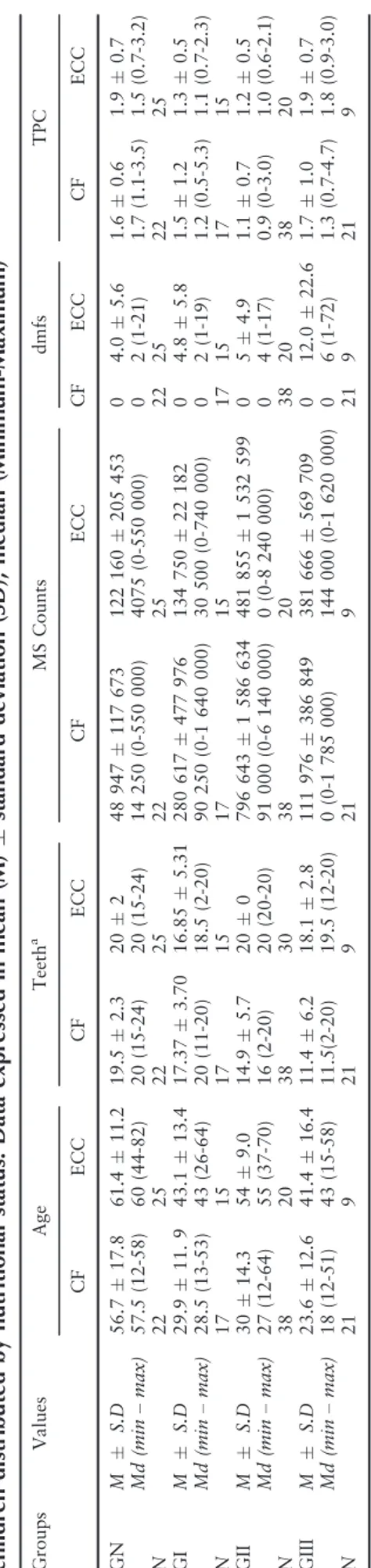
GIII children were the youngest among all groups (GI/GII, p= 0.006; GN, p =0.000). When comparing ages (12–70 month aged children) between genders in the different undernutrition levels, boys GIII were found to be significantly younger than girls (p= 0.003). However, TPC, MS levels and dmfs scores did not differ between genders in GN, GI, GII or GIII. Furthermore, no significant association was observed between genders and severity of ECC (p= 0.668). Comparison between ages among chil-dren who were caries-free, and chilchil-dren with and without severe ECC demonstrated a statistical signifi-cant difference (p= 0.000), for instance severe ECC was more often present among children at older ages. This finding did not depend on the child’s nutritional status. A statistically significant positive correlation was found between age and MS counts (p= 0.031); age and dmfs scores (p= 0.000); age and TPC (p= 0.008).
Caries experience and undernutrition
Forty-five undernourished (37.5%) and 25 (53.2%) normal-weight children presented a history of dental
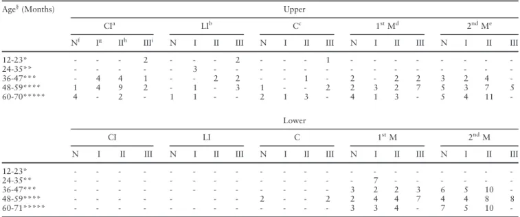
caries (dmfs>0). A total of seven undernourished chil-dren (15.5%) presented carious surfaces on the anterior teeth only, 22 (49%) children presented carious sur-faces on the posterior teeth only, and 16 (35.7%) par-ticipants had carious surfaces on both the anterior and posterior dentitions. In GN, carious surfaces affected only the anterior teeth in a total of three (12%) chil-dren, only the posterior teeth in 15 (60%), while seven (28%) children had carious surfaces affecting both the anterior and posterior dentitions (Table 1).
The second primary molar was the most affected pri-mary tooth by dental caries among undernourished and GN children, followed by the first primary molar and upper central incisor. Lower incisors were not affected by dental caries in any of the studied groups. White spot lesions were identified in only seven (15.9%) undernourished and six (24%) GN children. Only three (2.5%) undernourished children were identified as hav-ing enamel hypoplasia, one in GII (primary upper cen-tral incisors were affected) and two in GIII (upper canines and molars). In contrast, six (12.5%) GN chil-dren presented with 11 affected teeth, consisting of pri-mary molars, central incisors and canines.
Caries experience and mutans streptococci levels
MS was present in the saliva of 69 (57.5%) children in the undernourished groups (Table 2), of which 30 (25%) children were moderately and 39 (32.5%) were highly contaminated with MS. In GN, 31 (65.9%) children had detectable levels of MS. In this group, 12
Table 1. Number of carious teeth distributed by age group and nutritional status
Age§
(Months) Upper
CIa LIb Cc 1stMd 2ndMe
Nf Ig IIh IIIi N I II III N I II III N I II III N I II III
12-23* - - - 2 - - - 2 - - - 1 - - -
-24-35** - - - 3 - - -
-36-47*** - 4 4 1 - - 2 2 - - 1 - 2 - 2 2 3 2 4
-48-59**** 1 4 9 2 - 1 - 3 1 - - 2 2 3 2 7 5 3 7 5
60-70***** 4 - 2 - 1 1 - - 2 1 3 - 4 1 3 - 5 4 11
-Lower
CI LI C 1stM 2ndM
N I II III N I II III N I II III N I II III N I II III
12-23* - - -
-24-35** - - - 7 - - -
-36-47*** - - - 3 2 2 3 6 5 10
-48-59**** - - - 2 - - 2 2 4 4 7 4 4 8 8
60-71***** - - - 3 3 4 - 7 5 10
(38.7%) children presented high MS concentrations, whereas 19 (61.3%) children had moderate salivary MS levels. Comparisons between children without ECC (dmfs = 0), and children with caries showed a significant difference in MS counts (p =0.001) and caries experience (p =0.000). MS counts and dmfs scores were positively correlated (p =0.000). Hence, an increase in MS counts was observed when caries became more severe, regardless of the child’s nutri-tional status. Comparison of children with different levels of undernutrition (GI, GII, GIII) and the GN group showed no statistically significant differences in MS counts (p = 0.541) or caries experience (p = 0.609).
Total protein concentration
Comparisons between children without ECC (dmfs =
0), and children with caries showed no significant dif-ference in TPC between groups (p = 0.602). TPC did not correlate with caries experience (p = 0.565) or MS levels (p = 0.216) among healthy or undernour-ished children. TPC (p = 0.204) and caries experience (p = 0.395) did not differ between GI and GII. Never-theless, GIII children presented higher TPC than children in GI/GII groups (p = 0.000), but no statisti-cal difference in TPC was observed when comparing GIII and GN groups (p =0.531).
Multivariate analysis of caries experience
When using a binary logistic regression model for simultaneous analysis of all variables, gender (p = 0.709) and TPC (p= 0.272) did not significantly contribute with caries experience, but age (p = 0.000), MS count (p = 0.032) and nutritional status (p = 0.032) significantly contributed with caries expe-rience (Table 3). The risk of experiencing dental caries was significantly higher with an increase in age, as
Table 2. Age, MS counts, total protein concentra tion (TPC), dmfs scores and number of teeth of caries-free (CF) and Early Childhood Caries (ECC) children distributed by nutritional status. Data expressed in mean (M) standard deviation (SD), median (Minimum-Ma ximum) Grou ps V alues Age Teeth a MS C ounts dmfs TPC CF ECC C F ECC CF ECC C F ECC CF ECC GN M S.D 56.7 17 .8 61.4 11 .2 19.5 2. 3 20 2 48 947 11 7 673 12 2 160 20 5 453 0 4.0 5.6 1. 6 0.6 1.9 0.7 Md (m in – max ) 57.5 (12-58 ) 60 (44-82 ) 20 (15 -24) 20 (15-24 ) 14 250 (0-550 000) 40 75 (0-550 00 0) 0 2 (1-21 ) 1. 7 (1. 1-3.5) 1.5 (0.7-3 .2) N 22 25 22 25 22 25 22 25 22 25 GI M S.D 29.9 11 . 9 43.1 13 .4 17.3 7 3.70 16.85 5. 31 280 61 7 477 97 6 13 4 750 22 182 0 4.8 5.8 1. 5 1.2 1.3 0.5 Md (m in – max ) 28.5 (13-53 ) 43 (26-64 ) 20 (11 -20) 18.5 (2-20 ) 90 250 (0-1 640 000) 30 500 (0-740 00 0) 0 2 (1-19 ) 1. 2 (0. 5-5.3) 1.1 (0.7-2 .3) N 17 15 17 15 17 15 17 15 17 15 GII M S.D 30 14 .3 54 9.0 14.9 5. 7 20 0 796 64 3 1 586 63 4 48 1 855 1 53 2 599 0 5 4.9 1. 1 0.7 1.2 0.5 Md (m in – max ) 27 (12-64 ) 55 (37-70 ) 16 (2-20) 20 (20-20 ) 91 000 (0-6 140 000) 0 (0-8 240 00 0) 0 4 (1-17 ) 0. 9 (0-3. 0) 1.0 (0.6-2 .1) N 38 20 38 30 38 20 38 20 38 20 GIII M S.D 23.6 12 .6 41.4 16 .4 11.4 6. 2 18.1 2.8 111 97 6 386 84 9 38 1 666 56 9 709 0 12.0 22.6 1. 7 1.0 1.9 0.7 Md (m in – max ) 18 (12-51 ) 43 (15-58 ) 11.5 (2-20) 19.5 (12 -20) 0 (0-1 785 00 0) 14 4 000 (0-1 620 000) 0 6 (1-72 ) 1. 3 (0. 7-4.7) 1.8 (0.9-3 .0) N 21 9 21 9 21 9 21 9 21 9
Table 3. Analysis of different variables and their asso-ciation with caries experience using a Binary Logistic Regression Model (P <0 05)
Variables Statistics
B Wald DF P-value
Gender 0.142 0.139 1 0.709
Undernutrition – 8.831 3 0.031
GIa 1.857 8.190 1 0.004
GIIa 0.755 1.995 1 0.158
GIIIa 1.466 4.343 1 0.037
Age 0.081 29.099 1 0.000
MS Log Counts 0.071 4.593 1 0.032
TPC 0.265 1.207 1 0.272
well as with an increase in MS counts. Caries risk was also higher in GI (p = 0.004) and GIII (p= 0.037), but was not significantly elevated in GII (p= 0.158), when comparisons were made with GN.
DISCUSSION
In the present study, age played a major role in dental caries experience. Thus, the older the child, the higher the risk of caries development. An association between the prevalence and severity of ECC, and an increase in age has been recently described among children with different nutritional status.20 Among
undernourished children, this association has been for-merly linked to a delayed dental eruption. Alvarez
et al.21 while studying the association between chronic undernutrition and dental caries, described a shift to the right on the deft-versus-age curve. Further studies confirmed these findings.9 However, plotting dmfs scores as a function of age in the present study did not demonstrate the previously described shift to the right of the age distribution curve of caries among undernourished children. We believe the presently observed influence of age over caries experience could be partly explained by delayed eruption. Nevertheless, it is also a factor of a greater picture, where under-nourishment could lead to a deficit in the child’s abil-ity to immunologically defend against ECC. If this is a fact, the longer the child remains in an undernour-ished state, which happens with chronically under-nourished children at older ages, the higher the risk of dental caries experience.
A limitation of this study was its sample size. The possible influence of potential confounders determined the establishment of strict inclusion/exclusion criteria, which may explain the limited number of participat-ing children. For instance, children who breastfed past age 6 months, as well as those without very high consumption of sucrose-containing liquids/foods (≤3
sucrose-containing meals/day) were excluded from the study. Undernourished children consumed a great variety of junk foods, but insufficient foods/drinks with real nutritional value. Nutritionists’ recommen-dations focused on the need for dietary modification, and instructions to intensify consumption of milk or formula, rice, beans and oil (to increase caloric intake), allowing adequate growth and development. These children received free milk/formula every other week, in addition to rice, beans and oil to supplement their diets. This protocol was followed for all chil-dren, regardless of their level of undernutrition. Hence, this study population was constituted by children at very high risk for experiencing dental caries, and this risk factor was mainly attributed to: (1) diet; (2) socio-economic status; (3) parents’ level of education; and (4) nutritional status.
In the present study, the risk of experiencing dental caries was elevated among mildly and severely under-nourished children, but the moderately undernour-ished group was not at a significantly higher risk of developing the disease. We believe this phenomenon may be explained by two different factors: (1) GN children constituted by definition a high caries risk group; (2) GII children with caries were at a similar age group, when compared to GN, unlike GI and GIII-children who were at a younger age range. These similarities may have masked the high-risk category of GII in relation to GN, in the presence of other risk factors, in a multivariate statistical model. It must also be noted that the level of significance (p = 0.158) observed for GII does not exclude the possibility of observing a higher level of caries risk in a larger patient population. Oliveira et al.22 have investigated the association of nutritional status and dental caries in Brazilian preschool children. Their results showed an increased risk of developing dental caries in chil-dren with low Z-scores in body mass index-for-age and weight-for-height. The authors used the WHO Child Growth Standards Reference to evaluate nutri-tional status, but did not classify different levels of undernutrition. Therefore, differences in caries risk associated with mild, moderate and severe undernutri-tion could not be observed. An associaundernutri-tion between nutritional status and dental caries among elementary school children has been previously described;23 this
phenomenon was also observed in the permanent den-tition.24 Since a reduction in salivary flow rate,25 decreased buffering capacity and changes in salivary protein activity of undernourished children have been previously described,22 we believe future investigation of the biochemical aspects of saliva in undernourished populations may give a better understanding of the different systemic aspects involved in the caries risk mechanism of undernourished children during early childhood.
undernourished children had high levels of MS in sal-iva. In their study all participants were contaminated with MS, and no difference in salivary MS levels was found between groups. In the present study, mean lev-els of salivary MS were not found to be as elevated as the ones observed by Johansson et al.,20 since not all participants were contaminated with MS, and high concentrations of MS were only observed in approxi-mately 30% of the undernourished population. How-ever, in agreement with our findings, nutritional status alone did not influence MS contamination in any of these previous studies.
Since we have formerly observed an association between TPC and undernutrition (data not shown), we have measured TPC in saliva in an attempt to verify if higher or lower TPC in children with differ-ent grades of undernutrition was capable of influenc-ing the risk of experiencinfluenc-ing dental caries. A variety of factors are capable of influencing TPC measure-ments, such as age,27 geographic location, nutritional habits and the method of saliva sampling.28 Appar-ently, TPC is not influenced by dental eruption27 and no difference between genders has been previously described. Our results demonstrated a lack of correla-tion between TPC and ECC, as has been previously reported by Farias and Bezerra29 among healthy chil-dren. The authors found no differences in TPC among 12–47 month old children with or without dental caries. Conversely, Karg€ul et al.30 observed a
linear rise in TPC with an increasing number of cari-ous surfaces. We believe this difference was possibly due to the wide discrepancy in children’s age, since over 50% of their sample included 6–13 year old children and age factor may significantly influence TPC.28
Dental caries is a multifactorial disease, therefore simultaneous analysis of all possible risk factors was needed in order to establish the ability of each indi-vidual variable to significantly contribute with caries experience. This type of analysis revealed a significant contribution given by MS counts and the child’s nutritional condition in the development of caries. Furthermore, the presence of PEU was a relevant pre-disposing factor in the development and manifestation of the disease. Cleaton-Jones et al.31 studied the rela-tionship between nutritional status and dental caries in 2728 children aged 4–5 years. Although a statisti-cally significant trend in the increase of dmfs scores with increased wasting (low weight-for-height) was observed, no significant rise in caries prevalence was noted in this population. Despite demonstrating a sig-nificant association between wasting (low weight-for-height) and dmfs scores, the authors concluded that nutritional status was not a clinically significant factor in caries prevalence and experience. Nevertheless, in agreement with our data, previous work has suggested
a relationship between PEU and increased caries experience,9 and it has been proposed that a single mild to moderate undernutrition episode in the first year of life may result in higher caries rates in the pri-mary and permanent dentitions.9,10 Previously, the lack of agreement on the methods used to properly diagnose dental caries and undernutrition has been the main difficulty encountered in the comparison of our data and the data reported by others. Current understanding of dental caries as a sucrose-dependent disease, its associated risk factors, and the evidence-based malnutrition standards established in 2006 by the World Health Organization11 may allow future
studies to overcome these past limitations. In addition, very little is known on the salivary changes that inter-cept caries and undernutrition. Future studies should focus on identifying specific salivary characteristics that may be of interest in the development of preven-tive, diagnostic and/or therapeutic strategies against these diseases.
CONCLUSIONS
Our results suggest that nutritional status predisposes mildly and severely undernourished children to a higher risk of experiencing dental caries in early child-hood when other risk factors are considered, such as degree of undernutrition, age and MS levels in saliva.
ACKNOWLEDGEMENTS
We are grateful to the Fundacß~ao Cearense de Apoio ao Desenvolvimento Cientıfico e Tecnologico (FUNCAP) for supporting this work.
REFERENCES
1. Dutra-De-Oliveira JE, Marchini JS. Nutritional sciences. S~ao Paulo: Sarvier, 2003.
2. World Health Organization. Department of Nutrition for Health and Development 2010. Nutrition for health and devel-opment: a global agenda for combating malnutrition. URL: ‘http://whqlibdoc.who.int/hq/2000/WHO_NHD_00.6.pdf’. Accessed June 2011.
3. Schwartz S. A one year statistical analysis of dental emergencies in a pediatric hospital. J Can Dent Assoc 1994;60:959–968. 4. Ayhan H, Suskan E, Yildirim S. The effect of nursing or
rampant caries on height, body weight, and head circumference. J Clin Pediatr Dent 1996;20:209–212.
5. Low W, Tan S, Schwartz S. The effect of severe caries on the quality of life in young children. Pediatr Dent 1999;21:325–326. 6. American Academy of Pediatric Dentistry. AAPD 2010–11
defi-nitions, oral health policies, and clinical guidelines 2010. Policy on early childhood caries (ECC): classifications, consequences, and preventive strategies. Available at: http://www.aapd.org/ media/Policies_Guidelines/P_ECCClassifications.pdf.
8. Shishniashvili TE, Margvelashvili VV, Suladze NN, Kobakhidze KA. Correlation between the ecological risk factors and signifi-cant index of caries in young children. Georgian Med News 2012;206:30–33.
9. Alvarez JO, Eguren JC, Caceda J, Navia JM. The effect of nutritional status on the age distribution of dental caries in the primary teeth. J Dent Res 1990;69:1564–1566.
10. Alvarez JO. Nutrition, tooth development, and dental caries. Am J Clin Nutr 1995;61:410S–416S.
11. WHO Multicentre Growth Reference Study Group. WHO child growth standards based on length/height; weight and age. Acta Paediatr 2006;Suppl 450:76–85.
12. Fonteles CS, Guerra MH, Ribeiro TR,et al. Association of free amino acids with caries experience and mutans streptococci lev-els in whole saliva of children with early childhood caries. Arch Oral Biol 2009;54:80–85.
13. Ruoff KL, Whiley RA, Beighton D. Streptococcus. In: Murray PR, Baron EJ, Jorgensen JW, Pfaller MA, Yolken RH, eds. Manual of clinical microbiology. Washington DC: ASM Press, 2003:405–421.
14. Nikiforuk G. The caries process, morphological and clinical events. In: Understanding dental caries. Etiology and mecha-nisms, basic and clinical aspects. New York: Ed Karger, 1985:261.
15. Giro C. Enamel hypoplasia in human teeth: an examination of its causes. JADA 1947;34:310–317.
16. Sabel N. Enamel of primary teeth–morphological and chemical aspects. Swed Dent J Suppl 2012;222:1–77.
17. Gold OG, Jordan HV, van Houte J. A selective medium for Streptococcus mutans. Arch Oral Biol 1973;18:1357– 1364.
18. Jordan HV, Laraway R, Snirchi R, Marmel M. A simplified diagnostic system for cultural detection and enumeration of
Streptococcus mutans. J Dent Res 1987;66:57–61.
19. Bradford MM. A rapid and sensitive method for the quantita-tion of microgram quantities of protein utilizing the principle of protein-dye binding. Anal Biochem 1976;72:248–254.
20. Johansson I, Saelstrom A-K, Rajan BP, Parameswaran A. Sali-€
vary flow and dental caries in Indian children suffering from chronic malnutrition. Caries Res 1992;26:38–43.
21. Alvarez JO, Lewis CA, Saman C,et al. Chronic malnutrition, dental caries, and tooth exfoliation in Peruvian children aged 3–9 years. Am J Clin Nutr 1988;48:368–372.
22. Oliveira LB, Sheiham A, B€onecker M. Exploring the associa-tion of dental caries with social factors and nutriassocia-tional status in Brazilian preschool children. Eur J Oral Sci 2008;116: 37–43.
23. Lueangpiansamut J, Chatrchaiwiwatana S, Mukatabhant B, Inthalohit W. Relationship between dental caries status, nutri-tional status, snack foods, and sugar-sweetened beverages con-sumption among primary schoolchildren grade 4–6 in Nongbua Khamsaen school, Na Klang district, Nongbua Lampoo Prov-ince Thailand. J Med Assoc Thai 2012;95:1090–1097.
24. Delgado-Angulo EK, Hobdell MH, Bernabe E. Childhood stuting and caries increment in permanent teeth: a three and a half year longitudinal study in Peru. Int J Paediatr Dent 2013;23:101–109. 25. Psoter WJ, Spielman AL, Gebrian B, St Jean R, Katz RV. Effect
of childhood malnutrition on salivary flow and pH. Arch Oral Biol 2008;53:231–237.
26. Li Y, Navia JM, Caulfield PW. Colonization by mutans strep-tococci in the mouths of 3- and 4-year-old Chinese children with or without enamel hypoplasia. Arch Oral Biol 1994;39: 1057–1062.
27. Dezan CC, Nicolau J, Souza DN, Walter LR. Flow rate, amy-lase activity, and protein and sialic acid concentrations of saliva from children aged 18, 30 and 42 months attending a baby clinic. Arch Oral Biol 2002;47:423–427.
28. Ruhl S, Rayment SA, Scmalz G, Hiller KA, Troxler RF. Pro-teins in whole saliva during the first year of infancy. J Dent Res 2005;84:29–34.
29. Farias DG, Bezerra ACB. Salivary antibodies, amylase and pro-tein from children with early childhood caries. Clin Oral Inves-tig 2003;7:154–157.
30. Kargul B, Yarat A, Tanboga I, Emekli N. Salivary protein and€
some inorganic element levels in healthy children and their relationship to caries. J Marmara Univ Dent Fac 1994;2:434–440. 31. Cleaton-Jones P, Richardson BD, Granath L,et al. Nutritional
status and dental caries in a large sample of 4- and 5-year-old South African children. S Afr Med J 2000;90:631–635.
Address for correspondence: Dr Cristiane Sa Roriz Fonteles Unidade de Pesquisas Clınicas Universidade Federal do Ceara Laboratorio de Farmacologia Metabolica