H
OX GENES
C
ONTROL THE
S
PECIFICATION
OF
G
LOBAL
V
ERTEBRAL
D
OMAINS
Tânia Vinagre
Dissertação apresentada para obtenção do grau de doutor em
Biologia pelo Instituto de Tecnologia Química e Biológica da
Universidade Nova de Lisboa
!
!
!
!
!
!
!
!
!
!
To Scott, the best of friends.
"Science may set limits to knowledge, but should not set limits to
imagination."
A
CKNOWLEDGEMENTSI would like to, first and foremost, thank Moisés for accepting me in his lab and teaching me so much more than I tought possible. Thank you for coaching me into becoming a better scientist, for teaching me when to fight for an idea and when to let it go. I am truly grateful for all the ventures, both scientific and otherwise, I was allowed to pursue during my PhD, for they have certainly shaped the person I am today. I also thank Moisés for reading my thesis and helping me put together much better arguments. I would like to thank my thesis committee, Leonor Saúde and Rui Martinho, for their enthusiasm towards my project throughout the years, and for their sensible words of advice and patience.
I thank Instituto Gulbenkian de Ciência and its scientific community for the wonderful environment that makes going to work such an enjoyable habit. I would like to thank Fundação para a Ciência e a Tecnologia for the financial support (Apoio financeiro da FCT e do FSE no âmbito do Quadro Comunitário de apoio, BD nº SFRH / BD / 27306 / 2006).
I would like to thank Gage Crump for taking me into his lab and teaching all
about zebrafish transgenics and for the great discussions about my project.
I also thank the Crump lab for accepting me as one of them, and showing
me around LA. In particular I thank Megan Matsutani and Pablo Castillo for
their fantastic help with my fishes.
I would like to thank Peter Rigby and Jaime Carvajal for giving me the
opportunity to learn the BAC recombineering technique from them. I also
thank the members of the Rigby lab for their interest in my work.
I thank Rubina Caldeira, Sara Carvalho and Catarina Pereira for being my
friends at all times, for hearing me rant about work and never complaining.
I would like to thank Inês Manteigas for always being by my side and for
understanding me better than anyone else, and also for proofreading my
thesis. I thank Inês Vala for her friendship and for being an example of
courage and determination.
I would like to thank David Cristina for always having the right words to say
or a loving hug when everything else fails. His perseverance and talent are
an inspiration for me.
Lastly, I will thank my parents in Portuguese:
Agradeço aos meus pais todo o apoio e amor incondicionais e por
A
BSTRACTThe development of an animal from embryo to adult is an actively regulated process, largely controlled through differential gene expression. Hox genes are key modulators of embryonic development. Among other functions, they are essential for patterning the body plan by conferring identity to segments along the anterior-posterior axis. In vertebrates, Hox genes can specify the identity of both individual vertebrae and global vertebral domains in the axial skeleton. Hox group 10 is responsible for the layout of the lumbar region by inhibiting rib formation, while Hox group 11 defines the sacral domain of the skeleton. It was previously thought that ribs were set out by default. Hence, it was suggested that another Hox gene would have to inhibit rib formation in the cervical domain, similarly to Hox group 10 in the caudal part of the skeleton. We produced mice bearing ribs in every vertebrae by over-expressing Hoxb6 in the PSM under the control of the Dll1 promoter, showing that the thoracic area is formed through the activity of Hox group 6 genes that specifically induce rib formation, and that the cervical domain is defined as the area that precedes Hox group 6 expression. In this study, we used our two Hox over-expression mouse models with complementary rib phenotypes to study the molecular mechanisms of rib development. Our previously described Dll1-Hoxa10 transgenics are completely rib-less, while
the Dll1-Hoxb6 embryos form ectopic ribs throughout the whole length of
their skeletons. Interestingly, our findings indicate that the rib patterning cues provided by these two Hox genes are first interpreted in the muscle-forming myotomal compartment through the regional-specific hypaxial expression of Myf5/Myf6 genes. We believe this is a non-myogenic
Myf5/Myf6-specific function that is translated to the bone-forming
S
UMÁRIOO desenvolvimento de um animal, desde embrião até adulto, é um processo activamente regulado e controlado, em grande parte, através da expressão diferencial de genes. Os genes Hox são moduladores do desenvolvimento embrionário por excelência. Para além de outras funções, são essenciais para a padronização do plano axial, uma vez que conferem identidade aos segmentos ao longo do eixo anterior-posterior. Em vertebrados, os genes Hox especificam não só vértebras individuais, mas também grupos vertebrais completos do esqueleto axial. O grupo Hox 10 é responsável pela planificação da região lombar ao inibir a formação de costelas, ao passo que o grupo Hox 11 define o domínio sacral do esqueleto. Anteriormente pensava-se que as costelas eram determinadas por defeito, tendo sido sugerido que a zona cervical seria formada pela actividade repressora de costelas, conferida por um gene Hox com uma função idêntica à do grupo Hox 10 na parte caudal do esqueleto. Neste trabalho, produzimos murganhos que possuem costelas em todas as vértebras, através da sobre-expressão de Hoxb6 na mesoderme pré-somítica, sob o controlo do promotor Dll1. Os nossos dados demonstram que a região torácica é determinada pela acção de genes do grupo Hox 6 que induzem a formação de costelas, e que a zona cervical é definida como a área que precede a actividade do grupo 6. Utilizámos dois modelos de murganho com fenótipos complementares nas costelas para estudar os mecanismos moleculares do desenvolvimento das mesmas. Os nossos transgénicos Dll1-Hoxa10, anteriormente descritos, não desenvolvem costela alguma, enquanto que os embriões Dll1-Hoxb6 formam costelas ectópicas ao longo de todo o comprimento do seu esqueleto. Curiosamente, os nossos dados indicam que a informação providenciada por estes dois genes Hox é inicialmente interpretada pelo compartimento gerador de músculo, o miótomo, através da activação hipaxial de Myf5/Myf6
actividade de factores de crescimento FGF e PDGF. Os nossos resultados apoiam um modelo funcional para o desenvolvimento e evolução do sistema musculo-esquelético dos vertebrados.
T
ABLE OFC
ONTENTSAcknowledgements i
Abstract iii
Sumário v
Table of Contents vii
List of Figures x
List of Tables xii
Glossary xiii
CHAPTER I - INTRODUCTION 1
I.I HOW TO MAKE AN EMBRYO 4
I.II HOX GENES 4
HOX EXPRESSION AND REGULATION 8
HOX FUNCTION AND SPECIFICITY 10
I.III ABODY PLAN:THE VERTEBRATE AXIAL SKELETON 14
SOMITOGENESIS 15
SOMITE DERIVATIVES 20
MAKING SENSE OF SEGMENTS: HOX GENES AT WORK 27
I.IV MUSCLE AND BONE:THERE IS NO “I” IN TEAM 33
I.V OBJECTIVES 35
CHAPTER II –HOX SPECIFICATION OF GLOBAL VERTEBRAL
DOMAINS INVOLVES INTERACTIONS WITH MYOGENIC-RELATED FACTORS 37
II.I SUMMARY 39
II.II BACKGROUND 39
II.III RESULTS 40
HOX GROUPS 6 AND 10 CONTROL REGIONAL HYPAXIAL EXPRESSION OF
GENES IN THE MYF5/MYF6 PATHWAY 42
HYPAXIAL MYF6 EXPRESSION RESCUES THE RIB-LESS DLL1-HOXA10
PHENOTYPE 49
BINDING OF HOX GROUPS 6 AND 10 PROTEINS TO AN ENHANCER
THAT DRIVES HYPAXIAL EXPRESSION OF MYF5 51
IMPAIRED MYF5 HYPAXIAL EXPRESSION IN H1-MUTATED BAC
REPORTER LINE 52
ALTERED HYPAXIAL EXPRESSION OF LBX1 IN DLL1-HOXA10/B6
TRANSGENICS 57
II.IV DISCUSSION 59
II.V MATERIALS AND METHODS 67
II.VI ACKNOWLEDGEMENTS 75
CHAPTER III – MECHANISMS OF HOX FUNCTIONAL SPECIFICITY:
THE ROLE OF HOX10-SPECIFIC MOTIFS 77
III.I SUMMARY 79
III.II BACKGROUND 79
III.III RESULTS 81
SPECIFIC PEPTIDE MOTIFS ARE IMPORTANT FOR HOX GROUP 10’S FUNCTION 81
II.IV DISCUSSION 86
II.V MATERIALS AND METHODS 90
II.VI ACKNOWLEDGEMENTS 92
CHAPTER IV –FINAL CONSIDERATIONS ON HOW TO MAKE A SKELETON 93
GLOBAL SPECIFICATION OF VERTEBRAL DOMAINS 95
REFERENCES 103
APPENDIX I: MALLO ET AL. 2009 I
L
IST OFF
IGURESFigure 1: Classic examples of homeotic mutations.
Figure 2: Schematic representation of the fruit fly and mouse Hox clusters.
Figure 3: Schematic representation of somite differentiation into four
compartments.
Figure 4: Somitic sources of muscle progenitors for epaxial, hypaxial, and
limb muscles.
Figure 5: Schematic representation of vertebral domains of the axial
skeleton, aligned with somite levels of the mouse and expression of the Hox genes.
Figure 6: The role of Hox paralog group 10 in the patterning of the axial
skeleton.
Figure 7:Hoxb6 over-expression in the PSM induces ectopic rib formation.
Figure 8: Normal Hox group 10 expression in Dll1-Hoxb6 transgenics.
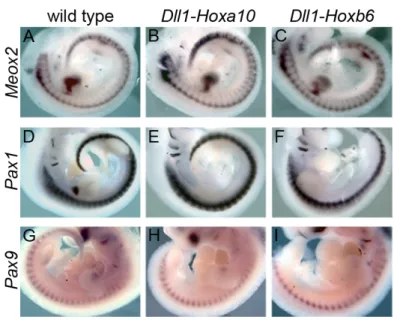
Figure 9: Normal sclerotomal patterning in Hox transgenics.
Figure 10: Hox groups 6 and 10 modulate regional expression Myf5 and
Myf6.
Figure 11: Hox groups 6 and 10 modulate regional expression of genes in
the Myf5/6 pathway.
Figure 12: Myotomal and muscle analysis of Hox transgenics.
Figure 13: Rescue of the Dll1-Hoxa10 phenotype with hypaxial Myf6.
Figure 14:Myf5/Myf6 as functional targets of Hox groups 6 and 10 genes.
Figure 15: Schematic representation of Myf5/6 BAC reporters.
Figure 16: !-galactosidase staining of B195-APZ L20 (control) and
Figure 17: Lbx1 regional expression is altered in Dll1-Hox transgenics.
Figure 18: Lbx1 expression in hypaxial interlimb somites results in mild rib
defects.
Figure 19:Hox groups 6 and 10 specify global vertebral domains.
Figure 20: Schematic representation of the two-stage PCR strategy used to
produce the BAC recombination cassettes.
Figure 21: Sequences of several Hox group 10 proteins of different
vertebrates, from Xenopus leavis to Homo sapiens.
Figure 22: Schematic representation of the Hoxa10 deletion mutants and
the chimeric proteins between Hoxa10 and Hoxa11.
Figure 23: Phenotypic analysis of the axial skeleton of two Hoxa10 deletion
mutants.
Figure 24: Phenotypic analysis of the axial skeleton of two transgenics of
L
IST OFT
ABLESTable 1: Comparison of the skeletal phenotype of Pax3pr-Myf6,
Dll1-Hoxa10 and Pax3pr-Myf6::Dll1-Hoxa10 fetuses.
Table 2: Skeletal phenotype of Pax3Pr-Myf6::Dll1-Hoxa10 fetuses.
Table 3: Summary of the expression profile for all BAC reporter lines.
Table 4: Details of primers used to genotype plasmid-based and BAC
transgenics.
Table 5: Details of the RNA probes used for in situ hybridization.
Table 6: Details of the primers used in ChIP experiments for the
amplification of the H1 enhancer and a negative control region.
Table 7: Details of the primers used to quantify the Hoxa10 cDNA.
Table 8: Sequence of primers used to genotype the different transgenics
G
LOSSARYA-P Anterior-posterior
BMP Bone Morphogenetic Protein
BIP2 Bric-à-brac interacting protein 2
Cdx Caudal type homeobox
Disco disconnected
Disco-r disco-related
Dll Delta Like
Exd Extradenticle
FGF Fibroblast Growth Factor
FGFR Fibroblast Growth Factor Receptor
Gdf11 Growth differentiation factor 11
HOM Homeotic
HOM-C Homeotic Complex
Hth Homothorax
Hox Homeobox genes
Lfng Lunatic fringe
Meox Mesenchyme homeobox
Mng Myogenin
MRF Myogenic Regulatory Factors
Pax Paired box
Pbx Pre B cell leukemia transcription factor
Pdgf Platelet derived growth factor
PdgfR Platelet derived growth factor Receptor
PSM Presomitic Mesoderm
RA Retinoic Acid
RBP Recombining Binding Protein
Scx Scleraxix
Shh Sonic Hedgehog
TALE Three Amino acid Loop Extension
Tbx T box
TGF! Transforming Growth Factor ! TrxG Trithorax Group
Tsh tea-shirt
C
HAPTERI
–
I
NTRODUCTION"In the beginning, there was nothing. Then God said, "Let there be light".
And there was still nothing, but now you could see it."
The development of a well-organized, fully functional animal from a single cell has amazed scientists throughout history. Most of the early embryological studies can be traced back to ancient Greek philosophers who studied reproduction, differentiation, growth and traits’ inheritance as part of a field known as generation. In the fourth century B.C., Aristotle (384-322 B.C.) published the first acknowledged study of comparative anatomical embryology, and he was also the first to use the chicken egg as a model to study development (Aristotle 350 B.C.).
For the longest time, from the sixteen hundreds to mid nineteen century, two opposing theories on the origin of life were intensely debated all over the world. The theory of preformation defended that since all creatures were originated at the same time as a result of God’s work of creation, each generation had to be present in a fully formed miniature version within its progenitors’ egg or sperm. The opposing theory of epigenesis, originally conjectured by Aristotle, favored that each embryo was formed de novo
through the gradual development of parts from an undifferentiated mass (Speybroeck et al. 2002; Pinto-Correia 1997; Gilbert 2003).
I.I
How to make an embryo
“Let the Sperm of a man by itself be putrefied in a gourd glass, sealed up,
with the highest degree of putrefaction in Horse dung, for the space of forty
days, or so long until it begin to be alive, move, and stir, which may easily
be seen. After this time it will be something like a Man, yet transparent, and
without a body. Now after this, if it be every day warily, and prudently
nourished and fed with the Arcanum of Mans blood, and be for the space of
forty weeks kept in a constant, equal heat of Horse dung, it will become a
true, and living infant, having all the members of an infant, which is born of a
woman, but it will be far less. This we call Homunculus”.
- Paracelsus, Of the Nature of Things.
There is one way of going from genotype to phenotype, and that is through development. Tomas Morgan realized this in a time when embryology and genetics were one and the same (Morgan 1926). Mechanistically speaking, the way we go from genotype to phenotype is through differential gene expression, the ultimate masterpiece of development.
When asking the question of how molecular changes in development lead to concrete morphological differences, one particular family of genes, the Hox genes, often stands out. The study of particular functions of this family of genes in the development of the mouse embryo is main subject of this thesis work.
I.II
Hox genes
“Here, there is one central field. Development. How the egg turns into the
organism. But development ultimately includes all of biology: and it will have
to be put on a molecular basis.”
Hox genes have captured the imagination of scientists like few others, due to their power to shape animal morphology and their implications in both developmental and evolutionary processes.
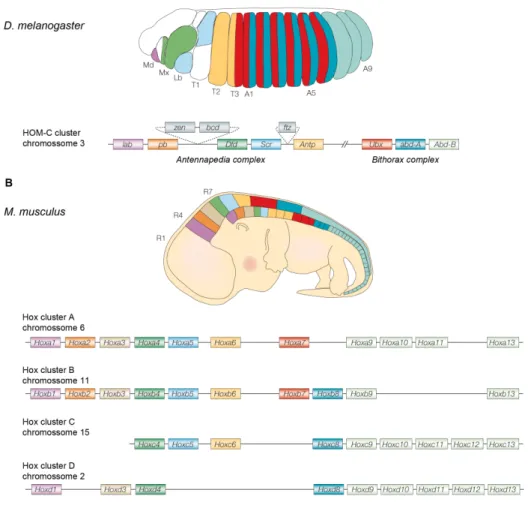
The concept of homeosis was first contemplated by William Bateson, who described it as “‘something that has been changed into the likeness of something else’’ (Bateson 1894). Two of the most paradigmatic homeotic transformations are observed in the Bithorax and Antennapedia mutants (Fig. 1). The genes in the origin of those remarkable phenotypes were informatively named Homeotic (HOM) genes, and extensive genetic and molecular analyses indicated they are part of a gene complex that controls segment identity in Drosophila (Lewis 1978; McGinnis and Krumlauf 1992).
The Drosophila Homeotic Complex (HOM-C) is composed of eight genes
organized in two loci (or complexes) in Drosophila’s chromosome three (Lewis 1978). The Antennapedia complex includes the Labial (Lab),
Proboscipedia (Pb), Deformed (Dfd), Sex combs reduced (Scr) and
Antennapedia (Antp) genes responsible for the specification of the head and
first thoracic segments (Wakimoto et al. 1984). The Bithorax complex contains the Ultrabithorax (Ubx) gene, which specifies the third thoracic segment, and the Abdominal A (Abd-A) and Abdominal B (Abd-B) genes, which are responsible for the identities of the abdominal segments (Casanova and White 1987; Sánchez-Herrero et al. 1985).
thoracic segment and one pair of halteres in the third thoracic segment. B. In the
Antennapedia mutant the antennae are converted into leg structures by a mutation in the regulatory region of the Antennapedia gene, which causes it to be expressed in the head. C. The Bithorax mutant is a four-winged fruit fly in which the third thoracic segment is transformed into another second thoracic segment (i.e., transformation of halteres into wings). This mutant results from three mutations in cis regulatory regions of the Ultrabithorax
gene (adapted from various sources).
Molecular analysis of the homeotic genes led to the discovery of the homeobox, a conserved 180 base pair long sequence that encodes a 60 amino-acid DNA binding domain called the homeodomain (McGinnis et al. 1984; McGinnis et al. 1984; Gehring et al. 1990; Gehring 1987). Although transcription-independent functions of Hox genes have been described (Brunet et al. 2005; Nédélec et al. 2004; Topisirovic et al. 2005; Prochiantz and Joliot 2003), the DNA-binding properties of their homeodomains conferred them the title of transcription factors (Gehring et al. 1990).
Over the past 20 years, Hox homologs have been found in virtually every billaterian animal (Garcia-Fernàndez 2005), including chordates, arthropods, platyhelminths, nematodes, and nemertines, where they consistently control axial identities and the formation of the body plan (Kmita-Cunisse et al. 1998; Salser and Kenyon 1994; Bayascas et al. 1998; Carroll 1995; Garcia-Fernàndez and Holland 1994; Krumlauf 1994; McGinnis and Krumlauf 1992).
Hox gene activation within the cluster follows a temporal and spatial sequence that correlates with their physical order in the chromosome, a phenomenon known as colinearity (reviewed in Kmita and Duboule 2003; Krumlauf 1994). In particular, the “temporal colinearity” manifested in mammals and short germ-band insects, represents the sequential activation of Hox genes, with 3’-located genes being expressed earlier than more 5’-located genes (reviewed in Kmita and Duboule 2003).
Figure 2: Schematic representation of the fruit fly Drosophila melanogaster, (A) and mouse,
Mus musculus, (B) Hox clusters. In both panels (A and B) embryos are represented and the
colors stand for the relative expression domains of corresponding Hox genes (top), which are represented according to their distribution in the chromosome (bellow). The Antennapedia
From an evolutionary perspective, the relationship between colinear Hox activation and morphogenesis has been suggested to work as a constraint to maintain the clustering of Hox genes (Ferrier and Minguillon 2003; Kmita and Duboule 2003). This view, however, has been recently challenged after the cloning and analysis of Hox genes in new model organisms (like urochordates), in which some level of coordination of Hox gene expression is achieved even in the absence of clustering (Lemons and McGinnis 2006; Monteiro and Ferrier 2006; Galliot 2005). This suggests that the precise mechanism regulating the expression of these genes is irrelevant, provided that the correct spatiotemporal pattern of expression is met (Kmita and Duboule 2003)
HOX EXPRESSION AND REGULATION
Hox genes are activated in the primitive streak when this structure is almost fully extended, and then in its caudal-most part, in a domain that contributes mostly to extra-embryonic mesoderm (Gaunt and Strachan 1994; Deschamps and Wijgerde 1993; Deschamps et al. 1999). The initial expression domains subsequently stretch anteriorly until they reach their definitive distribution in the three germ layers (Deschamps et al. 1999). Interestingly, there is evidence of a priming mechanism in the posterior section of the primitive streak that sets the conditions for Hox transcriptional activation 12 hours before it actually begins (Forlani 2003).
Recent work in chicken suggested that activation of Hox gene expression in the early epiblast is responsible for controlling the timing of ingression through the primitive streak (Limura and Pourquié 2006).
produces sharp anterior expression limits, this expression often gradually decrease towards the posterior end of the embryo. Overall, this expression scheme results in overlapping patterns of expression of different Hox genes along the AP axis (Kessel and Gruss 1990; 1991).
Long-term maintenance of Hox transcriptional states seems to be achieved through epigenetic marks. The trithorax group (trxG) is involved in the epigenetic maintenance of active Hox genes in the correct segments, whereas the polycomb group (PcG) is required for blocking their activation in the wrong spatial domains (Ringrose and Paro 2004). It has been suggested that the combined action of PcG and trxG proteins ensures that Hox gene expression is properly restricted to specific embryonic domains (Mahmoudi and Verrijzer 2001).
HOX FUNCTION AND SPECIFICITY
Hox genes are involved in a wide variety of developmental and cellular functions, including cell proliferation, cell death, cell adhesion, cell cycle regulation and cell migration (Pearson et al. 2005). At a higher morphological level, Hox genes help building organs either by modulating the positional identity of a given structure or by producing novel structures. In either case, in order to achieve their function, Hox genes must both control signaling pathways and establish complex regulatory networks (Hombríaa and Lovegrove 2003).
The extraordinary functional specificity of Hox genes in vivo contrasts with their indiscriminate binding specificity in vitro, which is due, to some extent, to the high degree of similarity and conservation of their homeodomains (Hoey and Levine 1988).
raises questions about how Hox proteins identify their correct target sites from the multitude of potential binding sites.
Hox specificity has been extensively connected to the interaction with other proteins, usually DNA-binding proteins themselves (Mann and Chan 1996; Mann and Affolter 1998; Mann 1995). The most common Hox cofactors comprise the PBC and MEIS classes of TALE proteins. The first class includes Exd in Drosophila and the Pbx proteins in vertebrates, whereas the MEIS class contains Hth in the fly and the vertebrate Meis and Prep homeodomain containing proteins (Moens and Selleri 2006). Similarly to Hox genes themselves, Hox partners were initially identified because their mutation led to morphological patterning defects in Drosophila. The first of these was Exd, which was indentified as a Hox cofactor because its loss-of-function produced homeotic transformations in specific segments without affecting Hox gene expression (Rauskolb et al. 1995; Rauskolb et al. 1993; Peifer and Wieschaus 1990). Pbx1 was independently identified in vertebrates through a chromosomal translocation that caused human preB cell leukemia (Nourse et al. 1990; Kamps et al. 1990; Rauskolb et al. 1993). Pbx/Exd primarly interact with Hox proteins through the three amino acid loop in their homeodomain, which binds a tryptophan-containing hexapeptide motif (NY/FP/DWMK/R), located N-terminal to the homeodomain and present in Hox proteins from paralog groups 1 to 8 (Piper et al. 1999; Chang et al. 1995; Knoepfler and Kamps 1995; Neuteboom et al. 1995; Passner et al. 1999; Phelan et al. 1995).
The second class of TALE cofactors, which includes Meis and Prep proteins, regulates Hox activity in several ways: by binding DNA together with Hox, by forming part of ternary Hox-PBC-MEIS DNA-binding complexes, and by modulating Pbx/Exd activity independently of DNA (Mann and Morata 2000; Moskow et al. 1995; Knoepfler et al. 1997; Chang
In addition to PBC and MEIS, other proteins have been reported to work as Hox partners in Drosophila. These include disco, disco-r and tsh zinc finger proteins, and more recently BIP2 (Prince et al. 2008). However, whether or not these proteins are also Hox cofactors in vertebrates remains to be discovered (Moens and Selleri 2006).
Despite extensive evidence indicating that cofactor cooperative binding helps improve Hox binding specificity, the in vivo effects on Hox function are still controversial, most particularly in vertebrates. Hox/PBC complexes can act either as transcriptional activators or repressors, depending on the target and the developmental context. This led to the suggestion that the Hox-mediated transcriptional outcome at a particular target gene is determined not by the presence of cofactors but rather by their ability to recruit co-activators or co-repressors to specific regulatory sequences (Saleh et al. 2000; Kobayashi 2003; Gebelein et al. 2004). Conversely, others have reported that some targets usually repressed by Hox genes, become activated when the hexapeptide is mutated (Merabet et al. 2003; Galant et al. 2002), suggesting that PBC binding alone could stipulate the transcriptional status of Hox proteins, at least in some situations. Considering the available information, it seems that Hox proteins obey very few rules when it comes to their functional activity, and even for any given Hox protein, specificity seems to be achieved differently depending on the particular set of downstream targets in question (Pearson et al. 2005; Merabet et al. 2009; Foronda et al. 2009). It is therefore critical that search for these targets is taken as a priority.
et al. 2001; Rohrschneider et al. 2007; Zhao and Potter 2001). The results of these studies suggest, as expected, that the expression of many genes involved in cellular functions is Hox-regulated. However, another interesting insight that stands out is that 63-69% of targets are unique for a specific Hox protein (Lu et al. 2003; Saleh et al. 2000; Shen et al. 2001), which is at odds with previous predictions that suggested a higher number of common targets. Moreover, Hox proteins can bind to chromatin-modifying proteins, such as histone deacetylases and acetyltransferases, possibly regulating downstream targets through epigenetic mechanisms (Lu et al. 2003; Saleh
et al. 2000; Shen et al. 2001).
The paradox of functional specificity has yet another side to it, that has to do with Hox protein structure. Sequence analysis of Hox proteins, led to the identification of key residues outside of the homeodomain that are critical for Hox function. The best characterized is the above-mentioned hexapeptide, which has also been called PBC interaction domain (PID) (Phelan et al. 1995; Morgan et al. 2000; Merabet et al. 2009; Johnson et al. 1995; Chang
et al. 1995). Interestingly, the area between this domain and the
homeodomain, called linker region, was shown to be relevant for Hox activity, particularly its size and amino-acid composition (Gebelein et al. 2002; In Der Rieden et al. 2004; Merabet et al. 2003). In addition to these, other protein domains seem to have a role in conferring functional specificity to Hox proteins, by interacting with other cofactors, by mediating specific DNA contacts or by promoting proper folding of the protein into its correct three-dimensional structure. These residues are usually located around the homeodomain, either N- or C-terminally (Lin and McGinnis 1992; Gibson et al. 1990; Furukubo-Tokunaga et al. 1993; Dessain et al. 1992; Berry and Gehring 2000; Passner et al. 1999; Zeng et al. 1993). Importantly, some of these motifs are paralog-specific signatures, which have been proven to be essential for the specific functional activity of Hox proteins in vivo (Merabet
understanding of the mechanisms by which Hox activity regulates specific developmental processes.
I.III
A Body Plan: The vertebrate axial skeleton
"Nature does nothing uselessly."
– Aristotle
The axial skeleton composed of vertebrae is the ultimate hallmark of vertebrates. It provides support, allows moment and offers protection to vital organs. The skeleton also plays a role in maintaining the body’s homeostasis, as it is involved in endocrine regulation, serves as storage of minerals and is the site of blood cell production.
as changes in this formula are often associated with increased incidence of cancer and neural problems (Galis 1999).
Both the cause and consequences of the diversity of axial formulas in the animal kingdom have provoked speculation from morphologists for centuries, and will be discussed later. For now, I will focus on the embryonic origin of the skeleton, the somites.
SOMITOGENESIS
One of the most noticeable illustrations of segmentation in vertebrates occurs during early embryonic development in the process of somite formation. Somites are paired mesodermal structures that form periodically from the unsegmented presomitic mesoderm, and represent the embryological origin of the skeletal muscle of the trunk and limbs, the axial skeleton and the dermis of the back (Brand-Saberi and Christ 2000).
Somites are part of the paraxial mesoderm (Jouve et al. 2002; Freund et al. 1996), and as such, are located symmetrically on both sides of the neural tube, and flanked laterally by the intermediate and lateral plate mesoderm. This position within the embryo is important for the establishment of the tissue interactions that control somite differentiation.
The process of somite formation is controlled at least at two different stages of development. It first relies on the emergence of paraxial mesodermal cells from the epiblast and later on the oscillatory activity of a variety of signaling processes in the PSM (Pourquié 2001). The formation of the paraxial mesoderm is closely associated with the morphogenetic movements of gastrulation. During the regression of the primitive streak, somitogenic stem cells are deposited and become a resident population first in the primitive streak and then in the tail bud. It is thought that the somitic mesoderm mostly derives form these progenitors (Stern 1992; Schoenwolf
et al. 1992; Selleck and Stern 1991; Hatada and Stern 1994; Psychoyos
It has been suggested that production of somitic mesoderm occurs in two consecutive phases. According to this hypothesis, anterior somites would derive from the paraxial mesoderm produced from the primitive streak, and posterior somites would originate from the tail bud, once it becomes the source of paraxial mesoderm after the closure of the neuropore (Catala et al. 1995; Tam 1984; Pourquié 2001). However, some authors consider the distinction between the two phases somewhat artificial because some lines of evidence suggest that paraxial mesoderm production, and consequently, somite formation is a continuous process (Pourquié 2001).
The generation of paraxial mesoderm requires that proliferation, specification and migration events take place in an orderly fashion. The molecular control of these processes is mostly provided by the coordinate activity of at least three main signaling cascades: the WNT, FGF and RA pathways. Disrupting any of these pathways has severe consequences to axis formation, typically producing axial truncations resulting from impaired paraxial mesoderm formation (Pourquié 2001). In particular, mutations in
the Wnt3a gene, the FGFR1! isoforms or the RA-producing enzyme
Raldh2, resulted in disrupted somitogenesis after the formation of the first few somites (Greco et al. 1996; Takada et al. 1994; Yamaguchi et al. 1994; Niederreither et al. 1999; Ciruna and Rossant 2001). Mutations in the T-box genes Brachyury or Tbx6, which are regarded as mediators of the WNT signaling pathway, produced similar phenotypes, further documenting the requirement of these signals to produce mesodermal precursors (Yamaguchi et al. 1999; Chapman et al. 1996). Additionally, in some of these mutants, the posterior somitic tissue was replaced by ectopic neural tubes, indicating that these signaling pathways are involved in the specification of the paraxial mesoderm vs. neuroectoderm fate in the tail bud (Chapman and Papaioannou 1998; Yamaguchi et al. 1999; Yoshikawa
Somitogenesis is a sequential, directional and synchronous process, where somites are formed at the anterior border of the PSM, resulting in the progressive addition of new somites posterior to older ones (Yamaguchi 1997; Pourquié 2001; Dubrulle and Pourquié 2004a). The pace of somite formation is a species-specific characteristic. In the mouse, each new somite sprouts from the PSM every 120 minutes, whereas this process takes 90 minutes in chick and only 30 minutes in zebrafish (Dequéant and Pourquié 2008). Histologically, the vertebrate PSM gives the impression of being a loose mesenchyme with no morphological organization. However, during somite formation, the PSM cells change their adhesion properties and become progressively more epithelized (Duband et al. 1987), culminating with the establishment of a new somitic border.
This process of spatial and temporal segment specification is tightly regulated, and thought to depend on an intrinsic molecular clock that sets the pace of somite formation, associated with a maturation front that determines the location of the new inter-somitic border (Baker et al. 2006a; Schnell et al. 2002; Baker et al. 2006b). The first experimental evidence of such a molecular oscillator came with the documentation of the dynamic expression of the gene Hairy1 in the PSM of chick embryos (Palmeirim et al. 1997). This gene is expressed as a wave that sweeps the PSM in a posterior to anterior direction once every 90 minutes (the time that takes to form a new somite in chick), to finally merge in a band that correspond to the anterior part of the next somite to be formed. Hence, the last site of
Hairy1 expression in each cycle labels the position of the next inter-somitic
boundary.
mutations in genes related to this pathway, namely, Notch1, Delta1, Delta3,
RBPjk, Mesp2 and Lfng, typically display impaired formation of caudal
somite due to incorrect segmentation of the PSM (Saga et al. 1997; Wong
et al. 1997; Oka et al. 1995; Zhang and Gridley 1998; Evrard et al. 1998;
Conlon et al. 1995). In addition, recent data from thorough microarray studies suggest that the clock might be more complex than initially imagined and involve at least three signaling pathways (Goldbetera and Pourquié 2008; Dequéant et al. 2006; Mallo 2007).
formation and in the failure to establish an anterior-posterior pattern within each somite (Morimoto et al. 2005; Takahashi et al. 2000). In the anterior PSM, Mesp2 modulates an on/off state of Notch signaling that corresponds to the future somitic boundary through the activation of Lfng transcription (Morimoto et al. 2005; Oginuma et al. 2008; Oginuma et al. 2010).
SOMITE DERIVATIVES
Once formed, the epithelial somites progressively differentiate into distinct compartments as a response to signals from surrounding tissues, giving rise to different cell lineages. The ventro-medial part of the epithelial somite undergoes an epithelial-to-mesenchymal transition to form the sclerotome, which is the source of the axial skeleton (Fig. 3). The dorso-lateral part, the dermomyotome retains its epithelial character and with further maturation of the somite, cells delaminate from its edges and migrate underneath to form the myotome that will originate the skeletal muscles of the trunk. Once the myotome is formed, the remaining part of the dermomyotome is called dermotome and will form the dermis of the back (Borycki and Emerson 2000; Brand-Saberi and Christ 2000) (Fig. 3). More recently, a fourth somitic compartment was indentified, the syndetome, that is generated from the dorsolateral border of the early sclerotome and gives rise to the axial tendons (Brent et al. 2003) (Fig. 3).
underneath to form the myotome. A fourth somitic compartment, the syndetome, is generated within the sclerotome (adapted from various sources).
THE SCLEROTOME
During differentiation, the sclerotome is further compartmentalized into rostral and caudal halves (Stern and Keynes 1987), and also medial and lateral domains (Freitas et al. 2001; Brent and Tabin 2002). These sub-domains, which can be identified by the expression of different molecular markers, originate distinct structures. The rostro-caudal compartmentalization of the sclerotome, underlies the phenomenon of resegmentation, which is crucial for the development of the vertebral column and has strong implications in the metamerization of the peripheral nervous system (Christ and Wilting 1992; Rickmann et al. 1985; Christ et al. 1979). Resegmentation of the sclerotome results in each vertebra being formed by the posterior half of one somite and the anterior half of the next (Aoyama and Asamoto 2000; Christ et al. 1998; Bagnall et al. 1988). The dorsomedial sclerotome generates the spinous process and contributes to the distal ribs; the ventromedial sclerotome originates the vertebral bodies, neural arches, intervertebral discs and proximal ribs; and the ventrolateral scleromome also contributes to the distal part of the ribs (Olivera-Martinez et al. 2000; Freitas et al. 2001; Huang et al. 2000).
Pax1, Nkx3.1 and Nkx3.2 are the first molecular markers activated when the
presumptive sclerotome is induced (Schneider et al. 2000; Kos et al. 1998; Tribioli and Lufkin 1997; Rodrigo et al. 2003; Peters et al. 1999; Müller et al. 1996; Ebensperger et al. 1995). In early somite development, activation of
Müller et al. 1996). In the Pax1/Pax9 double mutant mice, the development of the ventral part of the vertebra, but not of the neural arches, is defective (Peters et al. 1999). Pax1 is genetically upstream of Nkx3.2 because expression of this gene cannot be detected in Pax1/Pax9 double mutants (Rodrigo et al. 2003; Tribioli and Lufkin 1997). However, Pax1 is expressed normally in the somites of Nkx3.2 mutant embryos, although later steps of vertebral differentiation are severely defective in these animals (Tribioli and Lufkin 1997). Nkx3.1 expression is activated in newly formed somites but mice carrying null mutations in this gene do not have any skeletal defects (Schneider et al. 2000; Kos et al. 1998). Altogether, these results suggest that the activation of Pax1 is the key event that triggers sclerotome formation during development (Monsoro-Burq 2005).
It is well established that sclerotome formation requires signals from axial midline tissues. Shh and Noggin, secreted by the notochord, are important to induce and maintain Pax1 expression (Marcelle et al. 1999; Chiang et al. 1996; Hammerschmidt and McMahon 1998). Loss-of-function Shh mice fail to form the entire vertebral column (Chiang et al. 1996). However, in these mutants, the sclerotome is formed, although significantly smaller in size, and
sclerotomal formation clearly depends on the correct equilibrium between dorsalyzing and ventralizing signals.
THE DERMOMYOTOME
The dermomyotome generates myotomal muscle progenitor cells and the dermis of the back. It is composed of a central epithelial sheet that is surrounded by contiguous lips (Huang and Christ 2000; Ordahl and Le Douarin 1992). The dermomyotome is marked by the expression of Pax3, contrasting with the activation of Pax1 in the sclerotome (Birchmeier and Brohmann 2000). This transcription factor starts to be expressed in the entire somite, but with further differentiation, Pax3 expression becomes restricted to the dermomyotome and, finally, it is up-regulated in the lateral edge of the dermomyotome (Williams and Ordahl 1994; Goulding et al. 1994; Bober et al. 1994). Pax3 is required for the proper formation of both migratory and non-migratory muscle precursor cells. Indeed, in mice Pax3
mutant mice, migratory myogenic precursors cells fail to delaminate and, consequently, these mutants lack muscles in the limbs and diaphragm (Epstein et al. 1995; Tremblay et al. 1998; Daston et al. 1996).
While the formation of the sclerotome is dependent on the notochord, dermomyotome development is mostly controlled by signals from the dorsal neural tube and surface ectoderm. WNT signals seem to have a prominent role in this process (Fan et al. 1997; Fan and Tessier-Lavigne 1994; Dietrich
et al. 1997). However, notochord produced, long range acting Shh is
thought to be necessary for both survival and proliferation of myogenic precursors, in particular those in the epaxial compartment (Krüger et al. 2001; Duprez et al. 1998; Teillet et al. 1998).
dermis (Olivera-Martinez et al. 2000). The ventro-lateral dermomyotome behaves differently at different axial levels. At limb bud levels, cells delaminate and migrate to invade the lateral plate mesoderm, and differentiate into limb muscles (Chevallier et al. 1977). At the interlimb level, cells from the ventro-lateral lip (VLL) of the dermomyotome move underneath to produce the hypaxial myotome. During this process, the ventrolateral dermomyotome and hypaxial myotome invade the lateral plate mesoderm together as a somitic bud, which will contribute to the formation of the body wall muscles (Brent and Tabin 2002; Brand-Saberi and Christ 2000) (Fig. 4).
Figure 4: Somitic sources of muscle progenitors for epaxial, hypaxial, and limb muscles. Myogenic progenitors originate in the dorsal-medial and ventral-lateral lips of the dermomyotome. Cells of the dorsal-medial lip (DML) migrate ventrolaterally, differentiate, and give rise to epaxial back muscles. The ventral-lateral lip (VLL) cells migrate underneath to form the ventral hypaxial body wall muscles at interlimb levels (yellow); and migrate to form the limb musculature at limb levels (orange) (adapted from Pownall et al. 2002).
Limb and hypaxial progenitors activate a particular expression profile in order to coordinate their migration with the activation of the myogenic program, which should only occur upon progenitors’ arrival to their final sites of differentiation (Birchmeier and Brohmann 2000). These genes include:
Pax3 and Msx1 transcription factors, which are required for progenitor cell
(scatter factor/hepatocyte growth factor), which are essential for delamination of migratory precursor cells from the epithelial dermomyotome (Maina et al. 1996; Bladt et al. 1995; Birchmeier and Gherardi 1998; Dietrich
et al. 1999); and Lbx1, another Pax3 target, that is only expressed in
migratory precursor cells and is critical for the migration of muscle precursors into the limb bud (Gross et al. 2000; Mennerich et al. 1998; Brohmann et al. 2000; Jagla et al. 1995; Dietrich et al. 1998).
Muscle development from the myotome is under the control of myogenic regulatory factors (MRFs): MyoD, Myf5, Myogenin, and Myf6/MRF4. These are a family of conserved basic helix-loop-helix (bHLH) transcription factors (Weintraub et al. 1991), characterized by their ability to activate the skeletal muscle program in many non-muscle cell types. This property has bestowed them the title of master regulators of muscle progenitor cell specification and differentiation (Davis et al. 1987; Choi et al. 1990). In the mouse, Myf5
becomes active slightly before MyoD and despite some differences in their expression, these genes seem to compensate for each other because, while both individual mutants develop normal musculature, double mutants completely fail to form skeletal muscles (Pownall et al. 2002; Braun et al. 1992; Tajbakhsh et al. 1996). The accumulation of studies on single and compound loss-of-function mutants of the different MRF factors led to the current view that Myf5 and Myf6 are at the top of the myogenic cascade. MyoD seems to operate downstream of Myf5/Myf6 in some precursors and in parallel with them in others, and myogenin operates as the final effector in the pathway, controlling terminal differentiation (Carvajal and Rigby 2010; Kassar-Duchossoy et al. 2004).
cofactors, Eya and Dach, are also important regulators of myogenesis. They cooperate with Pax proteins and MRFs and form a complex regulatory network of muscle development (Grifone et al. 2005).
Cis Regulation of Muscle Development
The different functions of MRFs are reflected in how the transcription of each of these genes is regulated. Genes situated higher in the myogenic hierarchy, such as Myf5 and Myf6, respond to more signaling pathways than those acting at the bottom of the myogenic cascade, such as myogenin
(Carvajal and Rigby 2010). Relevant to this work is the remarkably complex arrangement of distinct enhancers that are necessary to drive particular aspects of Myf5 and/or Myf6 expression (Carvajal et al. 2008). However, while Myf6 transcriptional regulation seems to require at least four enhancer elements to specifically drive expression in the limbs, somitic bud, central thoracic myotome, ventral and dorsal myotome (Fomin et al. 2004; Carvajal
et al 2001; Pin et al. 1997), Myf5 regulation is much more complex. Myf5
expression is controlled by at least five elements in different compartments in the somites (Hadchouel 2003; Buchberger et al. 2003; Teboul et al. 2002; Teboul et al. 2003; Summerbell et al. 2000), at least three in the limbs (Buchberger et al. 2003; Hadchouel et al. 2000; Hadchouel 2003; Bajard et al. 2006), two in the developing central nervous system (CNS) (Summerbell
et al. 2000; Daubas et al. 2009; Daubas et al. 2000), and at least five in the
branchial arches (Summerbell et al. 2000; Carvajal et al 2001; Patapoutian
et al. 1993). Interestingly, while most regulatory regions of the Myf5/6 locus
for understanding the evolution of distal elements that control gene expression.
THE SYNDETOME
In order to function properly, the musculoskeletal system depends on the coordinated development of not only bone and muscle, but also tendons. However, whereas the formation of the first two somite derivatives has been extensively studied, tendon development has been overlooked until fairly recently.
The bHLH transcription factor scleraxis (Scx) is expressed both in mature tendons of the limbs and trunk, and in their progenitors in the developing somite (Schweitzer et al. 2001) (Fig. 3). It has been recently suggested that these tendon progenitors define a fourth somitic compartment, the syndetome. The syndetome originates from a domain at the dorsolateral edge of the early sclerotome that has the potential to activate either the tendon or cartilage programs (Brent et al. 2003). It has been shown that a myotomal signal, possibly involving FGF signaling, induces Scx expression in sclerotomal cells localized next to the myotome. Moreover, the analysis of
Sox5/Sox6 double mutants, in which the chondroprogenitors cannot
differentiate into cartilage, indicated that in order to form cartilage it is necessary to actively repress tendon development in the dorsolateral sclerotome (Brent et al. 2005).
MAKING SENSE OF SEGMENTS:HOX GENES AT WORK
One of the early challenges in the Hox field was to establish a correlation between Hox gene expression and function in the patterning of the axial skeleton. The expression of Hox genes is typically characterized by sharp anterior borders that have been correlated with skeletal phenotypes of several single Hox loss-of-function mutants and over-expression transgenics. These correlations led to the proposal of the “Hox code” model (Kessel et al. 1991; Burke et al. 1995; Burke 2000). This model states that the segment-specific morphological identities would result from combinatory “code” of Hox expression, which is distinctive in each segment along the AP axis (Kessel et al. 1991; 1990). An alternative model for Hox patterning activity was proposed on the basis of the phenomenon of “phenotypic suppression” described in Drosophila (Struhl 1983; Morata 1993) to explain the observation that individual Hox loss-of-function mutants often displayed phenotypes only in the most anterior regions of their expression domains, despite their broad expression patterns (Duboule and Morata 1994). This model, called “posterior prevalence”, postulates that posteriorly expressed Hox genes are functionally dominant over anterior genes. (Duboule and Morata 1994; Kmita and Duboule 2003). Recently, this subject was revisited and new data suggested that miRNAs could work as a mechanism to enforce the prevalence of posterior Hox genes by repressing anterior Hox
instructions (Yekta et al. 2008). Nevertheless, neither the “Hox code” or “the
posterior prevalence” models can entirely explain the phenotypes produced in several gain-of-function and loss-of-function studies of either individual Hox genes or whole paralogous groups (Mallo et al. 2010).
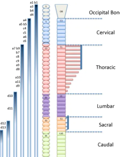
the prospective boundary between cervical and thoracic vertebrae in vertebrates with different numbers of cervical vertebrae. This transition is mapped to somites 18-19 in the goose, 16-17 in chick, 9-12 in the mouse, 2-3 in xenopus and 1-2 in zebrafish (Molven et al. 1990; Burke et al. 1995; Burke 2000). Interestingly, the early activation of Hox6 genes was later implicated as the cause of the reduction of the cervical region in pythons (Cohn and Tickle 1999), further suggesting that the Hox paralog group 6 is critical for the cervical-to-thoracic transition in vertebrates.
Figure 5: Schematic representation of vertebral domains of the axial skeleton, aligned with somite levels of the mouse and expression of the Hox genes. The anterior limit of expression of specific Hox genes is indicated as the top of the gradient, which represents the progressive decrease in expression anterior to posterior. OB: occipital bone; C: cervical; T: thoracic; L: lumbar; S: sacral; Cd: caudal (adapted from Burke et al. 1995; Favier and Dollé 1997).
SPECIFICATION OF GLOBAL VERTEBRAL DOMAINS
demonstrated by the precocious over-expression of Hoxa10 in the PSM of developing mouse embryos, which resulted in the overall inhibition of rib formation and, consequently, in completely rib-less mice (Carapuço et al. 2005) (Fig. 6 B). Together, these reports indicate that Hox group 10 is essential for the formation of the lumbar region by specifically blocking rib formation. Interestingly, the same studies showed that Hox group 11 is responsible for the development of the sacral domain. Whereas the loss-of-function of paralog group 11 results in the absense of the sacrum (Wellik and Capecchi 2003), over-expression of Hoxa11 in the PSM entails a “sacralization” of the lumbar region by promoting sacral-like fusions in adjacent vertebrae (Carapuço et al. 2005).
Figure 6: The role of Hox paralog group 10 in the patterning of the axial skeleton. The inactivation of all six alleles of Hox group 10 results in ectopic rib formation in the presumptive lumbar and sacral regions of the skeleton (A). Over-expression of a member of Hox group 10 in the PSM of mouse embryos driven by the Dll1 promoter results in complete blocking of rib development (B). Adapted from Wellik and Capecchi 2003 (A) and Carapuço
It is noteworthy that in the patterning of the ribcage, the generation of sternal versus floating ribs seems to be controlled by Hox group 9 (McIntyre
et al. 2007). These genes are specifically important to produce floating ribs,
since the global inactivation of this paralog group resulted in an increased number or sternal ribs (13-14 instead of the normal 7) (McIntyre et al. 2007). Much less is known about the patterning mechanisms of the neck region. Inactivation of members of the paralogous Hox groups 3-5 demonstrated that these genes have some role in specifying particular morphologies in the cervical vertebrae (Horan et al. 1995b; McIntyre et al. 2007; Condie and Capecchi 1994). However, a global transformation of the cervical region into the identity of another vertebral domain has not been achieved so far. It has been proposed that ribs are set out by default and, consequently, the cervical domain would be specified by a particular Hox gene or group that would inhibit rib formation in the neck similarly to Hox group 10 in the lumbo-sacral domain (Wellik and Capecchi 2003). However, this gene has not been identified yet and this hypothesis is somewhat at odds with the previously mentioned analysis of the anterior limits of expression of Hox group 6 in different vertebrates (Burke et al. 1995).
I.IV
Muscle and Bone: There is no “I” in Team
"Science... never solves a problem without creating ten more."
– George Bernard Shaw
A functional musculoskeletal system requires the coordinated development of muscle, bone and tendon. Increasing amount of evidence indicates that this coordination is, to a large extent, the consequence of a dynamic dialog between different somitic compartments.
Inactivation of Myf5 results in a fairly normal muscle development, but in strikingly severe rib defects (Tajbakhsh et al. 1996; Braun et al. 1992). Initially, the skeletal phenotype in Myf5 mutants was suggested to result from impaired production of myotomal inductive signals and/or from the disruption of patterning interactions necessary for rib formation (Braun and Arnold 1995; Braun et al. 1992). This idea was further supported by the generation of three different loss-of-function alleles of the Myf6 gene, located 8 Kb upstream of Myf5 on the mouse chromosome 10 (Patapoutian
et al. 1995; Zhang et al. 1995; Braun and Arnold 1995), which produced
variable defects in rib formation. However, the role of Myf5/Myf6 in rib development was later revisited and questioned when new Myf5 alleles were produced that showed no distinguishable rib defects (Kaul et al. 2000). This led to the suggestion that the rib abnormalities in Myf mutant mice resulted from some interference with the activity of another gene in the same genomic area as a consequence of the production of the mutant alleles. However, unexpected skeletal defects associated with genes involved in myogenic development are not exclusive of Myf5/Myf6. In addition to well-characterized skeletal muscle defects, myogenin mutant embryos present abnormal sternal formation (Nabeshima et al. 1993; Hasty
et al. 1993; Vivian et al. 2000). The splotch mouse mutants, which carry a
intercostal muscles and several rib deficiencies (Dickman et al. 1999; Henderson et al. 1999). Another interesting example is the loss-of-function
Six1 mutants. Mice lacking Six1 die at birth due to severe rib malformations and show extensive muscle hypoplasia specially in particular hypaxial muscles (Laclef 2003). Six1/Six4 double mutants show an aggravation of the Six1-null phenotype (Grifone et al. 2005), suggesting that these genes act redundantly in this function.
The above-mentioned studies demonstrate that alterations in the activity of several myogenic genes are often accompanied by impaired rib formation. There are a number of potential inductive signals are expressed in myotome that could potentially mediate these myotome-sclerotome interactions. Among these, FGFs and PDGFA seem to be prime candidates for such activity (Grass et al. 1996; Tallquist et al. 2000). PDGFR" starts to be expressed in the PSM and ephithelial somite, but later becomes restricted to the sclerotome and dermatome (Schatteman et al. 1992; Orr-Urtreger et al. 1992; Orr-Urtreger and Lonai 1992). PDGFR" loss-of-function mutants have a normal initial sclerotomal patterning, but later exhibit rib, sternum and vertebrae abnormalities (Soriano 1997). These mutants have perturbed myotomal patterning, which again suggests the skeletal defects could be due to a disruption of myotome-to-sclerotome signaling. Interestingly, PDGFA, a PDGFR" ligand normally expressed in the myotome, is absent in ribless Myf5 null mutants, and knocking-in PDGFA into the Myf5 locus resulted in a partial rescue of rib development (Tallquist et al. 2000). Similarly, Fgf4 and Fgf6 expression was shown to be down-regulated in
increases sclerotomal proliferation and enhances rib development, whereas inhibition of FGF signalling by SU5402 causes deletions in developing ribs (Huang et al. 2003).
Interestingly, chick-quail chimeras have shown the production of tendon progenitors form the dorsolateral sclerotome in response to FGFs secreted from the adjacent myotome (Brent et al. 2003). Thus, it seems that FGFs have a pivotal role in the communication between the somitic muscle and cartilage cell lineages involved both in the formation of tendons and ribs. All in all, these results demonstrate the importance of a correct communication between the different somite compartments, and that this dialog is likely achieved through the use of signaling molecules, such as FGFs and PDGFs growth factors.
I.V
Objectives
The overall goal of this work was to contribute to the general understanding of the function of Hox genes in the patterning of global domains of the vertebrate axial skeleton. Here we look at Hox function from two perspectives:
1. Which physiological mechanisms and pathways underlie Hox-mediated patterning of rib formation.
C
HAPTERII
–
H
OXS
PECIFICATION OFG
LOBALV
ERTEBRALD
OMAINSI
NVOLVESI
NTERACTIONS WITHM
YOGENIC-
RELATEDF
ACTORS*
*Adapted from Vinagre T, Moncaut N, Carapuço M, Nóvoa A, Bom J,
Mallo M. Evidence for a myotomal Hox/Myf cascade governing non-autonomous control of rib specification within global vertebral domains.
II.I
Summary
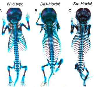
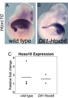
Hox genes are essential for the patterning of the axial skeleton. Hox group 10 has been shown to specify the lumbar domain by setting a rib-inhibiting program in the presomitic mesoderm (PSM). We have now produced mice with ribs in every vertebra by ectopically expressing Hox group 6 in the PSM, indicating that Hox genes are also able to specify the thoracic domain. We show that the information provided by Hox genes to specify rib-containing and rib-less areas is first interpreted in the myotome through the regional specific control of Myf5 and Myf6 expression. This information is then transmitted to the sclerotome by a system that includes FGF and PDGF signaling to produce vertebrae with or without ribs at different axial levels. Our findings offer a new perspective on how Hox genes produce global patterns in the axial skeleton and support a redundant non-myogenic role of Myf5 and Myf6 in rib formation.
II.II
Background
Hox genes have been classically described to be involved in the production of vertebrae with individual characteristics (R Krumlauf 1994; Mallo et al. 2009; Wellik 2007). More recently, it was discovered that Hox genes also play essential roles in defining global vertebral domains (Wellik and Capecchi 2003). In particular, it was shown that Hox group 10 is responsible for the layout of the less lumbar region by diverting it from a rib-containing thoracic identity (Wellik and Capecchi 2003; Carapuço et al. 2005). In addition, Hox group 11 was demonstrated to be required for the formation of the sacrum (Wellik and Capecchi 2003). However, it remains unclear whether or not Hox genes are involved in the global specification of the thoracic and cervical domains. Moreover, the mechanism by which Hox genes control these processes is completely unknown.
other Hox genes acting similarly to Hox group 10 in the lumbar region (Wellik and Capecchi 2003). However, this hypothesis is difficult to reconcile with published expression patterns for Hox genes (Burke et al. 1995), which instead suggest an alternative hypothesis. In particular, the anterior limit of expression of members of the Hox group 6 correlates with the cervical to thoracic transition in a variety of vertebrates bearing a different number of cervical vertebrae (Burke et al. 1995), indicating that this Hox group might have a role in promoting rib formation. Here we present evidence supporting this hypothesis, showing that Hox control of rib formation is mediated by regulation of Myf5 and Myf6 expression in the hypaxial myotome through the interaction with a relevant enhancer. Moreover, our transgenic analyses indicate that myotomal Myf5/Myf6 activation triggers a non-autonomous effect mediated by PDGF and FGF signaling, promoting rib formation in the adjacent sclerotome. Our data support a redundant non-myogenic role of
Myf5 and Myf6 in the processes leading to rib formation.
II.III
Results
OVER-EXPRESSION OF HOX GROUP 6 INDUCES ECTOPIC RIB FORMATION