UNIVERSIDADE NOVA DE LISBOA
THE ROLE OF
RIBONUCLEASE R IN BACTERIAL
ADAPTATION TO COLD SHOCK
CÁTIA BÁRRIA DA SILVA
DISSERTATION PRESENTED TO OBTAIN THE MASTER DEGREE IN MEDICAL MICROBIOLOGY
FROM FACULDADE DE CIÊNCIAS MÉDICAS UNIVERSIDADE NOVA DE LISBOA
UNIVERSIDADE NOVA DE LISBOA
THE ROLE OF
RIBONUCLEASE R IN BACTERIAL
ADAPTATION TO COLD SHOCK
CÁTIA BÁRRIA DA SILVA
DISSERTATION PRESENTED TO OBTAIN THE MASTER DEGREE IN MEDICAL MICROBIOLOGY
FROM FACULDADE DE CIÊNCIAS MÉDICAS UNIVERSIDADE NOVA DE LISBOA
Supervisor: Professora Doutora Cecília Maria Arraiano
Control of Gene Expression Laboratory
Instituto de Tecnologia Química e Biológica
i
List of Publications
Publications:
Bárria C, Malecki M, Arraiano CM (2011). Bacterial adaptation to cold shock. Microbiology. (Accepted)
Comunications:
ii
Acknowledgements
I would like to thank to the Comissão Científica e Organizadora da quarta Edição do Mestrado em Microbiologia Médica for its excellence and for providing the conditions to work.
First of all I would like to tank particularly to Professora Doutora Maria Teresa Marques for the organization of this Master and for the opportunity of developing my work in such a good conditions.
I would like to acknowledge all that supported me in this journey of my life. I could stay here describing all the important persons with whom I shared incredible moments but, I will try to express my gratitude briefly.
I would like to express my gratitude to Professor Cecilia Arraiano for supervising me and, for the opportunity to join her team and grow as a professional and as a person.
To my co-supervisor Michal Malecki for sharing his experience and support me with is knowledge as a student but also as a friend.
To all my Lab team: Andreia Aires, Rute Matos, Clementine Dressaire, Michal Malecki, Margarida Saramago, Inês Guinote, Inês Silva, Sandra Viegas, Susana Domingues, Ricardo Moreira, José Andrade, Vânia Pobre, Joana Pissarra e Filipa Reis. I apologize if I do not discriminate each one of you, my friends, but I suppose you know that you have a special place in my hearth!
To my wonderful family: Mum and Fernando for all the opportunities that you provided me throughout life! João, Bruno and Nadine my little and lovely brothers! To Mário, my best friend and also my husband! Thank you for support me every day of my life and for making me smile even when it’s so hard to do it!
And finally to my unconditional friends Eunice, Joana, Gabi, Rita e Pedro for being there even when I am not with a good mud…
iii
Abstract
Microorganisms react to the rapid temperature downshift with a specific adaptative response that ensures their survival in unfavorable conditions. Adaptation includes changes in membrane composition, in translation and transcription machinery. Cold shock response leads to overall repression of translation. However, temperature downshift induces production of a set of specific proteins that help to tune cell metabolism and readjust it to the new environmental conditions. For Escherichia coli
the adaptation process takes only about four hours with a relatively small set of specifically induced proteins involved. After this time, protein production resumes, although at a slower rate.
One of the cold inducible proteins is RNase R, one of the main E. coli
ribonucleases involved in RNA degradation. RNase R is an exoribonuclease that digest double stranded RNA, serves important functions in RNA maturation and turnover, release of stalled ribosomes by trans-translation, and RNA and protein quality control. The level of this enzyme increases about ten-fold after cold induction, and it is also stabilised in cells growing in stationary phase. The RNase R ability to digest structured RNA is important at low temperatures where RNA structures are stabilized but the exact role of this mechanism remains unclear.
Although specific bacterial cold shock response was discovered over two decades ago and the number of proteins involved suggests that this adaptation is fast and simple, we are still far from understanding this process.
iv
Resumo
Os microrganismos reagem à súbita descida de temperatura através de uma resposta adaptativa específica que assegura a sua sobrevivência em condições desfavoráveis. Esta adaptação inclui alterações na composição da membrana, na maquinaria de tradução e transcrição. A resposta ao choque térmico pelo frio induz uma repressão da transcrição. No entanto, a descida de temperatura induz a produção de um grupo de proteínas específicas que ajudam a ajustar/re-ajustar o metabolismo celular às novas condições ambientais. Em E. coli o processo de adaptação demora apenas quatro
horas, no qual um grupo de proteínas específicas são induzidas. Depois desde período recomeça lentamente a produção de proteínas.
A ribonuclease R, uma das proteínas induzidas durante o choque térmico pelo frio, é uma das principais ribonucleases em E. coli envolvidas na degradação do RNA.
É uma exoribonuclease que degrada RNA de cadeia dupla, possui funções importantes na maturação e “turnover” do RNA, libertação de ribossomas e controlo de qualidade de proteínas e RNAs. O nível celular desta enzima aumenta até dez vezes após exposição ao frio e estabiliza em células na fase estacionária. A capacidade de degradar RNA de dupla cadeia é importante a baixas temperaturas quando as estruturas de RNA estão mais estáveis. No entanto, este mecanismo é desconhecido.
Embora a resposta específica ao “cold shock” tenha sido descoberta há mais de duas décadas e o número de proteínas envolvidas sugerirem que esta adaptação é rápida e simples, continuamos longe de compreender este processo.
v
Table of Contents
List of Publications i Acknowledgements ii Abstract iii Resumo iv Table of Contents v List of Figures viii List of Tables xii Abbreviations xiv 1. Introduction 1 1.1 Cold shock response ... 11.2 Cold induced proteins ... 5
1.3 RNA metabolism under cold shock ... 8
1.3.1 Cold shock proteins – Csp ... 8
1.3.2 CspA ... 9
1.3.3 DeaD ... 11
1.4 RNA degradation ... 12
1.4.1 PNPase ... 13
1.4.2 The Degradosome ... 15
1.4.3 RNase R ... 16
1.5 Health and industrial implications of cold shock response ... 19
vi
2.2 Preparation of competent cells ... 20
2.3 Transformation ... 21
2.4 TAP tag purification ... 21
2.5 Acetone precipitation of proteins ... 22
2.6 DeaD amplification and cloning ... 23
2.7 SLIC reaction ... 26
2.7.1 Insert preparation ... 26
2.7.2 Vector preparation ... 28
2.7.3 T4 Treatment ... 29
2.7.4 Ligation ... 30
2.7.5 Transformation ... 30
2.8 Protein overexpression by IPTG induction ... 31
2.9 HIS tag purification ... 31
2.10 Total Proteins extraction ... 32
2.11 Protein migration on a gel filtration column ... 33
2.12 Western Blot ... 34
2.13 Silver staining protocol for SDS-PAGE gels ... 34
3. Results 36 3.1 Purification of E. coli ribonuclease R ... 36
3.1.1 Amplification and cloning of DeaD helicase ... 46
3.2 Studies of interaction between RNase R and DeaD helicase ... 50
3.2.1 Purification of overexpressed DeaD helicase ... 50
3.2.2 In vitro interaction between RNase R and DeaD proteins ... 53
3.3 RNase R behaviour after cold shock induction ... 55
vii
4.2 Studies of interaction between RNase R and DeaD helicase ... 64
4.3 RNase R behavior after cold shock induction ... 66
4.4 Final Conclusions ... 67
5. References 68
viii
List of Figures
Figure 1 – Representation of the protein expression pattern after cold shock induction
(Adapted from Kalbitzer et al. (73)). 2
Figure 2 – Representation of membrane fluidity according to the lipid composition. Palmitoleic acid has a double bond ligation that causes the bending of fatty acid molecules, while glyceryl laurate has only a single bond ligation. 4 Figure 3 - Overview of the nine conserved motifs (Q to VI) of the DEAD-box family of
E. coli. 11
Figure 4 – Representation of E. coli PNPase. A) Linear representation of E. coli PNPase
domains. B) View of PNPase crystal structure in the monomer organization (133). C)
PNPase trimer structure. 14
Figure 5 - Representation of the predicted E. coli RNase R 3D structure. The structural
model of RNase R was constructed based on the RNase II structure (16). It comprises two N-terminal Cold Shock Domains (CSD1 in orange and CSD 2 in yellow), a central RNB domain (in blue) and a C-terminal S1 domain (in green); the RNA molecule is
also represented (in red) (57) . 17
Figure 6 – Structure of the TAP tag consisting of protein A from Staphylococcus aureus
(ProtA), a cleavage site for the tobacco etch virus protease (TEV), a calmodulin-binding
peptide (CBP) and the target protein. 37
Figure 7 – Overview of the TAP tag purification method. Adapted from Rigaut et al
1999 (130). 38
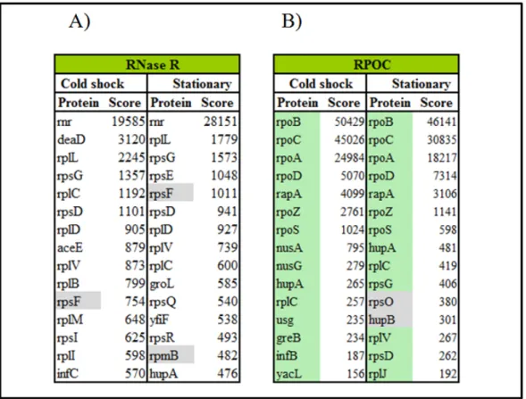
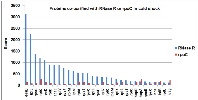
ix IgG – elution from immunoglobulin resin. RNase R and a protein detected during cold shock induction are indicated on the right side of the gel. 39 Figure 9 – Mass spectrometry results from TAP tag purification of RNase R or RPOC from cold shock or stationary growth phase cells. Graphs with scores of identified proteins purified with RNase R in cold shock (A) or stationary phase (B) and co-purified proteins with RPOC in cold shock (C) or stationary phase (D), from calmodulin
elution resin. 42
Figure 10 – Comparison of mass spectrometry results from TAP tag purification of
RNase R or RPOC from cold shocked cells. 45
Figure 11 – A) Visualization of DeaD amplification (1890 bps) by PCR on a 0.8% agarose gel. B) pUC18-DeaD digestion with SalI and NotI (1 - DeaD 1890 bps and
pUC18 2686 bps), visualized on a 0,8% agarose gel. The DNA size marker (M) is the commercial NZYDNA ladder III (NZYtech). In both gels, bands were visualized by
ethidium-bromide staining. 47
Figure 12 – Schematic diagram of pET28a plasmid. HIS, Kan and LacI regions are
represented by red arrows pointing the direction of transcription. Restriction sites are also indicated in the figure. This image was obtained by Clone Manager software
program. 48
Figure 13 – Representation of DeaD gene insertion in pET28a vector by homologous recombination and single strand annealing. Scheme adapted from Li & Elledge, 2007
(94). 49
Figure 14 – A – B) Restriction analysis of the recombinants derived from SLIC reaction. Plasmid DNA from one independent kanamycin resistant colony digested with
SalI (A), with SalI and NotI (2), or with EcorI (3). As a control (C) pET28a was
digested with SalI and NotI. Digestion products were separated on a 0,8% agarose gel
and visualized by staining with ethidium-bromide. The DNA size marker (M) is the
commercial NZYDNA ladder III (NZYtech). 50
x Soluble Proteins, HP – Proteins bound to Ni-NTA beads resin, M – Precision plus
Protein Standards (BioRad). 51
Figure 16 – Scheme of the investigation procedure used to study interaction between HIS tagged DeaD helicase and RNase R. A) Cells overexpressing HIS tag DeaD were purified using Ni-NTA agarose and B) HIS tag elutions were analysed in a 6% SDS-PAGE, transferred into a nitrocellulose membrane and Ponceau stained. The amount of RNase R was detected by Western blot (α-rnr). TP – Total protein fraction of purified
RNase R (R), SP – soluble protein fractions from cells overexpressing DeaD (D) or RNase II (II); HP – purified proteins fractions, M – Precision plus Protein Standards
(BioRad). 52
Figure 17 – A) Scheme of RNase R and DeaD purification by the HIS tag method. Purified proteins were mixed, separated on gel filtration column (Superdex 200) and monitorized by UV spectra (280nm). B) UV spectra (280nm) obtained after gel filtration of the mixture of RNase R with DeaD (red) or RNase R alone (blue). The green line represents the expected peak corresponding to RNase R protein (volumes
from 65 to 75ml). 54
Figure 18 – Representation of the experimental steps for total protein separation and then, RNase R monitoring by Western blot technique. Total proteins were extracted from cells in different growth conditions and extracts were subjected to the gel filtration procedure. Fractions collected were loaded on nitrocellulose membrane using dot blot device and proteins were detected using anti-RNase R antibodies. 56 Figure 19 – A, B) Protein spectra (280nm) obtained during filtration of E. coli total
protein extract on Superdex 200 column. Size of the markers used for column calibration is indicated by dashed lines and fraction numbers are indicated in the bottom part of the graph. UV spectra of protein extracts from E. coli after cold shock induction
xi Figure 20 – Representation of pUC18 plasmid, a commercial plasmid used as a cloning
vector. This image was obtained from “Fermentas”. 85
Figure 21 – Representation of pET28a plasmid, used as an expression vector. This
xii
List of Tables
Table 1 – Representation of the E. coli cold shock genes, products and respective
functions. 6
Table 2 – Master mix used in DeaD PCR amplification. 23
Table 3 - PCR Program used for DeaD gene amplification. 24
Table 4 - Ligation reaction of the DeaD sequence into the corresponding cloning sites of
pUC18. 24
Table 5 - Plasmid digestion of ligation products with SalI and NotI restriction enzymes. 26
Table 6 – Mix used for DeaD PCR amplification. 27
Table 7 - PCR Program used for DeaD gene amplification. 27
Table 8 - Digestion of pET28a vector with SalI and NotI restriction enzymes. 28
Table 9 - T4 DNA polymerase treatment. 29
Table 10 – Annealing reaction mix. 30
Table 11 – Digestion of pET28a:DeaD with SalI restriction enzyme. 31
xiii blue, and in purple are represented the proteins found in RNase R only in stationary
phase. 44
Table 14 – Culture media, solutions and gels used in the experimental procedures. 82
Table 15 – Strains used in the experimental procedures. 84
Table 16 – List of proteins detected to co-purify with RNase R or RPOC purification by TAP tag method during cold shock induction or in stationary growth cells. In green are represented the proteins previously related to interact with RNA polymerase (RNAP) and in gray are represented the proteins found in all preparations. 86 Table 17 – Primers used for DeaD amplification. Forward primers (Lprimer) include the
SalI restriction site on their sequence and the reverse primers (Rprimer) contained the NotIrestriction site (SalI and NotI restriction sites are underlined). 89
Table 18 – Primers used for DeaD amplification to perform the assembly of DeaD and pET28a fragments by homologous recombination using SLIC method. Forward primers (Lprimer) include the SalI restriction site and on their sequence and the reverse primers
(Rprimer) contained the NotI restriction site (SalI and NotI restriction sites are
xiv
Abbreviations
aa Amp APS ATP Bp BSA º C Cam CBB CDS CELUT CFU Csp ddH20DNA DNase dNTP ds dsRNA DTT E. coli EDTA h IPTG Kan kb kDa L Aminoacid Ampicillin Ammonium persulfate Adenosine triphosphate Base pairs
Bovine Serum Albumin Degree Celsius
Chloramphenicol
Calmodulin binding buffer Cold Shock Domain Calmodulin elution buffer Colony forming units Cold shock protein Bidistilled water Deoxyribonucleic Acid Deoxyribonuclease Deoxyribonucleotide triphosphate Double Stranded Double-stranded RNA Dithiothreitol Escherichia coli
xv LA LB LPS M Mg µg µl min miRNA ml mmol mol MQ mRNA ncRNA NEB ng Ni-NTA nm nts OD PAGE PCR PMSF PNPase RNA RNAP RNase II RNase III RNase E RNase R RNases Luria-Bertani Agar Luria-Bertani Broth Lipopolysaccharides molar/molarity (ml/L) Miligram microgram microliter minute microRNA milliliter millimolar mole Milli-Q water messanger RNA non coding RNA New England Biolabs Nanogram
Ni-nitrilotriacetic acid Nanometer
Nucleotides Optical Density
xvi rnb
rnr rpm RPOB RPOC rRNA SDS sRNAs ss TBE TEMED tmRNA Tris tRNA X-Gal wt
Ribonuclease II Ribonuclease R Rotations per minute RNA polymerase β subunit
RNA polymerase β prime subunit
Ribossomal RNA
Sodium dodecyl sulphate Small RNAs
Single stranded Tris-Borate-EDTA
Tetramethylethylenediamine Transfer-messenger RNA
Trishydroxymethyl aminomethane transfer RNA
1
1. Introduction
Microorganisms have to constantly adapt to different environmental changes. These changes include nutrient and oxygen availability, changes in temperature and osmotic stress. To survive and adapt to these changes, bacteria induce or repress the expression of certain genes leading to changes in cell physiology. Capacity of adaptation is one of the reasons why bacteria can survive under extreme conditions. Understanding mechanisms of bacterial adaptation can help developing strategies to avoid bacterial proliferation in unfavorable conditions like cold, with important applications in food and pharmaceutical industry.
1.1
Cold shock response
One of the environment changes that bacteria have to face is the temperature change. With a downshift of the temperature, bacteria react with a specific response called the cold shock. This mechanism is triggered by an abrupt shift of a culture growing exponentially from its optimum growth temperature (37°C) to a lower temperature (15°C). The cold shock response allows the cell to react and adapt to these changes.
After temperature decrease a number of changes occur in the cellular physiology. These effects include a decrease in the membrane fluidity, stabilization of secondary structures of nucleic acids, which leads to a reduced efficiency of mRNA translation and transcription, inefficient folding of some proteins and inhibition of ribosomal translation (68, 116, 117, 137).
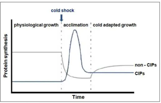
2 blocked (82) except for a group of cold inducible proteins (CIP) that are even induced. During this adaptation period the expression of the cold shock proteins increases. After acclimation phase cells become adapted to low temperature and resume growth at a rate that is slower than the one before the cold shock induction. The expression of the cold inducible proteins declines and the bulk protein synthesis restarts, adapted to the cold (Fig.1).
Figure 1 – Representation of the protein expression pattern after cold shock induction (Adapted from Kalbitzer et al. (73)).
Bacteria sense the change in temperature mainly at the level of cell membrane, nucleic acid and ribosomes (116).
3 promoters is modified affecting the recognition of some σ70 promoters such as the cold
inducible Escherichia colirecA promoter (145).
Bacteria also sense the temperature decrease at the level of ribosomes (141). Artificially inducing high levels of the guanosine 5’ triphosphate-3’ diphosphate and guanosine 5’ diphosphate-3’ diphosphate the expression of cold shock proteins decrease. However, at low concentration increases their production affecting the magnitude of the cold shock response (77).
The membrane composition is also affected by the temperature downshift. There is a decrease in the membrane fluidity which leads cells to lose viability. In Escherichia coli a rapid temperature downshift can induce phase separation of phospholipids. This
causes an increase in membrane permeability and possibly death (30).
The membrane of Gram negative cells is composed of lipopolysaccharides (LPS), which consist of a distal polysaccharide (O-antigen), a core polysaccharide and lipid A. E. coli Lipid A is required for growth (125, 126), and consists of two
glucosamine with attached acyl chains (fatty acids) that normally contain one phosphate group on each carbohydrate. Laurate (glyceryl laurate) is the fatty acyl chain of lipid A, usually detected in cells growing at 37°C. However, at low temperatures, there is a decrease in laurate counterbalanced by the appearance of palmitoleate (palmitoleate acid) (35). In contrast with laurate, that is a saturated fatty acid, palmitoleate is an unsaturated fatty acid. Unsaturated fatty acids increase membrane fluidity and lower its phase transition temperature. Palmitoleate has a double bond ligation, which causes the bending of fatty acid molecules and leads to a lose packing (Fig. 2).
LpxL is an acyltransferase that attaches laurate to the lipid A (134) and is important for lipid A synthesis at normal growth conditions. However, at low temperatures, LpxP (LpxL orthologue) is also expressed. LpxP is a cold inducible protein involved in cold-adapted replication. It is required for the production of lipid A adapted to the cold, because attaches palmitoleate to the Lipid A molecule instead of laurate (134, 143). LpxP was also found in Shewanella oneidensis and described to be
4 Figure 2 – Representation of membrane fluidity according to the lipid composition. Palmitoleic acid has a double bond ligation that causes the bending of fatty acid molecules, while glyceryl laurate has only a single bond ligation.
In Bacillus subtilis, adaptation to the cold shock also triggers changes in the
membrane. The adaptation of membrane fluidity to this stress condition involves a rapid desaturation of fatty acids in existing phospholipids. This happens by induction of fatty acid desaturase (Des) due to an increase of fatty acid. The induction of Des is regulated by the sensor kinase DesK and the response regulator DesR (4). The trans-membrane domain of DesK was described as a probable sensor of the membrane fluidity (5). With an abrupt shift to a lower temperature, DesK phosphorylates the transcriptional activator DesR which subsequently binds to the promoter of the des gene and activates
transcription of the D5-desaturase. This enzyme catalyzes the reaction to introduce a double bound into preexisting fatty acids tails of phospholipids inside the cellular membrane. This will increase the fluidity of the membrane bilayer, as a response to the decrease in temperature (3, 64). In Legionella pneumophila, during adaptation to cold
5
1.2
Cold induced proteins
After the cold shock induction, cells enter in the acclimation period in which a group of cold inducible proteins (CIP) are expressed. Most of these cold shock proteins are essential for the cell to survive at low temperatures and they are involved in different cellular processes (80, 82, 137). Proteins with described or presumptive functions during cold shock response are listed in table 1.
Gene Product Description/Function in cold shock References
aceE aceF cspA cspB cspE cspG cspI deaD dnaA gyrA hns hscA hscB hupB infA infB infC lpxP nusA otsA otsB pnp rnr rbfA recA AceE AceF CspA CspB CspE CspG CspI DeaD /CsdA DnaA GyrA H-NS Hsc66 HscB Huβ IF1 IF2 IF3 LpxP NusA OtsA OtsB PNPase RNase R RbfA RecA
Pyruvate dehydrogenase, decarboxylase.
Pyruvate dehydrogenase, dihydrolipoamide acetyltransferase.
Cold-inducible RNA chaperone and anti-terminator; transcriptional enhancer.
Cold shock inducible; Function unknown. RNA chaperone; transcriptional antitermination.
Cold shock protein homolog, cold-inducible; Function unknown. Cold shock protein, cold shock inducible; Function unknown.
ATP-dependent RNA helicase, facilitates translation of mRNAs with 5' secondary structures.
DNA binding and replication initiator, global transcription regulator. DNA gyrase, subunit A; DNA-binding/cleaving/rejoining subunit of gyrase. Nucleoid protein, transcriptional repressor, Repressor supercoiling. DnaK-like chaperone
DnaJ-like co-chaperone for HscA. Nucleoid protein, DNA supercoiling.
Protein chain initiation factor IF1, Translation initiation.
Protein chain initiation factor IF2, Translation initiation, fMet-tRNA binding, protein chaperone.
Protein chain initiation factor IF3, Translation initiation, stimulates mRNAs translation.
Lipid A synthesis; cold temperature inducible.
Transcription termination/antitermination/elongation L factor.
Trehalose phosphate synthase; cold- and heat- induced, critical for viability at low temperatures.
Trehalose phosphate phosphatase; cold- and heat- induced, critical for viability at low temperatures.
3'-5' exoribonuclease; component of RNA degradosome; cold shock protein required for growth at low temperatures.
3’ -5’exonucleases; increases 10-fold in cold shock.
Ribosome binding factor required for efficient processing of 16s rRNA; cold-shock adaptation protein.
General recombination and DNA repair; induction of the SOS response.
(78) (78)
(19, 20, 63, 76, 90, 132) (78, 119) (71, 119, 149) (105) (146) (17, 81, 109)
(11) (80)
(12, 51, 80, 90) (93)
(93) (61, 108) (49, 147) (29, 70, 78)
(69)
(35, 134, 143) (58, 78) (83)
(83)
(52, 78, 154, 156)
(9, 14, 28, 41) (24, 50, 79, 151)
6
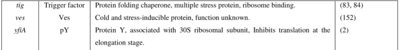
tig
ves
yfiA
Trigger factor Ves
pY
Protein folding chaperone, multiple stress protein, ribosome binding. Cold and stress-inducible protein, function unknown.
Protein Y, associated with 30S ribosomal subunit, Inhibits translation at the elongation stage.
(83, 84) (152) (2)
Table 1 – Representation of the E. coli cold shock genes, products and respective
functions.
The cold shock proteins can be divided in different groups according to their functions.
Several of these proteins are involved in RNA metabolism. A group of cold shock proteins is involved in transcription: the cold shock proteins (Csp) family, that can work as RNA chaperones but are also involved in transcriptional antitermination (19, 20, 63, 76, 90, 132), DeaD helicase, that works as RNA chaperone and facilitates translation of mRNAs with 5’ secondary structures due to its RNA unwinding activity (17, 81, 109, 139) , histone-like protein H-NS, a transcriptional repressor (12, 51, 80, 90), and the transcription factor NusA (58) that is involved in the transcription termination, antitermination and elongation of L factor (58, 78).
During the cold shock response two 3’ -5’ exonucleases are involved in the RNA degradation. Polynucleotide phosphorylase (PNPase) was shown to be required for growth at low temperature (13, 52, 78, 154, 156), selectively degrades cspA mRNA at
15°C and represses the CspA homologues production at the end of the lag phase (114, 154), and the ribonuclease R (RNase R) increases its level about 10-fold in the cold shock (9, 14, 28, 41).
7 stationary phase at a physiological temperature. pY is able to inhibit translation at the elongation stage by blocking the binding of aminoacyl-tRNA to the ribosomal A site and subsequently decline the protein synthesis, for example, during cold shock. When growth is resumed, pY is no longer detected in ribosomes which suggests that it function is to arrest translation in response to an environmental stress such as cold shock (2).
The cold shock proteins are also required for DNA metabolism. These proteins are: a DNA binding and replication initiator (DnaA) (11), a DNA gyrase subunit A (GyrA) (80), the DnaK-like chaperone (Hsc-66) (93) and the DnaJ-like co-chaperone (HscB) (93), the nucleoid protein Huβ involved in the DNA supercoiling (61, 108) and
the general recombination and DNA repair protein RecA (78, 144).
The cold shock proteins that are not grouped in the RNA or DNA metabolism are divided in two functional categories according to their targets: lipids or proteins. To the lipid group belong LpxP that is involved in the lipid A synthesis. In the protein group are described the OtsA and OtsB trehalose phosphates and the trigger factor. The OtsA and OtsB contain a cold box characteristic of the cold shock mRNAs, are critical for cell viability at low temperatures (83) and their induction is dependent on RpoS, increasing 8-fold upon temperature downshift (116). The trehalose mRNAs were also shown to enhance stability at low temperature (83). It was suggested that, at low temperatures trehalose acts by preventing denaturation and aggregation of proteins, functioning as a free radical scavenger in vivo, protecting against oxidative damage and
8 The induction levels of the cold shock proteins are different. In E. coli, CspA,
CspB, CspG, CspI, DeaD, RbfA, NusA, PNPase and RNase R are the most increased proteins comparing to the other CIP.
1.3 RNA metabolism under cold shock
Accordingly to the Central Dogma of Biology, the genetic information is processed from DNA to RNA to Protein. DNA is replicated while cell divides, is transcribed into RNA and subsequently translated into protein. The gene expression is determined by the efficiency of transcription of DNA to mRNA, the stability of mRNA, and the frequency of mRNA translation into proteins. In general the RNA population can be divided in four different categories: messenger RNA (mRNA), ribosomal RNA (rRNA), transfer RNA (tRNA) and small non-coding RNAs. However, these molecules can have different cellular functions, like working as enzymes (ribozymes) or as regulators of genetic expression.
The RNA metabolism at 37°C can be different from the one at low temperatures. With the decrease of the temperature, RNA secondary structures are more stable. Presumably, with RNA stabilization the transcription elongation, the ribosomal movement on RNA and then translation, slows down. Consequently, it requires proteins that destabilize these structures and allow these biologic processes to proceed.
1.3.1 Cold shock proteins – Csp
Csp is a family of small structurally related nucleic acid-binding proteins (115) which are composed of the typical cold shock domain (CSD). These proteins bind preferentially to single-stranded RNA or DNA (76, 101, 157). The Csp in E. coli family
9 The Csps have been functionally linked to the maintenance of chromosome structure and DNA replication (36) and also affect transcription by acting as transcription antiterminators. The majority of the Csp are able of binding DNA (76, 119, 153). At low temperatures, with the stabilization of RNA structures the protein translation is hampered. The Csp proteins function, as chaperones, is crucial due to mRNA stability in cold conditions. CspA, the major cold induced Csp, function as an RNA chaperone and destabilize the secondary structures (76). The CspE protein melts partially double stranded and hairpin structures (120). CspE was also shown to bind poly-A tails and thereby, stabilizing mRNA by reducing degradation by PNPase and RNaseE (56). CspE and CspC are expressed at both high and low temperatures (155) and have a critical impact on the stabilization of transcripts for a global stress response regulator (rpoS) and the universal stress response protein (uspA) (118).
In B. subtilis three proteins homologous to E. coli CspA were identified (CspB,
CspC and CspD), which are induced at low temperatures (67, 148). In many other bacterial microorganisms this type of cold shock proteins were also described (66, 68).
1.3.2 CspA
CspA was originally identified as the major cold shock protein (63). After the cold shock induction, its level increases up to 13% of the total protein content of the cells (63).
The regulation of CspA and its homologues expression during the cold shock induction occurs at the levels of transcription, mRNA stability and translation.
The induction of cspA, at low temperatures, does not require any additional
transcription factors (116). The cspA, cspB, cspG and cspI have an unusually long 5'
untranslated region (5'-UTR). The 5'-UTR contains a highly conserved unique 11-base sequence called the cold box (75, 150). The cold box is a presumed transcriptional pausing site and is involved in the repression of cspA expression (116). The cspA
5'-UTR is thought to be responsible for the extreme instability of cspA mRNA at 37°C,
10 The cspA mRNA is dramatically but transiently stabilized immediately
following cold shock. Its promoter is active at 37°C. However, CspA is hardly detected at 37°C due to the instability of its mRNA.
The cspAmRNA also contains a unique sequence located 14-bases downstream
of the initiation codon, the downstream box. It is present in CspB, CspG, CspI, CsdA and RbfA and is presumed to enhance translation initiation in the cold shock mRNAs by facilitating the formation of translation preinitiation complex through binding to 16S rRNA. However, the exact mechanism of the enhancing effect on translation initiation by the downstream box remains unknown (107, 110).
CspA and its homologues have been characterized as RNA chaperones, and this function is thought to facilitate translation at low temperatures (19, 65, 76, 78). These structures of RNA stabilize at low temperatures but CspA and its homologues presumably can destabilize the secondary structures and facilitate transcription and translation by acting as RNA chaperones. At low temperatures, the secondary structures of RNA stabilize, which presumably slows down the transcription elongation and the ribosomal movement on RNA and thus translation. CspA and its homologues presumably can destabilize the RNA secondary structures, and thus facilitate transcription and translation. The increased levels of CspA after cold shock may be important for compensating the higher stability of secondary structures in RNA at low temperatures (116). In E. coli, CspA and its homologues binds RNA without apparent
sequence specificity and with low binding affinity (76). CspB, CspC and CspEare able to more selectively bind RNA/ssDNA (116). The non-specific and weak binding of CspA homologues to RNA/ssDNA are also important for the chaperone function, as binding of the protein would not hamper ribosome movement on mRNA (116).
11
1.3.3 DeaD
The other group of proteins that can act as RNA chaperones are the DEAD-box-family of helicases (48). It is a DEAD-box-family of RNA dependent ATPases able to unwind double-stranded nucleic acids and characterized by 9 conserved motifs (81) (Fig 3).
Figure 3 - Overview of the nine conserved motifs (Q to VI) of the DEAD-box family of
E. coli.
Proteins from the DEAD-box family are involved in different processes of RNA metabolism such as translation initiation, ribosome biogenesis and assembly or RNA degradation (38, 55, 131, 136). E. coli has 5 identified DEAD-box proteins, SrmB,
DeaD, DbpA, RhlB and RhlE. SrmB and DeaD are essential at low temperature (39), (34) and DeaD is overexpressed in cold shock. DeaD (also named CsdA) is involved in many different processes in the cell. It was described that the deletion of DeaD results in the deficiency in free 50S subunits at low temperature. This results shows that this protein participates in the biogenesis of the 50S ribosomal subunit, probably by binding and changing the RNA structure of a 50S precursor (38). It is also required for initiation of translation of mRNAs with an extensive secondary structure (102), and it interacts with poly(A) polymerase (127). DeaD has the ability to unwind double-stranded RNA in the presence or absence of ATP (81). It is involved in the mRNA degradation of cold shock genes (154). It was proposed that DeaD can interact with RNase E, PNPase and other components of the RNA degradosome under cold shock conditions (123, 124).
12 with the RNA chaperone Hfq, but they did not reveal a physical interaction (129). In vitro assays showed that it associates directly with RNase E, but not with PNPase (123).
However we can not exclude the possibility to interact with other ribonucleases. This hypothesis remains to be solved and more work is required to clarify this question.
1.4 RNA degradation
RNA degradation is the major process that controls the RNA levels in the cell. Degradation is required for elimination of defective RNAs and superfluous transcripts whose expression is no longer required. Ribonucleases (RNases) are enzymes with the strongest role in this process. They can be selective and sensitive to specific elements of the RNA molecule and are involved in the quality control of all types of RNAs. They are involved not only in RNA degradation but also in RNA processing and maturation. There are other enzymes that act synergistically with ribonucleases within these processes, like helicases, polymerases and RNA binding proteins. RNases can act alone or can be part of RNA degradation complexes like the degradosome. Ribonucleases are divided in two main groups, endonucleases and exonucleases.
Endonucleases are the enzymes that cleave RNA internally by digesting phosphodiester bonds of the RNA molecule. In E. coli, eight endoribonucleases have
been identified, RNase E, RNase G, RNase III, RNase Z, RNase HI, RNase HII, RNase I and RNase P (7, 89). They play an important role in mRNA metabolism and in E. coli,
the major endoribonucleases are RNase E and RNase III.
Exoribonucleases are the enzymes that degrade RNA by removing terminal nucleotides in the 3’-5’ or 5’- 3’direction (9, 158). In prokaryotes, most RNases cleave the RNA in the 3’-5’direction. These proteins act releasing nucleotides that can be reutilized for the synthesis of new RNA molecules. In E. coli, seven exoribonucleases
13 RNase R, RNase II and PNPase are considering the three major 3’ -5’ processing exoribonucleases in E. coli. RNase R, together with RNase II belongs to the RNB
family (85), while PNPase is a member of the PDX family. RNase R (28) and PNPase (82, 156), are cold shock inducible and were suggested to be responsible for degradation of structured RNA in the cell at low temperatures (44, 95).
1.4.1 PNPase
Polynucleotide phosphorylase (PNPase) is an exoribonuclease encoded by the
pnp gene (128) and belongs to the PDX family of nucleases. In the genome it is located
downstream of the rpsO gene (122). Its expression is controlled both at transcriptional
and post-transcriptional levels (9, 74). PNPase binds to the 5' end of RNase III-processed pnp transcript, resulting in inhibition of translation and leading pnp mRNA
into the degradation pathway (60, 121). It is not an essential enzyme for the cell at 37°C however, it becomes extremely important at lower temperatures (13) . PNPase can be incorporated into the degradosome, a complex of multiple proteins, which also comprises RNase E (33, 99).
14 Figure 4 – Representation of E. coli PNPase. A) Linear representation of E. coli
PNPase domains. B) View of PNPase crystal structure in the monomer organization (133). C) PNPase trimer structure.
Most mRNAs that are already present in the cell when cold shock is induced, cannot be efficiently translated until specific cold shock factors like CspA, RbfA and DeaD, are produced (76, 79, 139). However, mRNA from cold shock genes at low temperatures are translatable and, like cspA transcripts, can become more stable (19,
62).
It was described that translation of pnp mRNA after cold shock may not be very
efficient (156). During the acclimation phase PNPase seems to restore intragenic transcription termination and regulate its own expression at the level of transcription elongation (156). With this autogenous regulation, pnp mRNA becomes more stable.
However, the levels of PNPase protein do not increase accordingly (104, 156).
15 has been implicated in virulence in several pathogens, namely in Salmonella, Yersinia, Campylobacter jejuni and Streptococcus pyogenes. Interestingly, while in some
microorganisms PNPase seems to act as a virulence repressor, in others this ribonuclease takes an important role in the establishment of virulence (10).
1.4.2 The Degradosome
The E. coli RNA degradosome has been described as an RNA decay “machine”
due to its function in mRNA degradation and RNA processing (33). This complex or its individual components can target specific gene products or affect the relative composition of different transcripts through differential decay rates (32).
The degradosome is mainly composed by RNase E, PNPase, RhlB (a DEAD-box RNA helicase) and the glycolytic enzyme enolase (33). The complex was discovered during the purification of RNase E (33), an endoribonuclease that mediates the principal pathway for the mRNA degradation in E. coli. This ribonuclease is
essential for bacteria. RNase E inactivates polyribosomal mRNA by endoribonucleolytic cleavage, producing mRNA fragments. These are subsequently digested to nucleotides by exonucleases (PNPase), helped by RhlB and poly(A) polymerase. In the presence of ATP, the RhlB helicase unwinds stem-loops to help PNPase digestion (86). The RNA degradosome is built on a region of RNase E that shows high sequence variation among closely related bacteria, the carboxy-terminal half, where PNPase, RhlB and enolase bind (88, 111). Only RNase E, PNPase, and RhlB are essential to reconstitute the activity of the degradosome in vitro (47). E. coli
mutants in which degradosome assembly is disrupted have a slow-growth phenotype and altered metabolic profiles (22). This results from the incapacity to degrade a number of degradosome substrates (88, 111) and reveals that this protein complex is essential in gene regulation.
16 distinct RNase E binding site (87, 123). To explain the association of DeaD with RNase E two theories were proposed (123). RhlB and DeaD associate with RNase E at the same time but each one bind to a separate site (87) or, cold shocked cells have heterogeneous population of the degradosome, some that can contain both DeaD and RhlB helicase and others with only one of the two helicase proteins (123). Structured mRNA is more difficult to degrade at low temperatures and it may require two RNA helicases (33). These findings showed that the degradosome is capable to adapt depending on the environmental conditions (123).
1.4.3 RNase R
Ribonuclease R (RNase R) belongs to the RNB family of enzymes. The members of RNB family are widely conserved in both prokaryotes and eukaryotes and serve several important functions. In eukaryotes they can be developmentally regulated (25) and mutations in its genes have been related with mitotic control, and cancer (98). In prokaryotes, this family of enzymes is important in stress responses, RNA and protein quality control, and it was shown to be required for virulence in different organisms (8, 28, 40, 44, 45).
17 Figure 5 -Representation of the predicted E. coli RNase R 3D structure. The structural
model of RNase R was constructed based on the RNase II structure (16). It comprises two N-terminal Cold Shock Domains (CSD1 in orange and CSD 2 in yellow), a central RNB domain (in blue) and a C-terminal S1 domain (in green); the RNA molecule is also represented (in red) (57) .
In contrast to RNase II, RNase R is able to digest RNA secondary structures without the help of a helicase. This function can be especially needed at low temperatures when RNA is highly structured. RNase R can also unwind double stranded structures in absence of RNase activity which lead to a suggestion that this protein has “helicase like” activity (14).
In E. coli, RNase R is responsible for the degradation of substrates such as
18 compared to the wild-type strain (28). RNase R has also been shown to be involved in virulence mechanisms (54, 91, 138, 140).
After cold shock induction, RNase R levels increase about ten-fold (41). The increased amount of RNase R was shown to be stable for many hours revealing that slow growth conditions at low temperatures leads to RNase R stabilization (42). A recent study revealed that the level of RNase R after addition of chloramphenicol was also elevated when compared with RNase R level from cells grown at 37°C. It was suggested that the regulatory processes that lead to this elevation during cold shock are a consequence of an increase in the amount of rnr message, that is substantially
stabilized in cold shock (28, 42). Recent results suggests that acetylation regulates RNase R stability (97). The Lys544 interacts with acidic residues within the C-terminal region of the protein. By this interaction, the C-terminal region stays in a position that is poorly accessible to tmRNA-SmpB, whose binding is essential for RNase R instability (96). Addition of the acetyl group would remove the positive charge on lysine, breaking the interaction with tmRNA-SmpB. RNase R in stationary phase cells is not acetylated, position 544 is positively charged so, the C-terminal region would not be accessible. Then tmRNA-SmpB binds weakly, resulting in a stable RNase R (97). These results revealed that the acetylation of one specific lysine residue in RNase R increases binding of the tmRNA-SmpB complex, resulting in protein instability (97). This direct effect of acetylation on the regulatory process of stability of RNase R in the stationary phase can also occur at low temperatures. However, further studies are required to understand it.
RNase R can complement the CsdA cold shock function. It was suggested that it is due to RNase R ability to degrade secondary structures and its presumable helicase activity (14).
It is clear that RNase R plays an important role in RNA degradation at low temperature but further studies are required to clarify how RNase R is involved with other cold shock proteins in the cold shock response.
19
1.5 Health and industrial implications of cold shock response
E. coli is a microorganism that is commonly found in the lower intestine of
warm-blood organisms. They are usually harmless but some serotypes can cause serious health complications. The presence of these bacteria in food or pharmaceutical products is indicative of fecal contamination.
Although E. coli is known as a usual pathogen, this bacterium is still the
microorganism of choice for most gene cloning experiments and has been widely used in biotechnology industry to overproduce determinate components.
Understanding cold shock response can be of the extreme importance. Food storage is commonly made using refrigeration to avoid proliferation of bacteria such as
Listeria or Campylobacter. Knowing how to block cold shock protein synthesis will
allow us to reduce/inhibit the cold shock response and that can be lethal to bacterial cells at low temperatures. Investigators revealed that E. coli growth can be inhibited by
the combination of rapid chilling with nisin, a polypeptide produced by lactic acid bacteria which is recognized as natural antimicrobial food preservative (46), reducing more than 6-log of the bacterial population (31). Combining the use of antimicrobial products with bacterial exposition to low temperatures can thus extend the efficiency of drugs to decrease bacterial proliferation.
On the other hand, by controlling the cold shock response, proteins that are not produced efficiently at 37°C can be overproduced at low temperatures using cold-inducible promoters (142).
A common bacteria used in industry is Lactobacillus (37). This bacterium also
has cold shock proteins that are induced at low temperatures. During the different industrial processes, cells have to face several environmental changes. Bacteria face changes in pH, salt concentration or temperature. By inducing the cold shock response,
Lactobacillus increases the probability to survive in low temperatures, which can be
extremely useful in this industry.
20
2. Materials and Methods
All materials and reagents are described on Annex I (table 14).
2.1 Bacterial strains
Bacterial strains used in this work are listed in Annex I (table 15) and were grown in LB medium at 37ºC supplemented, when required, with appropriate antibiotic. Bacterial strains were stored in 10% glycerol at -80ºC.
2.2 Preparation of competent cells
An overnight culture of BL21 (DE3) or DH5α was diluted in LB to a final
OD600nm of 0.05. BL21 (DE3) cells are used for protein overexpression and DH5α cells
for mutant constructions. Cells were incubated with agitation (180 rpm) at 37ºC.
21
2.3 Transformation
DNA (ligation mixture/plasmid) was added to 100 µL of E. coli competent cells.
The cells were incubated on ice for 30 minutes, heat shocked at 42ºC for 1 minute and immediately placed back on ice for 1 minute. LB medium was added (1ml) and the tubes were incubated at 37ºC for 45 to 60 minutes. After incubation, cells were plated on LA medium plates with the respective antibiotic and incubated overnight at 37ºC.
2.4 TAP tag purification
The TAP tag purification was performed following these steps:
• Three cultures were started from three single colonies of E. coli (BW113) with
RNase R protein fused with TAP tag. Colonies were inoculated in LB medium with kanamycin (50 µg/ml), and incubated overnight at 37ºC.
• The three pre-inoculums were diluted in 1L of LB to a final concentration of OD600 of 0,05 containing kanamycin (50 µg/ml) and incubated at 37ºC until cells reach exponential growth phase (OD600 of approximately 0.5).
• After that, the three cultures were processed in different ways:
o one of the three cultures was centrifuged at 5000 rpm for 8 minutes at 4ºC and the pellet was stored at -80ºC (exponential growth phase);
o one of the other cultures was incubated for 3h at 15ºC, centrifuged at 5000 rpm for 8 minutes at 4ºC and stored at -80ºC (cold shock growth phase);
o the third culture remained at 37ºC for 3h, was centrifuged at 5000 rpm for 8 minutes at 4ºC and stored at -80ºC (stationary growth phase).
• The pellets were unfrozen and each one was resuspended in 8 ml of Lysis buffer.
• Suspensions were lysed by two passages in French press.
• 0.5µl of benzonase (250 U/µl) was added to degrade the nucleic acids in the
22
• Samples were incubated on ice for 10 minutes and centrifuged at 35000 rpm, for 45 minutes at 4ºC in ultracentrifuge.
• Supernatants were filtered (0.45 µm) and an aliquot (soluble fraction) was taken for further SDS-PAGE gel analysis.
• 200 µL of Rabbit IgG agarose (beads) were added into a column (Poly-Prep Chromatography Columns from Biorad).
• The column was washed with 5ml of IPP150A with Triton X-100.
• Triton X-100 detergent (final 0,1%) was added to each supernatant and the mixture was loaded into the column.
• Columns remained shaking at 4ºC for 1.5h.
• After incubation, columns were washed twice with 10ml of IPP150 and subsequently two times with 10ml of TEVClevBuf.
• 200µL of TEVClevBuf and 35µL of TEV protease (approximately 100 units) were added to the column and incubated for 75 minutes, shaking at room temperature.
• After TEV cleavage, the supernatant was collected into an eppendorf by eluting twice with 250µL of CBB.
• An aliquot (IgG fraction elution) was taken for mass spectrometry analysis.
• CaCl2 (4µl of a 0,2M stock) was added to the mixture.
• The mix was transferred into an eppendorf with 300µL of CBB beads (previously washed 4 times with 500µL of CBB).
• Incubation was performed for 45 minutes shaking at 4 °C.
• After incubation the sample was transferred into a new column and washed twice with 5ml of CBB.
• As a final step, we elute with 600µL of CELUT into an eppendorf.
2.5 Acetone precipitation of proteins
23 After incubation each sample was centrifuged at 15000 rpm for 15 minutes at 4ºC. Supernatant was discarded and the pellet was washed with 500µL of 80% of cold acetone (-20°C). Supernatant was discarded, pellet was dried and stored.
2.6 DeaD amplification and cloning
The DeaD helicase gene (1,9 kb) from E. coli was amplified by PCR using a
high fidelity phusion polymerase which produces blunt end PCR products (Finnzymes). We used E. coli genomic DNA has a template and the primers described in Annex II,
table 17. Forward primers included SalI restriction site on their sequence and the
reverse primers contained the NotI restriction site. The PCR reaction mixture and the
program used are detailed in the table 2 and 3, respectively.
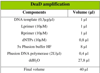
Table 2 – Master mix used in DeaD PCR amplification.
DeaD amplification
Components Volume (µl)
DNA template (0,3µg/µl) Lprimer (10µM) Rprimer (10µM) dNTPs (10µM) 5x Phusion buffer HF Phusion DNA polymerase (2U/µl)
ddH2O
1 µl 1 µl 1 µl 0,8 µl
8 µl 0,4 µl 27,8 µl
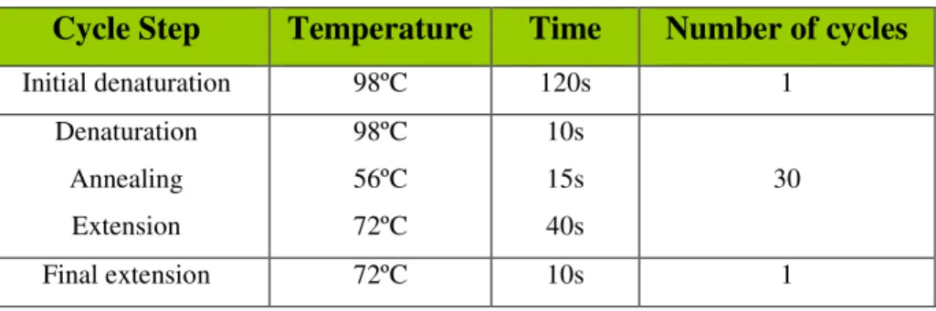
24 Table 3 - PCR Program used for DeaD gene amplification.
Cycle Step Temperature Time Number of cycles
Initial denaturation 98ºC 120s 1
Denaturation Annealing Extension
98ºC 56ºC 72ºC
10s 15s 40s
30
Final extension 72ºC 10s 1
After DeaD amplification, the PCR product (1,9 kb) was visualised in the 0,8% agarose gel.
Subsequently, a ligation was performed using the DeaD sequence (directly from the PCR amplification reaction) and the pUC18 (2,7kb) previously digested with
SmaI (Fermentas). This restriction enzyme cleaves the DNA generating blunt ends. The
ligation reaction was performed by T4 DNA ligase following the supplier protocol (Fermentas), at 22ºC in a water bath for 20 minutes as described in the following table:
Table 4 - Ligation reaction of the DeaD sequence into the corresponding cloning sites of pUC18.
Ligation
Components Volume (µl)
PCR product (100 ng/µl) pUC18 (100 ng/µl) 10x Buffer T4 DNA ligase
50% PEG (PEG 4000) T4 DNA ligase (10U/µl)
H2O
5 µl 1 µl 2 µl 2 µl 0,7 µl 9,3 µl
25 After incubation, enzymatic inactivation of T4 DNA ligase was performed at 65ºC for 10 minutes.
The ligation product was transformed into DH5α E. coli competent cells
following the protocol described above (2.3 section).
Transformants were selected on the LB plates supplemented with Ampicillin (100µg/ml), IPTG and X-Gal (See Annex I). The presence of ampicilin allows selecting
the colonies with the recombinant DNA by the presence of the resistance gene in the vector. The alpha-complementation allows determining whether a transformed bacterial colony has the ligation product or not. The lac-Z gene product (β-galactosidase) is a
tetramer and each monomer is made of two parts - lacZ-alpha, and lacZ-omega. If the alpha fragment is deleted, the omega fragment is non-functional. This alpha fragment functionality can be restored in-trans via plasmid. pUC18 plasmid has the deletion of the lac Z-alpha so, if ligation and transformation works, cell should express non-functional β-galactosidase (lac Z-alpha will be disrupted with the insertion of the gene
of interest). In the selection media (with IPTG to induce the lac repressor), the X-gal, a chromogenic substrate that yields a blue product when cleaved by β-galactosidase
colonies of interest should appear white. The blue colonies represent the cells that contain the unaltered vector.
The plasmid was then extracted from the positive clones (white colonies) using the ZR Plasmid Miniprep kit. The plasmid concentration was determined using the ND100 Spectrophotometer from NanoDrop.
Subsequently, pUC18 DeaD was digested with SalI and NotI restriction enzymes
26 Table 5 - Plasmid digestion of ligation products with SalI and NotI restriction enzymes.
Digestion reaction
Components Volume (µl)
DNA (350 ng/µl ) 10x Fast digest buffer
SalI (10U/µl) NotI (10U/µl)
H2O
30 µl 6 µl 2 µl 2 µl 20 µl
Final volume 60 µl
2.7 SLIC reaction
2.7.1 Insert preparation
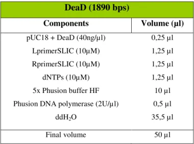
DeaD sequence (1.9 kb) from pUC18 DeaD vector was amplified by PCR using a high fidelity enzyme and the primers with 30 bp of overlapping sequence described in Annex III, table 18. The resultant fragment was used to ligate into pET28a using SLIC recombination method.
27 Table 6 – Mix used for DeaD PCR amplification.
DeaD (1890 bps)
Components Volume (µl)
pUC18 + DeaD (40ng/µl) LprimerSLIC (10µM) RprimerSLIC (10µM)
dNTPs (10µM) 5x Phusion buffer HF Phusion DNA polymerase (2U/µl)
ddH2O
0,25 µl 1,25 µl 1,25 µl 1,25 µl 10 µl 0,5 µl 35,5 µl
Final volume 50 µl
Table 7 - PCR Program used for DeaD gene amplification.
Cycle Step Temperature Time Number of cycles
Initial denaturation 98ºC 120s 1
Denaturation Annealing Extension
98ºC 56ºC 72ºC
10s 15s 40s
30
Final extension 72ºC 10s 1
After PCR amplification, the four PCR reactions were joined. We add 2U of
DpnI and incubate at 37ºC for 1 hour. DpnI cleaves methylated DNA from E. coli
(template DNA), whereas unmethylated DNA (in vitro synthesized DNA) is not cleaved
by these enzyme. By using DpnI after the PCR we are able to eliminate the plasmidic
28 Enzymatic inactivation was performed at 80ºC for 20 minutes. The DeaD insert was separated in a 0.8% agarose gel and the corresponding band was extracted from the gel. DeaD insert was cleaned using the Zymo's DNA Clean and Concentrator.
2.7.2 Vector preparation
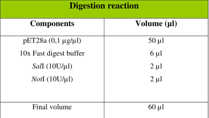
The expression vector, pET28a (5,4 kb), was digested with SalI and NotI
restriction enzymes (Fermentas). To perform this enzymatic restriction we prepared a digestion reaction as described in table 8.
Digestion reaction was incubated at 37ºC for 2 hours and then, enzymatic inactivation was performed at 80ºC for 10 minutes.
Table 8 - Digestion of pET28a vector with SalI and NotI restriction enzymes.
Digestion reaction
Components Volume (µl)
pET28a (0,1 µg/µl) 10x Fast digest buffer
SalI (10U/µl) NotI (10U/µl)
50 µl 6 µl 2 µl 2 µl
Final volume 60 µl
Cleavage products were separated in a 0,8% agarose gel and the appropriate size band (5368bps) was cut.
29
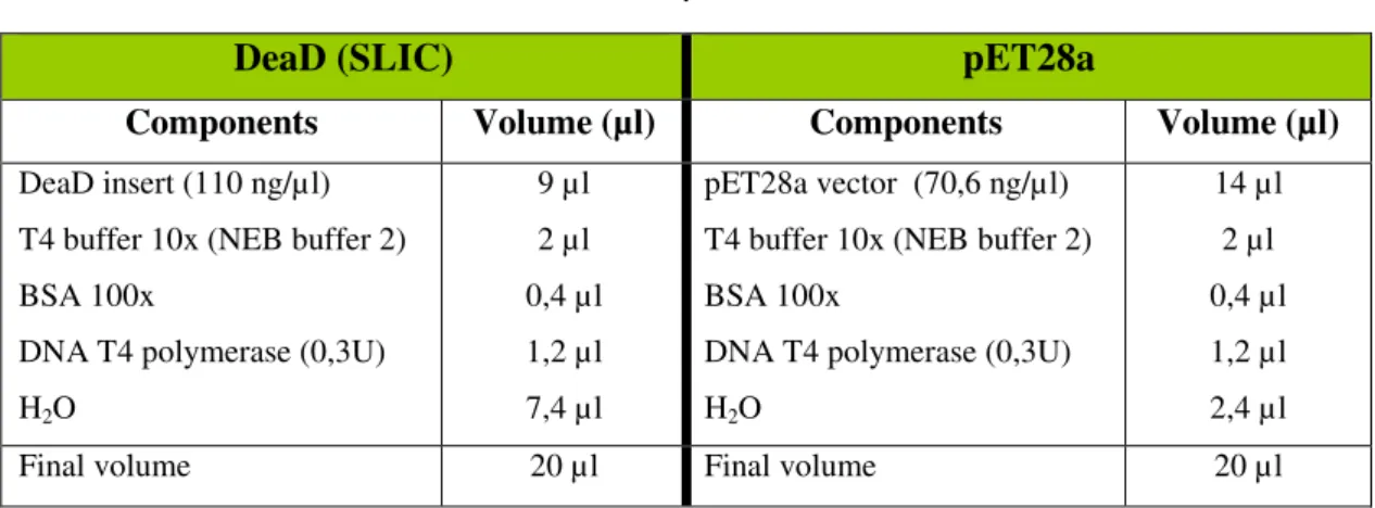
2.7.3 T4 Treatment
To perform SLIC recombination the prepared products (DeaD insert and pET28a vector digested with SalI and NotI) were subjected to T4 polymerase (NEB) treatment.
The T4 DNA polymerase catalyzes the synthesis of DNA in 5’ to 3’ direction but requires the addition of dNTPs. Without dNTPs it can create single stranded regions that will anneal to the prepared insert. The DeaD insert was used directly from PCR amplification, and pET28a was used after digestion and “gel cleaning” as described in 2.7.2 section. T4 Treatment was followed as described in table 9.
Table 9 - T4 DNA polymerase treatment. .
DeaD (SLIC) pET28a
Components Volume (µl) Components Volume (µl)
DeaD insert (110 ng/µl) T4 buffer 10x (NEB buffer 2) BSA 100x
DNA T4 polymerase (0,3U) H2O
9 µl 2 µl 0,4 µl 1,2 µl 7,4 µl
pET28a vector (70,6 ng/µl) T4 buffer 10x (NEB buffer 2) BSA 100x
DNA T4 polymerase (0,3U) H2O
14 µl 2 µl 0,4 µl 1,2 µl 2,4 µl
Final volume 20 µl Final volume 20 µl
30
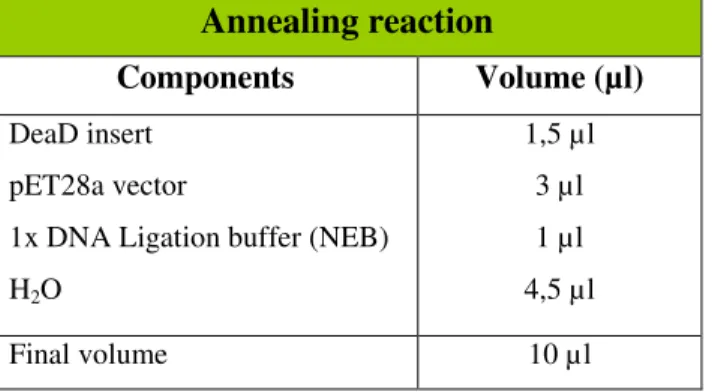
2.7.4 Ligation
The ligation of the products was performed using a 1:1 molar ratio insert to vector with 150ng of the digested vector (pET28a). The reaction was prepared as described in table 10 and incubated at 37ºC for 30 minutes.
Table 10 – Annealing reaction mix.
Annealing reaction
Components Volume (µl)
DeaD insert pET28a vector
1x DNA Ligation buffer (NEB) H2O
1,5 µl 3 µl 1 µl 4,5 µl
Final volume 10 µl
2.7.5 Transformation
The transformation was followed as described in 2.3 section, using 5 µL of the previous reaction and 100 µl of DH5α E. coli competent cells. Plasmid extraction was
performed from 10 of the obtained transformants using the ZR Plasmid Miniprep kit. Subsequently, pET28a:DeaD was digested with SalI restriction enzyme
31 Table 11 – Digestion of pET28a:DeaD with SalI restriction enzyme.
Digestion reaction
Components Volume (µl)
pET28a:DeaD (60 ng/µl) 10x Fast digest buffer
SalI (10U/µl) H2O
4 µl 1 µl 0,5 µl 4,5 µl
Final volume 10 µl
2.8 Protein overexpression by IPTG induction
To allow the expression of proteins upon IPTG induction, constructions of pET15b:rnr (pABA-RNR) (6) , pET15b:rnb (pFCT6.1) (27) or pET28a:DeaD were transformed into BL21 (DE3) competent cells as described in 2.3 section.
A single colony of each strain was inoculated in LB medium with kanamycin (50µg/ml) (for pET28a:DeaD) or ampicilin (100µg/ml) (for pET15b:rnr and pET15b:rnb), and incubated overnight at 37ºC. The pre-inoculum was diluted in 250 ml of LB to a final OD600 of 0.05 containing kanamycin (50 µg/ml) or ampicilin (100 µg/ml) and incubated at 37ºC. After cells reached exponential growth phase (OD600 of approximately 0.5), the protein expression was induced by addition of IPTG (0,25 mM final concentration) to the culture. Cultures remained 3h shaking at 180 rpm. The cultures were then centrifuged at 5000 rpm for 10 minutes at 4°C and the pellet was stored at -80ºC.
2.9 HIS tag purification
32 1M, 400 µl PMSF 0,2M and 500 µl imidazol 20mM). Cells were lysed with one of this two methods: 1) suspensions were lysed by two passages in French press or, 2) suspensions were added to glass beads (1/3 of the final volume) and cell lysis was made using FastPrep instrument in a speed of 6.0 M/s during 45s. In both cases, supernatant was collected and the pellet was discarded. A fraction from total proteins (20 µl) was separated for further SDS-PAGE gel analysis.
After lyses 0.5µl of benzonase (250 U/µl) was added to degrade the nucleic acids
in the sample. Samples were incubated on ice for 10 minutes and centrifuged at 17000 rpm, 30 minutes at 4ºC. 20 µl of the soluble proteins fraction were separated for further SDS-PAGE gel analysis. The HIS tag recombinant proteins were purified by affinity chromatography columns. The supernatant was loaded in a column equilibrated with 1ml of HIS tag agarose beads (Ni-NTA) previously washed with 1ml of IPP150-A. The column remained at 4ºC with gentle agitation for 1h. After incubation, the column was washed twice with 5ml of IPP150-B. As a final step, elution was performed with 1ml of IPP150-C into an eppendorf.
2.10 Total Proteins extraction
To allow the protein extraction, BW25113 E. coli cells were grown in two
33 after addition of glass beads to the suspension (1/3 of the volume). Samples were collected, centrifuged at 17000 rpm for 30 minutes at 4ºC. After centrifugation, supernatant was filtered (0.45 µm) and protein separation was performed by gel filtration column.
2.11 Protein migration on a gel filtration column
In order to visualize the protein pattern in different growth conditions proteins were separated using a gel filtration column (Superdex 200) and the AKTA FPLCTM System (GE Healthcare). Gel filtration is a chromatographic method that allows
separating protein according to their size.
The column was selected according to the fractionation range so that the expected molecular weight of our proteins falls approximately in the middle of the range for this column. The Superdex 200 column has a high resolution in the separation of proteins, peptides or other biomolecules according to the size, with a range from 10 kDa up to 600 kDa.
The equilibration of the column was performed following the supplier recommendations (GE Healthcare). Since the column has been stored in 20% ethanol,
we started to wash first with 1 column volume of distilled water. Subsequently, we equilibrated the column with 1 column volume of the buffer IPP150 A (Annex I, table 14). We monitored the changes in the conductivity to confirm that the buffer filled all the column and that were no more water in the column.
To determine in which column volume a protein is expected to be detected we analyzed proteins with known sizes: Ferritin (440 kDa), Aldolase (158 kDa), RNase II (72 kDa) and Chymotripsin (25 kDa).The protein migration profile was monitored by UV spectra at 280nm.