377
Spermatic characteristics and sperm evolution on the subfamily
Stevardiinae (Ostariophysi: Characiformes: Characidae)
Clarianna Martins Baicere-Silva
1,4, Katiane M. Ferreira
2, Luiz R. Malabarba
3,
Ricardo C. Benine
4and Irani Quagio-Grassiotto
4The monophyly and phylogenetic relationships among the members of Clade A characids (sensu Malabarba & Weitzman), later redefined and named as the Stevardiinae (sensu Mirande), have been primarily supported by traditional morphological and molecular data. Herein were examined, described and compared spermiogenesis and sperm ultrastructure of 12 species of the genera Boehlkea, Bryconacidnus, Bryconamericus, Creagrutus, Cyanocharax, Hemibrycon, Knodus, Odontostoechus, Piabina, and Rhinobrycon in order to evaluate possible phylogenetic signals and their potential use in recovering relationships of the Stevardiinae. All examined species demonstrated a nuclear rotation equal or less than 95º resulting in a lateral position of the double nuclear fossa and flagellum. In all species, sperm nuclei are slightly elongate toward the flagellum, the proximal centriole is partially inside the nuclear fossa and lies anterior and oblique to the distal centriole, and the midpiece is short and strongly asymmetric. All species analyzed herein and other species previously examined for these systems in the Stevardiinae share homologous sperm characteristics as evidenced by spermiogenesis, further supporting the monophyly of this clade. Spermatozoa of the Stevardiinae further show three morphotypes (M1, M2, M3) of arrangement of centrioles, flagellum, nucleus and midpiece, hypothesized as successively derived in a series of transformation from the most basal morphotype (M1).
A monofilia e filogenia dos membros do Clado A (sensu Malabarba & Weitzman), mais tarde redefinido e nomeado Stevardiinae (sensu Mirande), é suportada por dados morfológicos e moleculares. Aqui são examinadas, descritas e comparadas a espermiogênese e ultraestrutura do espermatozoide de 12 espécies dos gêneros Boehlkea,Bryconacidnus,Bryconamericus, Creagrutus,Cyanocharax,Hemibrycon,Knodus,Odontostoechus,Piabina e Rhinobrycon, a fim de avaliar possíveis sinais filogenéticos e seu uso potencial no estudo de relações filogenéticas em Stevardiinae. Em todas as espécies examinadas observa-se uma rotação nuclear igual ou menor que 95º, resultando em uma posição lateral da fossa nuclear dupla e do flagelo. Em todas as espécies o núcleo do espermatozoide é alongado em direção ao flagelo, o centríolo proximal é anterior e oblíquo ao centríolo distal e localiza-se parcialmente inserido na fossa nuclear, e a peça intermediária é pequena e fortemente assimétrica. Todas as espécies de Stevardiinae analisadas aqui e outras analisadas previamente compartilham características homólogas dos espermatozoides evidenciadas por sua espermiogênese, corroborando a monofilia deste clado. Os espermatozoides de Stevardiinae apresentam ainda três morfotipos (M1, M2, M3) de acordo com o arranjo dos centríolos, flagelo e peça intermediária, considerados como sucessivamente derivados em uma série de transformações a partir do morfotipo mais basal (M1).
Key words: Clade A, Phylogeny, Spermiogenesis, Sperm morphotype.
1Programa de Pós-Graduação em Biologia Geral e Aplicada, Instituto de Biociências, Universidade Estadual Paulista “Júlio de Mesquita
Filho” (UNESP). Rubião Júnior, s/n, 18618-970 Botucatu, SP, Brazil. [email protected]
2Museu de Zoologia da Universidade de São Paulo. Av. Nazaré, 481 Ipiranga, São Paulo, SP, Brazil. [email protected] 3Departamento de Zoologia, Instituto de Biociências, Universidade Federal do Rio Grande do Sul. Av. Bento Gonçalves, 9500, 91570-970
Porto Alegre, RS, Brazil. [email protected]
4Departamento de Morfologia, Instituto de Biociências, Universidade Estadual Paulista “Júlio de Mesquita Filho” (UNESP). Rubião
Spermatic characteristics and sperm evolution on the subfamily Stevardiinae 378
Introduction
Although current knowledge regarding relationships within the Characiformes has been inferred mainly from traditional morphological characters, other types of data have been shown to be potentially useful in the study of the group. Weitzman & Malabarba (1998) emphasized the need to use new characters, in addition to the traditional ones in cladistic analyses. Indeed, analyses of gene sequences, color pattern, mechanisms of swimming, features derived from the special mechanism of feeding associated with miniaturization, and the sexual system, are helping us to resolve questions concerning the interrelationships within Characidae (Burns et al., 1995; Oliveira, 2007; Menezes & Weitzman, 2009; Javonilloet al., 2010).
Recently, some sexually dimorphic characters, such as the presence of hooks on the rays of the anal, dorsal, pelvic, pectoral, and caudal fins, glandular tissue on the caudal and anal fins, gill glands, and ultrastructural characteristics of male germ cells have been used in addition to traditional morphological characters in the study of relationships in the Characidae (Burns et al., 1995, 1998; Burns & Weitzman, 1996; Malabarba, 1998; Weitzman & Menezes, 1998; Malabarba & Weitzman, 2003; Weitzman et al., 2005; Azevedo, 2004; Oliveira, 2007; Menezes & Weitzman, 2009).
Malabarba & Weitzman (2003) elaborated a hypothesis of relationships for the family Characidae plus Gasteropelecidae based on the presence of hooks on the anal, dorsal and pelvic fin rays of males, and other osteological features and external morphology. In that study some of the genera incertae sedis in Characidae (sensu Lima et al., 2003) were united in a presumed natural group therein named as Clade A. This group included the subfamily Glandulocaudinae (later split into Glandulocaudinae and Stevardiinae by Weitzman et al., 2005), the genus Cyanocharax, and the incertae sedis genera Attonitus, Boehlkea, Bryconacidnus, Bryconamericus, Caiapobrycon, Ceratobranchia, Creagrutus, Hemibrycon, Hypobrycon, Knodus, Microgenys, Monotocheirodon, Odontostoechus, Othonocheirodus,Piabarchus, Piabina, Rhinobrycon, and Rhinopetitia. The other incertae sedis genera remain in an unresolved polytomy. Currently, many authors recognize the Clade A sensu Malabarba & Weitzman (2003) as a monophyletic unit (Calcagnotto et al., 2005; Weitzman et al., 2005; Mirande, 2009, 2010; Javonillo et al., 2010).
Weitzman et al. (2005) have expanded Clade A to include a new genus described therein, Bryconadenos, and based on histological analysis of the glandular structure of the caudal fin have limited the subfamily Glandulocaudinae only to the tribe Glandulocaudini. The tribes Corynopomini, Diapomini, Hysteronotini, Landonini, Phenacobryconini, and Xenurobryconini previously classified to the Glandulocaudinae were joined in the subfamily Stevardiinae. Subsequently, Menezes & Weitzman (2009) in a revision of the subfamily Glandulocaudinae added reproductive characters in their analysis of relationships within this
subfamily, such as the mode of insemination and histology of the glandular tissues present in the anal fin of males. These studies show the importance and applicability of reproductive characters in the phylogenetic analysis of the Characidae, particularly in Clade A (sensuMalabarba & Weitzman, 2003). Menezes et al. (2009a, 2009b) described Phallobrycon adenacanthus and Bryconadenos weitzmani within Clade A. Mirande (2009, 2010) redefined Clade A of Malabarba & Weitzman (2003) to include Aulixidens and Nantis, and elevated the rank of the Stevardiinae to correspond to Clade A, instead of a taxon within it. The internal relationships within this group remain however unclear since the author has restricted his analysis to only fourteen of the forty two included genera (Acrobrycon, Attonitus, Aulixidens, Bryconamericus, Creagrutus,Cyanocharax,Diapoma, Hemibrycon,Knodus, Mimagoniates, Nantis, Odontostoechus, Piabina, and Pseudocorynopoma). No further information was provided regarding the tribes Corynopomini, Diapomini, Glandulocaudini, Hysteronotini, Landonini, Phenacobryconini, Stevardiini, and Xenurobryconini, or to which tribes the non-inseminating genera should be referred. Because monophyly of these clades has not been rejected, we will continue to consider them as internal monophyletic clades within Stevardiinae (sensu Mirande, 2009, 2010).
Sperm characteristics show large diversity and have been studied in many groups of fishes (Jamieson, 2009). The usefulness of these kinds of data to identify patterns of relationships, particularly among subfamilies in the Ostariophysi, including the Characidae, has been widely recognized (see Burns et al., 2009 for review). In addition to morphological characters of the spermatozoa, sperm ontogeny, viz. the type of spermatogenesis and spermiogenesis, is also variable and constitutes another interesting tool that should be applied in phylogenetic analysis or inference.
In light of the potential presence of phylogenetic signals in the reproductive system in the Characidae, herein were described and compared spermiogenesis and the ultrastructure of the male germ cells of some non-inseminating representatives of the subfamily Stevardiinae (sensu Mirande, 2009, 2010) in an attempt to discover patterns of informative spermatic characteristics shared by non-inseminating and/or inseminating species belonging to this subfamily.
Material and Methods
Examined material
Eigenmann, 1907 (LBP 4628), Ceratobranchia obtusirostris Eigenmann, 1914 (MUSM 22306), Cyanocharax alburnus (Hensel, 1870) (MCP 1950), Creagrutus meridionalis (Vari & Harold, 2001) (LBP 3972), Hemibrycon surinamensis Géry, 1962 (MZUSP 30530), Knodus meridae Eigenmann, 1911 (MZUSP 99146), Odontostoechus lethostigmus Gomes, 1947 (MCP 13657),Piabina anhembi da Silva & Kaefer, 2003 (LBP 4622), Piabina argentea Reinhardt, 1867 (MZUSP 88433), and Rhinobrycon negrensis Myers, 1944 (MZUSP 27100).
Preparation of specimens for observation of sperm characters Freshly collected specimens were anaesthetized with 0.1% benzocaine and killed (according to the institutional protocols and approval) for the removal of the testes. Gonad fragments from newly sacrificed fish were fixed overnight in 2% glutaraldehyde and 4% paraformaldehyde in 0.1 M Sorensen phosphate buffer, pH 7.4. The material was post-fixed in the dark for 2 h in 1% osmium tetroxide in the same buffer, stained in block with aqueous solution of 5% uranyl acetate for 2 h, dehydrated in acetone, embedded in araldite, and sectioned and stained with a saturated solution of uranyl acetate in 50% ethanol and lead citrate. Electron micrographs were obtained using a Phillips - CM 100 transmission electron microscope.
Testes from specimens of ichthyological collections previously fixed in 10% formalin and preserved in 70% ethanol were gradually rehydrated. Rehydration was done in a decreasing ethanol concentration. Once rehydrated, the material was re-fixed and prepared as with the freshly collected specimens above.
Measurement of nuclear rotation
The angle of nuclear rotation (toward the flagellum) that occurs during spermiogenesis was measured as follows (Fig. 1): two perpendicular lines were drawn, one along the longitudinal axis of the flagellum passing through the central region of the axoneme and the second line being transverse and bordering the tip of the distal centriole as illustrated in the early spermatid at Fig. 1A. In the late
spermatid or spermatozoon, a tangent line was drawn passing through the point of intersection of the previous perpendicular lines and touching the edge of the nucleus at the point closest to the proximal centriole. The degree of rotation is given by the angle between the transverse (horizontal) line bordering the tip of the distal centriole and the tangent line (Fig. 1B,C).
Results
Spermiogenesis (Fig. 2A-F; and Fig. 3)
Spermiogenesis in Boehlkea fredcochui,Bryconacidnus ellisi, Bryconamericus exodon, Ceratobranchia obtusirostris, Cyanocharax alburnus, Creagrutus meridionalis, Knodus meridae, Odontostoechus lethostigmus, Piabina anhembi, Piabina argentea, and Rhinobrycon negrensis is quite similar. In early spermatids the cytoplasm symmetrically encircles the nucleus, which displays diffuse homogenous chromatin, and has a circular outline. The centriolar complex lies medial to the nucleus and is anchored to the plasma membrane (Fig. 3A). The proximal centriole is anterior and oblique relative to the distal centriole. The distal centriole differentiates into the basal body and forms the flagellum (Fig. 3A). Chromatin starts to condense in the nucleus. A shallow double depression, the nuclear fossa, is formed in the nuclear outline at the level of the centriolar complex. The nucleus rotates up to 95 degrees toward the developing flagellum (Fig. 3B-D). Consequently, the nuclear fossa, centriolar complex and flagellum lie in a strongly eccentric or lateral position relative to base of the nucleus. During nuclear rotation the cytoplasm also moves towards the flagellar axis giving rise to the midpiece. Due to the distal centriole being anchored to the plasma membrane, as the cytoplasm shifts toward the flagellar axis, it encircles the initial segment of the flagellum thereby forming the cytoplasmic canal. Most of the cytoplasm concentrates at the base of the nucleus which is now strongly eccentric relative to the flagellar axis.
Spermatic characteristics and sperm evolution on the subfamily Stevardiinae 380
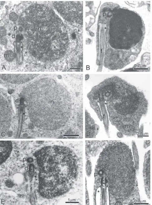
Fig. 2. Spermiogenesis in the subfamily Stevardiinae. Longitudinal sections from early (A,C, E) to late spermatids (B,D, F) of Creagrutus meridionalis (A,B), Bryconamericus exodon (C,D), and Piabina anhembi (E,F). Note from A to F the nuclear rotation toward the flagellar axis. (d): Distal centriole; (p): Proximal centriole; (n): Nucleus; (m): Mitochondria; (f): Flagellum, and (*): Cytoplasmic canal.
Consequently, the forming midpiece is asymmetric. The midpiece contains elongate mitochondria and vesicles. The progressive condensation of nuclear chromatin continues and results in a highly condensed, granular pattern.
Overview of Spermatozoa (Figs. 4-15)
In the spermatozoon of Boehlkea fredcochui, Bryconacidnus ellisi,Bryconamericus exodon,Ceratobranchia obtusirostris,Cyanocharax alburnus, Creagrutus meridionalis, Knodus meridae, Odontostoechus lethostigmus, Piabina anhembi,Piabina argentea, and Rhinobrycon negrensis the nucleus is spherical to ovoid and contains highly condensed granular chromatin and is surrounded by a narrow strip of
no organelles (Fig. 5A). Nuclear rotation toward the flagellar axis was about 80 degrees (Fig. 5A). The midpiece has a conical shape, measures approximately 0.5 m in length and contains some elongate mitochondria. The endomembrane system is composed by small vesicles with a lacy aspect and connected to the plasma membrane (Fig. 5B-E).
Bryconamericus exodon (Fig. 6A-L)
In the spermatozoon of Bryconamericus exodon the ovoid nucleus measures 1.5 m in the longitudinal axis and 1.2 m in the transverse axis. The surrounding strip of cytoplasm has no organelles (Fig. 6A). Nuclear rotation toward the flagellar axis was about 80 degrees (Fig. 6A-B). The midpiece has a conical shape, measures about 1.17 m in length and contains some slightly elongate mitochondria (Fig. 6A-E).The endomembrane system is composed by vesicles with different sizes connected to clump of smaller vesicles with a lacy aspect (Fig. 6F-L).
Ceratobranchia obtusirostris (Fig. 7A-D)
In the spermatozoa of Ceratobranchia obtusirostris the nucleus measures 1.3 m in the longitudinal axis and 1.1 m in the transverse axis. The surrounding strip of cytoplasm has no organelles (Fig. 7A). Nuclear rotation toward the flagellar axis was about 70 degrees (Fig. 7A-B). The midpiece has a conical shape, measures about 0.6 m in length, and contains some slightly elongate mitochondria (Fig. 7A-D). Information on the endomembrane system is not available, since the specimens of C. obtusirostris have been obtained from ichthyological collections and the gonads were not appropriately preserved for ultrastructural analysis.
Creagrutus meridionalis(Fig. 8A-K)
In the spermatozoon of Creagrutus meridionalis, the ovoid nucleus measures 1.5 m in the longitudinal axis and 1.13 m in the transverse axis. The surrounding strip of cytoplasm has no organelles (Fig. 8A-C). Nuclear rotation toward the flagellar axis was about 75 degrees (Fig. 8A). The midpiece has a conical shape, measures about 1.47 m in length, and contains some slightly elongate mitochondria (Fig. 8A-C). The endomembrane system is composed by vesicles with different sizes connected to clump of smaller vesicles with a lacy aspect (Fig. 8D-K).
Cyanocharax alburnus (Fig. 9A-G)
In the spermatozoon of Cyanocharax alburnus, the ovoid nucleus measures 1.7 m in the longitudinal axis and 1.4 m in the transverse axis. The surrounding strip of cytoplasm has no organelles (Fig. 9A-B). Nuclear rotation toward the flagellar axis was about 70 degrees (Fig. 9A-B). The midpiece has a conical shape, measures about 0.9 m in length and contains some slightly elongated mitochondria (Fig. 9C-G). The endomembrane system is composed of interconnected vesicles of different sizes (9A-G).
Hemibrycon surinamensis (Fig. 10A-F)
In the spermatozoon of Hemibrycon surinamensis the ovoid nucleus measures 1.52 m in the longitudinal axis and 1.2 m in Fig. 3. Scheme of spermiogenesis in the subfamily
Stevardiinae. Schematics drawings of longitudinal sections from early to late spermatids. Note from A to D the nuclear rotation toward the flagellar axis, the position of the proximal (p) and distal (d) centrioles, and the dislocation of the mitochondria (m) and cytoplasm to the flagellar portion, forming the midpiece of the spermatozoa. (n): Nucleus; (f): Flagellum; (v): Vesicles, and (*): Cytoplasmic canal.
system, composed of tubules and/or vesicles, is found throughout the major portion of the midpiece with the exception of the region occupied by the mitochondria. The flagellum contains a classic axoneme (9+2) and lacks lateral fins.
Details of sperm morphology
Boehlkea fredcochui (Fig. 4A-K)
In the spermatozoon of Boehlkea fredcochui the ovoid nucleus measures 2.0 m in the longitudinal axis and 1.5 m in the transverse axis. The surrounding strip of cytoplasm has large and small vesicles (Fig. 4A-C). Nuclear rotation toward the flagellar axis is about 60 degrees (Fig. 4A-B). The midpiece has a conical shape and a length of about 1.6 m, and contains some elongate mitochondria (Fig. 4A-G). The endomembrane system is composed of larger vesicles connected to a clump of smaller vesicles with a lacy aspect (Fig. 4H-K).
Bryconacidnus ellisi(Fig. 5A-E)
Spermatic characteristics and sperm evolution on the subfamily Stevardiinae 382
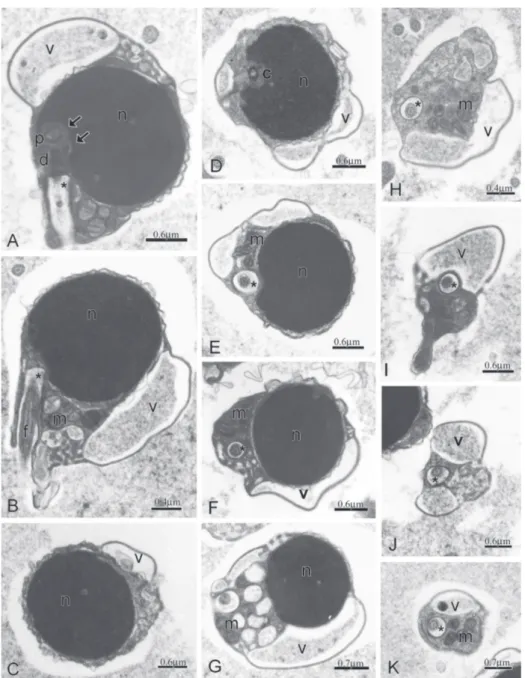
Fig. 4. Spermatozoon of Boehlkea fredcochui.A-B: Longitudinal sections of spermatozoa. Note the position of the nucleus (n) in relation to the flagellar axis and to proximal (p) and distal (d) centrioles, the position of the double nuclear fossa (double arrow), the midpiece and the cytoplasmic canal (*) containing the initial segment of the flagellum (f). The lacy aspect of an endomembrane system and a few vesicles (v) peripherally distributed from the tip of nucleus to the midpiece end. C-K:Cross sections from the middle to the base of the nucleus and at different levels of the strongly asymmetric midpiece exposing the lateral position of the centriole (c), cytoplasmic canal (*) and of the flagellum (f), the distribution of the few elongate mitochondria (m) accumulated in the larger portion of the midpiece.
the transverse axis. The surrounding strip of cytoplasm has no organelles (Fig. 10A). Nuclear rotation toward the flagellar axis was about 95 degrees (Fig. 10A).The midpiece has a conical shape, measures about 0.9 m in length and contains some slightly elongate mitochondria (Fig. 10A). A possible endomembrane system seems to be composed of a clump of small vesicles (Fig. 10B-F).
Knodus meridae (Fig. 11A-E)
In the spermatozoon of Knodus meridae, the ovoid nucleus measures 1.3 m in the longitudinal axis and 1.1 m
Odontostoechus lethostigmus(Fig. 12A-G)
In the spermatozoon of Odontostoechus lethostigmus, the ovoid nucleus measures 1.7 m in the longitudinal axis and 1.1 m in the transverse axis. The surrounding strip of cytoplasm has no organelles (Fig. 12A-B). Nuclear rotation toward the flagellar axis was about 60 degrees (Fig. 12A-B). The midpiece has a conical shape, measures about 0.9 m in length and contains some elongated mitochondria (Fig. 12A-C). The endomembrane system is composed of small vesicles connected to one another and to the plasma membrane, being found peripherally throughout the midpiece (Fig. 12A-F).
Piabina anhembi (Fig. 13A-K)
In the spermatozoon of Piabina anhembi the ovoid nucleus measures 1.94 m in the longitudinal axis and 1.64m in the transverse axis. The surrounding strip of cytoplasm has no organelles (Fig. 13A). Nuclear rotation toward the flagellar axis was about 80 degrees (Fig. 13A). The midpiece has a conical shape, measures about 1.13 m in length and contains some large and slightly elongated mitochondria (Fig. 13A). The endomembrane system is composed of vesicles with different sizes. The smaller are sparse, whereas the larger ones are connected to one another and to the plasma membrane (Fig. 13B-K).
Piabina argentea(Fig. 14A-E)
In the spermatozoon of Piabina argentea the ovoid nucleus measures 1.52 m in the longitudinal axis and 1.2 m in the transverse axis. The surrounding strip of cytoplasm has no organelles (Fig. 14A). Nuclear rotation
toward the flagellar axis was about 85 degrees (Fig. 14A). The midpiece has a conical shape, measures about 0.6 m in length and contains some elongate mitochondria (Fig. 14A,C-E). Information on the endomembrane system is not available, since the specimens of P. argentea have been obtained from ichthyological collections and the gonads were not appropriately preserved for ultrastructural analysis.
Rhinobrycon negrensis (Fig. 15A-E)
In the spermatozoon of Rhinobrycon negrensis, the spherical nucleus measures approximately 1.5 m in diameter. The surrounding strip of cytoplasm has no organelles (Fig. 15A). Nuclear rotation toward the flagellar axis was about 50 degrees (Fig. 15A-B). Large vesicles are found laterally to the nucleus (Fig. 15B-C). The midpiece has a rhomboid shape, measures about 1.1 m in length and contains some mitochondria (Fig. 15A-C). The endomembrane system is composed of a few large vesicles (Fig. 15B-E).
Discussion
Spermiogenesis
Available data on spermiogenesis in the Characiformes (see review in Burns et al., 2009) show that spermiogenesis in this group of fish is primarily Type I or a variation of that type. In Type I spermiogenesis (sensu Mattei, 1970), the centrioles are initially close to the spermatid plasma membrane and have a lateral position relative to the nucleus. They then migrate towards the nucleus as the spermatid flagellum forms.
Spermatic characteristics and sperm evolution on the subfamily Stevardiinae 384
During the migration, the attachment causes the plasma membrane to infold alongside the developing flagellum forming an invagination called the cytoplasmic canal. The developing flagellum is located in the interior of the canal as it develops. Coincidentally, the nucleus rotates 90 degrees in relative to the flagellar axis, changing from a lateral to a medial position relative to the nucleus. In the region of the nucleus that faces the centrioles, a nuclear fossa develops, which totally or partially encompasses the centrioles. The resulting spermatozoa are referred to as Type I spermatozoa. However, among the Characiformes, incertae sedis in Characidae, spermiogenesis of the Type I with a complete nuclear rotation has to date been reported only in Hemigrammus erythrozonus (Pecio et al., 2007). Nuclear rotation, however, may be incomplete. In this case, the flagellum is eccentric to the nucleus, and spermatozoa will be of an intermediate type when compared to Type II spermiogenesis. In Type II spermiogenesis nuclear rotation does not occur and the flagellar axis remains in a lateral position relative to the nucleus (Mattei, 1970). Among characids, this form of spermiogenesis has been reported in Mimagoniates barberi, M.microlepis (Pecio & Rafiñski, 1994, 1999; Burns et al., 1998), Diapoma speculiferum, Diapoma sp., Pseudocorynopoma doriae, Scopaeocharax rhinodus, Tyttocharax tambopatensis, T.cochui(Burnset al., 1998; Pecioet al., 2005), Bryconadenos tanaothoros (Weitzman et al., 2005) and Brittanichthys axelrodi (Javonilloet al., 2007). Despite the reporting of Type II spermiogenesis in these species, often the images provided were not clear enough for a definitive determination of spermiogenesis type, leaving some doubt concerning the true type of spermiogenesis in these species.
Another type of spermiogenesis is Type III (Quagio-Grassiotto & Oliveira, 2008). In that type, at the beginning of
spermiogenesis the centriolar complex is close to the plasma membrane, lies in a medial position relative to the nucleus and remains there throughout the spermiogenesis process and also in the sperm (Quagio-Grassiotto & Oliveira, 2008).
Spermiogenesis, in the species of the Stevardiinae studied herein, differs from that of other characid fishes and cannot be classified as Type I or Type II as described above, but appears to be a variation of Type III. The differences are related to (1) the centriolar complex that lies medial instead of lateral to the nucleus in the earliest spermatids. In these spermatids, (2) the nucleus rotates from a medial position toward the flagellar axis, instead of from lateral toward a medial position. As a consequence, (3) the forming nuclear fossa, the centriolar complex and the flagellum become strongly eccentrically lateral relative to the nucleus. This sequence of events during spermiogenesis is unique among those observed in characiforms and allows the recognition of the resulting spermatozoa as homologous among the following externally fertilized species of Stevardiinae: Boehlkea fredcochui,Bryconacidnus ellisi,Bryconamericus exodon, Ceratobranchia obtusirostris, Cyanocharax alburnus, Creagrutus meridionalis,Knodus meridae,Odontostoechus lethostigmus, Piabina anhembi, Piabina argentea and Rhinobrycon negrensis (current study).
Taking in account the final form of the sperm described in the following inseminating species of the Stevardiinae, Bryconadenos tanaothoros (Weitzman et al., 2005), Chrysobrycon sp. (Burns et al., 2009), Corynopoma riisei (Pecio et al., 2007), Mimagoniates barberi (Pecio & Rafiñski, 1999), Mimagoniates microlepis (Burns et al., 1998), Gephyrocharax atracaudata, Gephyrocharax intermedius (Burns et al., 2009), Pseudocorynopoma doriae (Burns et al., 1998), Tyttocharax cochui, Tyttocharax tambopatensis, Scopaeocharax rhinodus
Spermatic characteristics and sperm evolution on the subfamily Stevardiinae 386
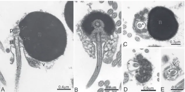
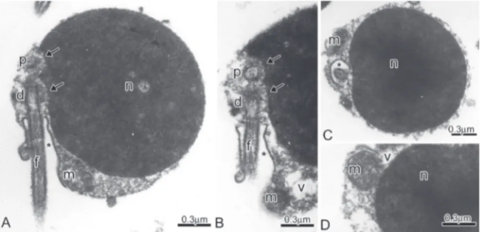
Fig. 8. Spermatozoa of Creagrutus meridionalis.A-C:Longitudinal sections of spermatozoa. Note the lateral position of the nucleus (n) in relation to the flagellar axis and to proximal (p) and distal (d) centrioles, the position of the double nuclear fossa (double arrow), the midpiece and the cytoplasmic canal (*) containing the initial segment of the flagellum (f). D-K: Cross sections through the middle of the nucleus and at different levels of the strongly asymmetric midpiece exposing the lateral position of the cytoplasmic canal (*) and of the flagellum (f), the distribution of the few elongate mitochondria (m) accumulated in larger portion of the midpiece, the lacy aspect of the endomembrane system and a few vesicles peripherally distributed in the midpiece (v).
(Pecio et al., 2005), Xenurobrycon macropus, Xenurobrycon polyancistrus, and Xenurobrycon heterodon (Burns et al., 2008), it allows the suspicion that they share a homologous mode of spermiogenesis with those of the externally fertilized species of Stevardiinae analyzed herein. An analysis of spermiogenesis in these taxa, however, is pending to support or refute this hypothesis.
In the tribe Xenurobryconini, the sperm have the centriolar complex positioned in an anterior position relative on to the nucleus that, in turn, can be more or less elongated toward the flagellar axis depending on the genus (see Burns et al., 2009, for review). This position of the centriolar
complex allows the supposition that during spermiogenesis in these species, nuclear rotation reaches the maximum degree, thereby displacing the nuclear fossa and the centriolar complex to an anterior position from their original medial and posterior original position to the nucleus to an anterior position. The Xenurobryconini are recognized (see Weitzman et al., 2005) as the more derived taxa within Stevardiinae.
Fig. 9. Spermatozoa of Cyanocharax alburnus.A-C: Longitudinal sections of spermatozoa. Note the lateral position of the nucleus (n) in relation to the flagellar axis, the eccentric position of the double nuclear fossa (double arrow), the midpiece and the cytoplasmic canal (*) containing the initial segment of the flagellum (f). D-G: Cross sections through the middle of the nucleus and at different levels of the strongly asymmetric midpiece exposing the lateral position of the centriole (c), cytoplasmic canal (*) and of the flagellum (f), the distribution of the few long mitochondria (m) accumulated in larger portion of the midpiece, a possible endomembrane system and a few vesicles peripherally distributed (v).
Spermatic characteristics and sperm evolution on the subfamily Stevardiinae 388
Elongation towards the flagellar axis is not the only type of nuclear elongation found among the inseminating Characidae. In the Cheirodontinae (Oliveira, 2007) nuclear elongation during spermiogenesis can occur both towards or forward of the flagellar axis, suggesting divergent patterns of evolution of the sperm shape in inseminating species (Azevedo, 2004). A comparative analysis of spermiogenesis demonstrates that sperm ontogeny is a key to the detection of different processes involved in the final form of the spermatozoon, allowing the
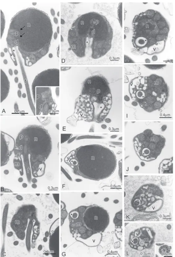
determination of homologies among spermatozoa of different taxa. The correct understanding of potential homologies in final shape of the sperm is crucial to formulating and supporting hypotheses of relationships among studied taxa. Therefore, considering sperm ontogeny, the derived characters shared by the members of the subfamily Stevardiinae sensu Mirande (2009, 2010) during spermiogenesis supports the recognition of that subfamily as monophyletic and different from other incertae sedis characid genera (see Burns et al., 2009 for review). Fig. 11. Spermatozoa of Knodus meridae.A-D: Longitudinal sections of spermatozoa. Note the lateral position of the nucleus (n) in relation to the flagellar axis and to proximal (p) and distal (d) centrioles, the eccentric position of the double nuclear fossa (double arrow), the midpiece and the cytoplasmic canal (*) containing the initial segment of the flagellum (f). E: Elongate mitochondria (m) and vesicles (v) are located in larger portion of the midpiece.
Sperm
A comparative analysis of sperm characters, taking into account nuclear rotation and consequently the position of the centriolar complex and flagellum in relation to the nucleus, the arrangement between the centrioles, the level of nuclear elongation, and the form of the midpiece, brought to light the existence of three distinct morphotypes of sperm within the Stevardiinae.
Spermatic characteristics and sperm evolution on the subfamily Stevardiinae 390
Fig. 14. Spermatozoa of Piabina argentea.A: Longitudinal section of spermatozoon. Note the position of the nucleus (n) in relation to the flagellar axis and to proximal (p) and distal (d) centrioles, the eccentric position of the double nuclear fossa (double arrow), the midpiece and the cytoplasmic canal (*) containing the initial segment of the flagellum (f). B: Detail of the centriolar complex. Note the position of the centrioles to one another. C-E: Cross sections at the middle of the nucleus (n) and at different levels of the strongly asymmetric midpiece exposing the lateral position of the centrioles, cytoplasmic canal (*), the flagellum (f), and the distribution of the few elongate mitochondria (m) accumulated in larger portion of the midpiece.
sleeve, and in most species the mitochondria are elongate. This morphotype is shared by Boehlkea fredcochui, Bryconacidnus ellisi, Bryconamericus exodon, Ceratobranchia obtusirostris, Cyanocharax alburnus, Creagrutus meridionalis,Knodus meridae,Odontostoechus lethostigmus, Piabina anhembi, Piabina argentea, and
Rhinobrycon negrensis. Weitzman et al. (2005) as modified by Menezes & Weitzman (2009) proposed that these genera are situated in a basal polytomy, distinct from all inseminating genera of the tribes Glandulocaudini, Diapomini, Phenacobryconini, Hysteronotini, Stevardiini and Xenurobryconini.
In the second morphotype (M2), the nucleus is strongly elongated towards the flagellum. M2 is characterized by some synapomorphies as the proximal centriole positioned anterior and perpendicular to the distal centriole, and only part of the proximal centriole lies inside the nuclear fossa; the midpiece located at the base of the nucleus, is slightly to moderately elongate. This sperm pattern is shared by all inseminating genera of the tribes Glandulocaudini as described in Mimagoniates barberi (Pecio & Rafiñski, 1999) and Mimagoniates microlepis (Burns et al., 1998); Diapomini as described in Diapoma speculiferum (Burns et al., 1998); Hysteronotini as described in Pseudocorynopoma doriae (Burns et al., 1998); Corynopomini as described in Gephyrocharax atracaudata and Gephyrocharax intermedius(Burnset al., 2009) and Corynopoma riisei (Pecio et al., 2007); and Xenurobryconini as described in Chrysobrycon sp. (Burns et al., 2009), plus Bryconadenos tanaothoros (Weitzman et al., 2005).
The third morphotype (M3) is characterized by some synapomorphies as sperm with a nucleus strongly elongated toward the flagellar axis; the centriolar complex located in an anterior position relative to the nucleus; the proximal centriole oblique at an obtuse angle to the distal centriole; the nuclear fossa and the midpiece absent; and the mitochondria very long and located along the nucleus. This pattern is shared by the members of the tribe Xenurobryconini (sensu Weitzman et al., 2005), Tyttocharax cochui, T. tambopatensis,Scopaeocharax rhinodus (Pecio et al., 2005), Xenurobrycon heterodon,X. macropus, and X.polyancistrus (Burnset al., 2008).
According to Weitzman et al. (2005) external fertilization is a plesiomorphic character. The inseminating mode is likely to have evolved once only within Stevardiinae and may constitute a synapomorphy shared by the most derived species of this subfamily. Spermiogenesis allows us to consider the details of the sperm of all stevardiines as homologous, and considering the M1 morphotype of the externally fertilized stevardiines as the primitive morphotype of the sperm form in this subfamily, it is possible to tentatively recognize M1, M2 and M3 as successive apomorphic spermatozoa in a transformation series, being M1 synapomorphic to Stevardiinae, M2 synapomorphic to all the inseminating species of the Stevardiinae, and M3 as synapomorphic to the Xenurobryconini. M2 constitutes an intermediate morphotype and is present in most of the inseminating taxa of this subfamily, likely having evolved early within the inseminating species. The sperm characteristics found within the species with morphotype M2 deserve further study and comparison, since within this sperm morphotype on finds great morphological variability in nuclear size and elongation.
Acknowledgements
We thank the E. M. Laboratory of IBB-UNESP for allowing the use of their facilities. Study supported by the following Brazilian Agencies: FAPESP (Fundação de Apoio à Pesquisa do Estado de São Paulo); CNPq (Conselho Nacional de
Desenvolvimento Científico e Tecnológico); CAPES/PROEX (Coordenação de Aperfeiçoamento de Pessoal de Nível Superior). We also thank Marco A. Azevedo by the suggestions to the manuscript.
Literature Cited
Azevedo, M. A. 2004. Análise comparada de caracteres reprodutivos em três linhagens de Characidae (Teleostei: Ostariophysi) com inseminação. Unpublished Ph.D. Dissertation. Universidade Federal do Rio Grande do Sul, Porto Alegre, 238p.
Burns, J. R., A. Pecio & S. H. Weitzman. 2008. Sperm and spermatozeugma structure in Xenurobrycon (Teleostei: Characidae: Stevardiinae: Xenurobryconini). Copeia, 2008(3): 656-660.
Burns, J. R., I. Quagio-Grassiotto & B. G. M. Jamieson. 2009. Ultrastructure of spermatozoa: Ostariophysi. Pp. 287-388. In: Jamieson, B. G. M. (Ed.). Reproductive biology and phylogeny of fish (Agnatha and Osteichthyes). Science Publishers, Enfield, NH, USA, 768p.
Burns, J. R. & S. H. Weitzman. 1996. Novel gill-derived gland in the swordtail characin, Corynopoma riisei (Teleostei: Characidae: Glandulocaudinae). Copeia, 1996: 627-633.
Burns, J. R., S. H. Weitzman, H. J. Grier & N. A. Menezes. 1995. Internal fertilization, testis and sperm morphology in glandulocaudine fishes (Teleostei: Characidae: Glandulocaudinae). Journal of Morphology, 224: 131-145. Burns, J. R., S. H. Weitzman, K. R. Lange & L. R. Malabarba.
1998. Sperm ultrastructure in characid fishes (Teleostei, Ostariophysi). Pp. 235-244. In: Malabarba, L. R., R. E. Reis, R. P. Vari, Z. M. S. Lucena & C. A. S. Lucena (Eds.). Phylogeny and Classification of Neotropical Fishes. Porto Alegre, Edipucrs, 603p.
Calcagnotto, D., S. A. Schaeffer & R. DeSalle. 2005. Relationships among characiform fishes inferred from analysis of nuclear and mitochondrial gene sequences. Molecular Phylogenetics and Evolution, 36: 135-153.
Jamieson, B. G. M. 2009. (Ed.). Reproductive biology and phylogeny of fish (Agnatha and Osteichthyes). Science Publishers, Enfield, NH, USA, 768p.
Javonillo, R., J. R. Burns & S. H.Weitzman. 2007. Reproductive morphology of Brittanichthys axelrodi (Teleostei: Characidae), a miniature inseminating fish from South America. Journal of Morphology, 368: 23-32.
Javonillo, R., L. R. Malabarba, S. H. Weitzman & J. R. Burns. 2010. Relationships among major lineages of characid fishes (Teleostei: Ostariophysi: Characiformes), based on molecular sequence data. Molecular Phylogenetics and Evolution, 54: 498-511.
Lima, F. C. T., L. R. Malabarba, P. A. Buckup, J. F. P. Silva, R. P. Vari, A. Harold, R. Benine, O. T. Oyakawa, C. S. Pavanelli, N. A. Menezes, C. A. S. Lucena, R. E. Reis, F. Langeani, L. Casatti, V. A. Bertaco, C. R. Moreira & P. H. F. Lucinda. 2003. Genera
incertae sedis in Characidae. Pp. 106-169. In: Reis, R. E., S. O. Kullander & C. J. Ferraris, Jr. (Eds.). Check List of the Freshwater fishes of South and Central America. Porto Alegre, Edipucrs, 729p.
Spermatic characteristics and sperm evolution on the subfamily Stevardiinae 392
Malabarba, L. R. & S. H. Weitzman. 2003. Description of a new genus with six new species from southern Brazil, Uruguay and Argentina, with a discussion of a putative characid clade (Teleostei: Characiformes: Characidae). Comunicações do Museu de Ciênci-as e Tecnologia da PUCRS. Série Zoologia, 16(1): 67-151. Mattei, X. 1970. Spermiogenése des poisson. Pp.57-72. In: Baccetti,
B. (Ed.). Comparative Spermatology, New York, Academic Press, 573p.
Menezes, N. A., K. M. Ferreira & A. L. Netto-Ferreira. 2009a. A new genus and species of inseminating characid fish from the rio Xingu basin (Characiformes: Characidae). Zootaxa, 2167: 47-58.
Menezes, N. A., A. L. Netto-Ferreira & K. M. Ferreira. 2009b. A new species of Bryconadenos (Characiformes: Characidae) from the rio Curuá, rio Xingu basin, Brazil. Neotropical Ichthyology, 7(2): 147-152.
Menezes, N. A. & S. H. Weitzman. 2009. Systematics of the Neotropical fish subfamily Glandulocaudinae (Teleostei: Characiformes: Characidae). Neotropical Ichthyology, 7(3): 295-370.
Mirande, J. M. 2009. Weighted parsimony phylogeny of the family Characidae (Teleostei: Characiformes). Cladistics, 2: 574-613. Mirande, J. M. 2010. Phylogeny of the family Characidae (Teleostei: Characiformes): from characters to taxonomy. Neotropical Ichthyology, 8(3): 385-568.
Oliveira, C. L. C. 2007. Análise comparada da ultraestrutura dos espermatozóides e morfologia da glândula branquial em espéci-es de Cheirodontinae (Characiformespéci-es: Characidae). Unpublished Ph.D. Dissertation. Universidade Federal do Rio Grande do Sul, Porto Alegre, 136p.
Pecio, A., J. R. Burns & S. H. Weitzman. 2005. Sperm and spermatozeugma ultrastructure in the inseminating species
Tyttocharax cochui, T. tambopatensis and Scopaeocharax rhinodus (Pisces: Teleostei: Characidae: Glandulocaudinae: Xenurobryconini). Journal of Morphology, 263: 216-226.
Pecio, A., J. R. Burns & S. H. Weitzman. 2007. Comparison of spermiogenesis in externally fertilizing Hemigrammus erythrozonus
and the inseminating Corynopoma riisei (Teleostei: Characiformes: Characidae). Neotropical Ichthyology, 1(1): 35-45.
Pecio, A. & J. Rafiñski. 1994. Structure of the Testes, Spermatozoa and Spermatozeugmata of Mimagoniates barberi Regan, 1907 (Teleostei: Characidae), an Internally Fertilizing, Oviparous Fish. Acta Zoologica, 75: 179-185.
Pecio, A. & J. Rafiñski. 1999. Spermiogenesis in Mimagoniates barberi (Teleostei, Ostariophysi, Characidae), an oviparous, internally fertilizing fish. Acta Zoologica, 80: 35-45.
Quagio-Grassiotto, I. & C. Oliveira. 2008. Sperm ultrastructure and a new type of spermiogenesis in two species of Pimelodidae, with a comparative review of sperm ultrastructure in Siluriformes (Teleostei: Ostariophysi). Zoologischer Anzeiger - A Journal of Comparative Zoology, 247: 55-66.
Weitzman, S. H. & L. R. Malabarba. 1998. Perspectives about the phylogeny and classification of the Characidae (Teleostei: Characiformes). Pp. 161-170. In: Malabarba, L. R., R. E. Reis, R. P. Vari, Z. M. S. Lucena & C. A. S. Lucena (Eds.). Phylogeny and Classification of Neotropical Fishes. Porto Alegre, Edipucrs, 603p. Weitzman, S. H. & N. A. Menezes. 1998. Relationships of the tribes and genera of the Glandulocaudinae (Ostariophysi: Characiformes: Characidae), with a description of a new genus, Chrysobrycon. Pp. 171-192. In: Malabarba, L. R., R. E. Reis, R. P. Vari, Z. M. S. Lucena & C. A. S. Lucena (Eds.). Phylogeny and Classification of Neotropical Fishes. Porto Alegre, Edipucrs, 603p.
Weitzman, S. H., N. A. Menezes, H.-G. Evers & J. R. Burns. 2005. Putative relationships among inseminating and externally fertilizing characids, with a description of a new genus and species of brazilian inseminating fish bearing an anal-fin gland in males (Characiformes: Characidae). Neotropical Ichthyology, 3(3): 329-360.