ARGENTINE HEMORRHAGIC FEVER:
CURRENT KNOWLEDGE
by
Norma E. Mettier, M.D.
PAN AMERICAN HEALTH ORGANIZATION Pan American Sanitary Bureau, Regional Office of the
WORLD HEALTH ORGANIZATION
ARGENTINE HEMORRHAGIC FEVER:
CURRENT KNOWLEDGE
by
Norma E. Mettier, M.D.
Scientific Publication No. 183
PAN AMERICAN HEALTH ORGANIZATION Pan American Sanitary Bureau, Regional Office of the
NOTE
One of the commitments of the Pan American Health Organization, carried out through its Department of Research Development and Co-ordination, is to improve communication among biomedical scientists. In view of the scientific and public health importance of Argentine hemorrhagic fever---a disease that has been under study for over ten years by diferent teams of investigators-it seemed that a summary of current knowledge in this field would be of considerable interest to physicians and epidemiologists throughout the Americas. The present monograph, which is completed by an exhaustive bibliography, was pre-pared by Dr. Norma E. Mettler, formerly of the Department of Micro-biology and Parasitology of the School of Medicine, University of Buenos Aires, Argentina, and at present at the Department of Epidemiology and Public Health of the Yale University School of Medicine, New Haven,
CONTENTS
Page
1. Historical Background ... 1
2. General Ecology of Argentina ... ... 5
3. Epidemiological Observations ... 11
4. The Disease ... 15
5. Junín Virus ... 22
6. Immunological Investigation ... 30
7. Isolation and Identification of Junín Virus ... ... 33
8. Special Study of the 1963 Outbreak ... 36
9. Recent Extension of AHF to New Areas ... 42
10. Final Considerations ... 43
Figures and Tables
Page
Figure 1 Province of Buenos Aires, Argentina: Northwestern Parishes in Which AHF Was First Recorded ... 2 Figure 2 Argentina: Major Political Subdivisions; Principal Rivers, Roads, and
Rail-roads ... 6 Figure 3 Argentine Hemorrhagic Fever: Morbidity and Mortality Curves, 1959, 1960,
and 1961 ... 12 Figure 4 Curves Comparing Registered Cases of AHF with Serological Results, 1963 36
Table 1 Clinically Diagnosed Cases of AHF: Geographical and Chronological Dis-tribution, 1963 ... 37 Table 2 Cases of AHF with Serological Conversion for Junín Virus as Against All AHF
Cases Serologically Studied, 1963 ... 38 Table 3 AHF Cases Serologically Negative with Respect to Junín Virus: Cases with
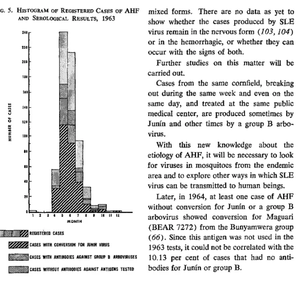
Antibodies for Group B Arboviruses as Against Total Junín-Negative Cases Tested for Group B, 1963 ... 40 Figure 5 Histogram of Registered Cases of AHF and Serological Results, 1963 ... 41 Table 4 Presence of Junín Virus CF Antibodies in Persons Who Had AHF from
1. HlISTORICAL BACKGROUND
The endemo-epidemic disease that is the subject of this essay has been observed for a great many years in Argentina, in the northwestern part of the Province of Buenos Aires. Currently named Argentine hemor-rhagic fever (AHF), after the country in which it was first described, it has been known in the past by a variety of other
names-enfermedad del sello, mal de los rastrojos ("stubble disease"), mal de O'Hig-gins ("O'Higgins' disease"), enfermedad de Junín ("Junín disease"), gripón ("severe flu"), and so on.
It is difficult if not impossible to deter-mine the exact year in which this malady was first observed. According to the data collected by Martínez Pintos (54), epi-demics of variable intensity, reaching their peak in the fall of the year (April and May) and sometimes showing a high rate of mor-tality, have been recorded since 1943. Pre-liminary observations were made at the Julio de Vedia Hospital in Nueve de Julio Parish, Province of Buenos Aires (Fig. 1), where the clinical symptoms were thought to be due to "malignant grippe."
The first mention of the disease in the literature comes from Arribalzaga (5), who reported "a new epidemic disease of un-known etiology: nephrotoxic, leukopenic, and [characterized by] enanthematous hyper-thermia." The first cases were observed in the city of Bragado, Province of Buenos Aires, in 1953 and 1954. The incidence was highest among rural laborers, and the first patients were potato harvesters who had been working in the outskirts of Bragado and Alberti. Later the epidemics spread to the
urban areas. The 1953 epidemic started with sporadic cases early in the year, rapidly came to a peak in April and May, and tapered off slowly, to disappear after Au-gust. The following year morbidity was lower but the characteristics were basically the same. Cases did not break out simul-taneously in the same household or in insti-tutions. Males were more frequently affected than females, and only a few cases in chil-dren, 10 to 12 years old, were observed. At the time of the epidemics, Arribalzaga looked, without success, for epizootic dis-eases in the afflicted area and for changes in weather or sanitary conditions that might ac-count for the outbreak. Attempts to isolate viruses, Leptospira, or bacteria from patients were also unsuccessful. He concluded, on the basis of differential diagnosis, that a virus was probably responsible for the dis-ease. It was postulated that the source of contamination was something in the field, since all the cases but two failed to provide evidence of direct human-to-human trans-mission (5).
In 1956, Domingo Duva published a re-port (34) on 25 patients whom he attended personally from February to September 1953 and from April to August 1954 in Mechita, a small town in Bragado Parish. Mechita, with a population of 2,150, is a typical rural area with unpaved streets and idle land even in the center of town. Of the 25 patients, 5 were youngsters between 12 and 16, and the remaining 20 were
FIG. 1. PROVINCE OF BUENOS AIRES, ARGENTINA: NORTHWESTERN PARISHES IN
WHICH AHF WAS FIRST RECORDED
61° 60°
DE MAYO
¡ 340
35°
61° o60
(REPRODUCED FROM PIROSKY, et al, 19591
ALEM
and an unusual number of rats were found nearby the patients' homes. Remarking on the lack of interhuman contagion, Duva con-sidered the possibility of a leptospiral in-fection.
Though there are no publications or offi-cial records on cases during 1955, 1956, or 1957, physicians in the endemic area claimed that indeed there had been cases every year and that their attempts to report them to public authorities had met with un-successful results. The situation came to a head, however, in 1958. It so happened that the Minister of Public Health, a physician with knowledge of the problem, had land-holdings in the endemic area. The epidemic that year was more severe than ever, and it received wide publicity through newspa-pers, radio, and television. All these cir-cumstances combined to press the public health authorities into action.
At last local physicians had support from the public and the government. Work could start on looking for the etiology of the dis-ease and developing preventive measures. Commissions were organized. One, headed by Dr. Pirosky, was appointed by the Na-tional Ministry of Public Health; another, under Dr. Martínez Pintos, was set up by the Provincial Ministry of Public Health; a third was organized by the Medical School of Buenos Aires University at the invitation of local medical groups in the endemic area. This last team was made up of personnel from the Department of Infectious Diseases, then headed by Dr. Humberto Ruggiero, and the Department of Microbiology and Parasi-tology, under Dr. Daniel Greenway. The present author was engaged in a full-time program of teaching and research at the lat-ter department during this period.
Field trips from the city of Buenos Aires, where the laboratories were located, to the endemic area, especially the Junín Regional Hospital, were conducted to collect mate-rials for the isolation of infectious agents. Blood, spinal fluid, urine, feces, and pieces
of organs from fatal cases were inoculated in experimental animals and in media for growing bacteria and fungi. The search was centered on pathogenic microorganisms, viruses and Leptospira in particular, that would be most likely to be involved because of the epidemiological pattern.
Guinea pigs proved to be the most sus-ceptible of the animals used. They developed a pathological picture similar to the human disease, with exaggerated hemorrhagic man-ifestations.
From three patients the Buenos Aires University team isolated three strains of an agent-in one instance from the blood, in another from the urine (a case with hema-turia), and in the third from organs of a fatal case (81). The agent was named "Junín virus," because the work had been performed with materials collected from the Regional Hospital in the city of Junín. Inde-pendently, Dr. Pirosky's team isolated a virus with which it was possible to reproduce the disease in man (93). Even though the agent isolated by the Buenos Aires Univer-sity group was also capable of producing the disease in humans (108), it was not until 1961 that the viruses isolated by both teams were compared to determine whether they were one single agent or different viruses producing similar clinical manifestations
(69).
Studies directed toward finding Junín virus in nature were started, and successful isola-tions were achieved with wild mice (79) and mites (85). The agent's presence in wild mice and in an arthropod, plus its sensitivity to sodium desoxycholate and the lack of in-terhuman infection, gave reason to place it in the arthropod-borne group of viruses.
Junín was tested against all the other viruses in the collection of The Rockefeller Foundation Virus Laboratories and found to bear an antigenic relationship to a strain of Tacaribe virus isolated from bats in Port of Spain, Trinidad, in 1956 (32). Since
Tacaribe had primacy, a new complex with this name was born. Further isolations of Junín from patients and wild animals in na-ture by different laboratories resulted in the collection of some 100 or more strains.
Shortly after the isolation of Junín virus, an epidemic with clinical symptomatology similar to that of AHF appeared in Bolivia. Serological study showed the presence of antibodies against Junín virus in convales-cents (129). The following year, in a sim-ilar outbreak in San Joaquín Valley, Bolivia, another virus was isolated and named Machupo. This agent showed a relationship to Junín in the complement-fixation test but not in the serum neutralization test. (46).
Still another agent, designated Portillo virus, was isolated at the Children's Hospital in the city of Buenos Aires in 1963 in the course of an etiological study of a children's disease known as uremic-hemolytic syn-drome. This virus, shown to be related to Tacaribe and Junín by complement-fixation
(70), is currently under study at Yale
University to fully determine its relationship to the other members of the Tacaribe group. Shortly after the designation of Portillo virus, the Belém Laboratories in Brazil an-nounced that their team had isolated a virus related to the Tacaribe group from wild rodents. This agent was recognized as a new member and given the name Amaparí (87).
In 1966, Pichinde virus, also related to the group, was isolated from wild rodents in Colombia. This agent is currently under
study at the World Reference Center for Arboviruses at Yale University (122). And finally, not long ago the Communicable Dis-ease Center in Atlanta, Georgia, isolated two strains from wild rodents trapped in Tampa, Florida, and found the virus to be related to Amaparí and Pichinde. It has been named Tamiami (Chamberlain, personal communi-cation).
2. GENERAL ECOLOGY OF ARGENTINA
Geography
Argentina owes its wide range of climate to sharp variations in altitude and a latitudi-nal extension of over 33 degrees, from 21°55'S to 55°3'S. The wedge-shaped con-tinental territory, 3,700 km long and 1,700 km across at its widest point, covers ap-proximately 2,800,000 km2 (Fig. 2).
The Andean mountain chain, which ex-tends the entire length of South America, forms a natural division between Argentina and Chile. In the north, the Andean ranges extend east through approximately one third of the Argentine territory, but farther south the width of the mountainous border dimin-ishes sharply. All the territory east of this chain, comprising by far the greater part of the country, has the character of a plain, rising from sea level at the Atlantic coast to the Andean foothills. The highest elevation in the Argentine Andes is the Aconcagua, a
7,021-meter peak north of the Río Negro. In the Province of Córdoba there are three short parallel ranges belonging to another formation older than the Andes.
In all parts of the republic, January is the warmnest month of the year, and June and July are the coolest.
The main watersheds belong to the La Plata system, which is made up of the Paraná, Uruguay, Pilcomayo, Bermejo, Para-guay, and La Plata rivers. Other important rivers are the Colorado, the Negro, the Chubut, and the Deseado, which form the Patagonian system and flow into the Atlantic. In the interior there is the Andean snow-fed Desaguadero system. And finally, there is
the Dulce river, which starts in the highlands, undergoes various name changes, and finally flows into the Mar Chiquita lagoon in the Province of Córdoba.
The railroad system, which has a total of 44,000 km of track, stems outward from the port of Buenos Aires and connects with lines in neighboring Chile, Bolivia, and Paraguay. The main roads also fan outward from the capital. Neither the railroads nor the highways seem to bear any direct rela-tion to the spread of AHF, though there are numerous fences along these routes, and also between fields, that constitute an im-portant shelter for rodents.
There is aviation service to all the mniajor cities of the interior. Exchanges with inter-national service are made at the heavily trafficked Ezeiza Airport, which is situated a few miles from the city of Buenos Aires.
Administratively, Argentina is divided into a federal district, 22 provinces, and a terri-tory. The Federal District (city of Buenos Aires) is the political, economic, and cul-tural capital of the country. The provinces most affected by AHF are Buenos Aires, Santa Fe, La Pampa, and Córdoba.
per-FIG. 2. ARGENTINA: MAJOR POLITICAL SUBDIVISIONS; PRINCIPAL RIVERS, ROADS, AND RAILROADS
CORRIENT
ARGENTINA
· CITY RIVER ROAD t: ;:;aii :
RAILROAD-... PROVINCE BOUNDARY
sons per square kilometer, but there is a high concentration in Greater Buenos Aires, where some 7,000,000 people have their homes.
Economy
Argentina is essentially an agricultural country. Around 5,220,000 tons of corn are harvested each year from 3,420,000 hectares, mainly in the AHF endemic area. Wheat is cultivated on about 5,250,000 hectares and yields over 5,000,000 tons a year. Nearly half of this produce-2,570,000 tons of corn and 2,500,000 tons of wheat-is ex-ported annually. Flax is abundant, and Ar-gentina is the first producer of this oleaginous plant in the world. Oats, barley, and rye are
also cultivated on a broad scale.
The bulk of the nation's agricultural prod-ucts, with certain exceptions, come from the AHF endemic area. The exceptions are sugar cane, from Tucumán, Salta, and Jujuy; cotton, from Chaco, Formosa, Corrientes, and Santiago del Estero; grapes, from Men-doza, San Juan, and the Río Negro Valley; and citrus fruits, apples, and pears from Río Negro, Entre Ríos, Corrientes, Misiones, Salta, Tucumán, and Jujuy.
Forests cover more than 100,000 hec-tares. Quebracho extract and timber are the most important forest products, and they come mainly from the Province of Misiones. Argentina's fishing industry is greatly un-derexploited. Though the country has a "marine pampa" extending along the At-lantic coast from the La Plata river to the Beagle canal and covering an area of 1,000,000 km2, there are only a few fishing centers-among the most important, Mar del Plata, Necochea, and Rawson (Chubut). The country also has vast mineral wealth potential, but so far the only important ef-forts at exploitation have been limited to petroleum and iron, mainly in Patagonia and in the northwestern provinces-Salta, Jujuy, and Mendoza.
Meat, grapes, wine, sugar, oil, yerba mate (Paraguayan tea), and textile manufactures are old and characteristic Argentine in-dustries. Heavy and light industries, such as the manufacture of cars, refrigerators, tele-vision sets, and petrochemical products-plastics, synthetic detergents, india rubber, insecticides, synthetic fibers, and the like-have been developed since World War II.
Cattle raising, which already thrived dur-ing the Spanish colonial period, is the oldest and most important rural activity. Argentina is the world's number one exporter of meat. Of approximately 43,500,000 head of cattle, most are concentrated in the provinces of Buenos Aires, Santa Fe, Corrientes, and Entre Ríos. Sheep raising is extensive mainly in the southern provinces. In all, the country has about 50,000,000 sheep, and its wool exports place it internationally in third place, after Australia and New Zealand. There are about 7,300,000 horses, which are raised mainly in the provinces of Buenos Aires,
Santa Fe, and La Pampa.
Endemic Area
General description
The advent of European civilization in Argentina produced a marked change in the flora of this region. Useful trees and plants from every part of the world-cereals, alfalfa, grasses, and all varieties of fruit-were introduced. The Australian eucalyptus, in particular, thrives in the Pampa. Other varieties that have been planted extensively include the acacia, the sycamore, the para-dise tree, and several types of evergreens. The trees are especially seen close to homes in the rural areas.
In winter the land is plowed in prepara-tion for spring planting. In the spring there are fields of flax and wheat, and in the sum-mer, corn. Alfalfa and pasture are seen at all times of the year. The fields are fre-quently from 50 to 80 hectares in size. Dur-ing summers with humidity and abundant rain-for example, the summer of 1964 grassland vegetation is luxuriant. This con-dition is accompanied by an increase in the wild rodent population, because the vegeta-tion provides abundant food as well as pro-tection against predators such as owls and hawks. If the rodent population gets too high, epizootics and intra- and interspecific fighting-similar to the 1958 Microtus crash in Oregon-develop among these animals by the end of the winter (30).
Most of the towns in the endemic area are small. Generally the streets are unpaved and there is no running water or sewage disposal system. Distances between large cities with proper sanitary facilities are usually quite great.
The rural dwellings, scattered over the large areas between the towns, are typically made of adobe, rarely of concrete. The floors and patios are made of brick or plain earth; roofs are of wood and zinc; and the insulation usually consists of adobe, straw, or sometimes wattle and daub. A home generally has one or two bedrooms and an adjoining room that serves as kitchen and dining room. Tin sheds, chicken coops, and
privy installed over a cesspool only a few feet from the house completes this unit. Drinking water is obtained from a pump, well, spring, or cistern. The boundaries be-tween these dwellings and the open fields are wire fences, reed grass, or shrubs and do not form a barrier for wild rodents.
The corn and sunflower harvest coincides with the autumnal peak of the AHF epi-demic cycles (April and June). The manual harvesting of the crops employs a great number of laborers from the town or from other provinces, and usually an entire fam-ily, including women and children, par-ticipates. During this time the laborers, most often coming from other places, build huts of maize stems and straw on the ground over the field where the crop is being harvested. Thus, both their working and leisure time is spent in intimate proximity with the wild rodents.
Fauna: wild and domestic
The Pampa has a great variety of bird life, including hawks, falcons, owls, herons, storks, swans, partridge, plovers, ducks, chajas, and many other species known by native names. Frogs, toads, and ants are ubiquitous.
The search for the etiological agent of AHF in the animal reservoir was concen-trated mainly on wild rodents because of their high population density in the corn fields.
Field rats and mice (suborder Myomorpha, family Cricetidae) are present not only in the cornfields but also in the untilled lands surrounding the towns in the endemic area. There are many South American species in the Myomorpha suborder, among them
Akodon arenicola hunteri, Akodon areni-cola beatus, Hesperomys murillus murillus,
Hesperomys laucha, and Oryzomys flavescens
(54). Akodon arenicola hunteri stays away
in captivity or in a wild environment. ,Hesperomys laucha seems to be present only in the eastern Pampa in the Province .of Buenos Aires (A. Cabrera and J. Yepes). Hesperomys bimaculatus bonaerensis is con-sidered a geographic form of H. bimaculatus bimaculatus (A. Cabrera et al.). It is found almost exclusively in the northeastern part of the Province of Buenos Aires. Hesperomys murillus murillus is a species of the Argen-tine Pampa that inhabits the Province of Buenos Aires and eastern Córdoba. In the central provinces and toward the southwest, another subspecies, H. murillus cordobensis, is recognized (A. Cabrera and J. Yepes). Oryzomys flavences is present in a large area of Argentina and the southern part of Uruguay, except at high altitudes.
In addition to Cricetidae and native South American mice, there are also rats and mice imported from Europe. These latter rodents reproduce in such numbers in the endemic area that they are considered real pests.
The Rattus genus is represented by two species, the black rat (Rattus rattus rattus) and the Norwegian rat (Rattus norvegicus). These species are enemies and never live together. They migrate en masse during the summertime and are not even stopped by rivers.
With respect to naturalized exotic species of Muridae, Mus musculus musculus has been reported to be a plague in the Province of Buenos Aires (R. Ringuelet and R. H. Aramburu, 1957). It lives close to man, invading human habitations and nesting in the furniture or in quiet places of the house. It eats food found in people's homes-fruit, cheese, candy, and the like, and it also chews on materials such as wood, paper, and clothing to make a powder for its nest. It is also found away from houses in big nests in the stubble. Two other species of the genus Mus were described by José Yepes in 1935: M. musculus Linn. and M. mus-culus brevirostis Waterh. The first is found
throughout the entire country, and the sec-ond in the eastern part, close to La Plata river.
Other rodents abundant in the area in-clude those of the Caviidae family, Cabiinae subfamily. Cavia pamparum is a plague in the Pampa. It is similar to the common guinea pig, but lives in the wild state. Other members of this family are Microcavia australis and Galea musteloides littoralis; both shun man.
As a consequence of the discovery of sylvatic plague in Argentina by Dr. de la Barrera, many studies dealing with the dis-tribution of wild animals in the country were conducted. These studies are very useful, especially in showing the distribution of the rodents from which Junín virus has been isolated (119).
Other wild animals very common in the endemic area are Ctenomydae porteonsi porteonsi and the skunk, Conepatus sulfo-cans gibsoni, usually seen along the road-sides. From the order Marsupialia there are opossums, of which the Didelphis para-guayensis parapara-guayensis and the red
opos-sum, or Lutreolina crassicaudata bonari, are real pests. Among the bats, Myotis albes-cens, Histiotus montanus, Lasiurus borealis blosseoillii, Lasiurus cinereus villosissius, and Tadarida brasiliensis have been recog-nized in Argentina. The hare, order Lagomorfos, family Leporidae, Lepus euro-paeus euroeuro-paeus is extremely abundant, and so is the partridge. Both species are avidly hunted as food delicacies. In the process, there is ample opportunity for the hunters to come in contact with reservoirs of AHF.
(Pseudalopex gymnocercus), and the minor ferret (Galictis cuja huronax). The "cat of the straw" (Lynchailurus pajeros pajeros)
and the Oncijelis geoftroyi have almost dis-appeared (54).
Dogs and cats, the good friends of man, are always present in the home.
As stated before, cattle, horses, sheep, and pigs exist in large numbers and consti-tute one of the area's major sources of wealth. The poultry usually found close to
homes are chickens, hens, ducks, turkeys, and geese.
As a rule, both wild and domestic animals have a great number of ectoparasites-mites, fleas, and the like. In the Province of Buenos Aires at least 6 families, 8 subfam-ilies, 12 genera, and 19 species of fleas have been identified (20).
3. EPIDEMIOLOGICAL OBSERVATIONS
The high incidence of AHF among corn harvesters is so well recognized that the affliction is called "stubble disease." Though the present trend in the area is toward mechanized picking, there are times when it is impossible to harvest corn with machines. This is the case, for example, when the plants have fallen over because of strong winds.
Although AHF bears an apparent re-lationship to the rural environment, fre-quently there are patients who have not left the towns or cities in the epidemic area. These urban areas, however, are not free of rodents.
It is noteworthy that the annual period of the epidemics coincides with the rainy sea-son and that, in a given outbreak, weekly and monthly fluctuations in the number of patients interned at the public medical centers appear to be related to climatic fac-tors, especially temperature and rainfall (54). The morbidity data since 1958 show con-siderable fluctuation in the annual incidence of the disease:
Year Number Mortality
of cases (% of cases)
1958 283 19
1959 1,027 6.31
1960 335 6.27
1961 816 7.84
1962 362 ?
1963 653 4.42
1964 3,026 ?
1965 148 ?
1966 643 ?
1967 until Sept. 6 1,059 8.69
Mortality, however, has shown a tendency to decline, thanks to improved diagnostic and therapeutic measures that permit early recognition of the disease and timely treat-ment of its severe and typical forms. De-layed diagnosis of severe cases accounts for the high mortality reported during the first large epidemic in 1958. Also, in these earlier years, cases of medium and mild severity may have escaped detection. Thus, in more recent years, as the total number of reported cases increased, the proportion of deaths became lower.
In general, the first cases occur late in March, the epidemic reaches its peak in May and June, and after that the cases de-crease steadily during July and remain very low in August and September, when the morbidity curve drops to nearly zero (Fig. 3). It should be kept in mind that this dis-tribution corresponds to the fall and winter months in the Southern Hemisphere. Only sporadic cases have been observed during the rest of the year.
pos-FIG. 3. ARGENTINE HEMORRHAGIC FEVER: MORBIDITY
1959, 1960, AND 1961
AND MORTALITY CURVES,
1961
MORBIDITY 816 ... MORTALITY 64
1960
MORBIDITY 335... MORTALITY 21
1959 MOR
... MOR
RBIDITY 1027
RTALITY 65
.
DAYS 1 10 |O 20 1 |0lO 20 31 2 20 2 0 | 20 | 31 10 | 20 1 30 lo1 20 1 1 1 20 | 31 lO 20 2
M.IONTHS JANUARY FEBRUARY 1 MARCH APRIL1 MAY JUNE JULY AUGUST SEPTEMUER OCTOSER NOVEIBER
Z ,I E 2 | 6 8I 1 | 2 4 4 6 82 10 125 10 68 40 52 64 42 39 23 1 13 II 1
TOTAL D.ATHS - I ' I " I 10_ lo I 22 1" 5 2 , _
(Reproduced from Martínez Pintos, 1962)
U,4
U
ug
u
AM
z
M,I
U
M.
o
eg rU
z
=E
Zj
40
20
120
#A
VW vi u U-LUJ
Z
z
10080 60
40 20
sible role of food or drinking water in the transmission of AHF. When the disease appears in an area, it becomes an endemic focus for several years thereafter, suggesting that the source of infection is something that remains in the environment once it is estab-lished.
The endemic nature of AHF in certain localities in the Province of Buenos Aires is well explained by the presence of rodents in-fected with the virus (79, 84, 112). The same is true for the Province of Córdoba
(26, 124).
There is a definite parallel between the density of rodents and the incidence of AHF. Seasonally, the rodent population is highest in autumn and winter, which is when the AHF epidemics are at their peak, and lowest in summer, when only sporadic cases are observed (30). Also, the lands directly associated with AHF infection are those de-voted to field crops, where rodents are most populous, rather than cattle-raising lands. The great majority of cases occur among people living in primitive conditions out in the open, where contact with rodents is facilitated. Disease-afflicted homes are fre-quently located on the border of uninhabited grasslands or open fields. The few cases observed among people living in cities may be accounted for by brief trips to the countryside.
Repeated isolations of Junín virus from wild rodents (26, 79, 84, 91, 112), as well as several isolations from their ectoparasites
(4, 85, 89), point convincingly to a
connec-tion between the appearance of AHF in humans and the presence of rodents in-fected with a virus from the zoonoses group, the disease probably being trans-mitted through the animals' excreta or their blood-sucking ectoparasites.
In 1959, 449 wild rodents were trapped in the endemic area. Of these, 52.8 per cent were Mus musculus; 24.4 per cent,
Hesperomys laucha (Dumarest); 11.1 per
cent, Akodon arenicola; 6.2 per cent,
Oryzomys fiavences (Waterhouse); and 5.3
per cent, Rattus sp. Ten strains of Junín virus were isolated from the Mus musculus,
one from the Hesperomys laucha, and one from the Akodon arenicola (79). In the Province of Córdoba, 46 strains were iso-lated from Calomys laucha alone, and addi-tional strains were taken from Akodon azarae, Akodon obscurus, Calomys
muscu-linus, and Oryzomys flavences (26). Other
workers have isolated Junín virus from wild guinea pigs, or Cavia pamparum (cuis), and from hares (Lepus europaeus) as well
(4, 22). There may be an even wider
distribution in nature, but not many attempts to isolate virus from other sources have been made to date.
With mouse ectoparasites, it was demon-strated by Pirosky and co-workers in 1959 that mites (Mesostigmata without further identification) were able to transmit the Junín virus infection experimentally (89).
Further investigations in mites yielded Junín virus from Echinolaelaps echidninus
(Berlese) taken from nests of naturally in-fected Mus musculus (85). Another suc-cessful isolation from mites was made in 1965, this time from Eubrachilaelaps
rotundus taken from Akodon azarae (4).
from an environment contaminated with the urine of infected rodents, without any inter-vention of arthropods, the lack of infection in summertime could be explained by the natural sterilization of the field by ultraviolet radiation from the sun.
Although there are few well-documented instances of human-to-human transmission, there is a theoretical possibility that with in-creased adaptation to man and the develop-ment of persistent chronic infection the virus may propagate by human contact in the absence of an arthropod vector.
4. THE DISEASE
Clinical Description
AHF is an acute infectious disease of 7
to 15 days' duration followed by a long
convalescent period of approximately one
month; with few exceptions, there are no
sequelae.
For the most part, the persons who
con-tract the disease are newcomers in the
endemic area who have contact with the
field. The patients are usually males-corn
harvesters or people involved in other rural
activities. The disease is unkown or
un-recognized in infants, and only few cases in
children have been recorded. Morbidity is
higher in males than in females, the ratio
being approximately 5 to 1.
The clinical symptoms are not always
uniform, but the typical case is diagnosed
readily; the local physicians were so
im-pressed by the signs and symptoms that it
was named for its hallmark
(enfermedad del
sello).
In a fully developed, "classical" set
of symptoms and laboratory findings, the
complex includes involvement of the renal,
cardiovascular, hematic, and nervous
sys-tems.
This essay synthesizes the observations on
the clinical characteristics of the disease
made by different teams of investigators
from 1955 up to the present
(3, 5, 35, 40,
57, 62, 74, 90, 91, 105, 106, 109, 110,
113, 114),
plus the personal observations of
the author as part of a team and/or as an
individual physician.
In general, the disease is considered to
have mild, moderate, and severe forms,
though sometimes the characteristics
over-lap. After an asymptomatic incubation period of 7 to 16 days, three stages can be differentiated: the initial period, the acute phase, and convalescence. In 3 to 30 per cent of the patients, depending on the severity of the epidemic, death occurs be-fore the convalescent period. Although these three states are not found in all cases of AHF, the phasic course of the disease is beyond doubt, and the suggested division in distinct periods should lead to a better understanding of the essence of the path-ological changes and their sequential appear-ance.
In the first stage-the initial or invasion period-the usual onset is vague with grippe-like symptoms, such as malaise, fatigue, chills, and lumbar backache. In around 5 per cent of the cases, however, the first symptoms appear suddenly with severe prostration and lumbar and limb pains. Fever and accentuated asthenia are always present regardless of the type of onset
(113). The bulbar and palpebral
conjunc-tivas are injected without any secretion or tearing. The face becomes edematous and flushed, as in measles. Headache, postorbital pain on moving the eyeballs, and sub-palpebral edema are almost constant. Stiff-ness is present in the neck and the muscles of the costovertebral canals. The patient sometimes complains of feeling as if he were strapped down to a pale.
The patient is dull, drowsy, confused, and often apathetic. He does not respond im-mediately to questions. Tremor of the tongue during its protrusion is accentuated,
patient's gait is unsteady, and active move-ment is slow. Frequently there is tremor of the upper extremities when the arms are extended forward at shoulder level, as in hyperthyroidism. Another conmmon symptom is noninflammatory painless generalized iymphadenopathy.
Enanthemas are present on the buccal mucosa. At the juncture of the soft and hard palate one can usually see a number of small hemorrhages, petechiae, and/or some twenty to thirty microvesicles of clear content, without color change in the surrounding area. They are not painful; they are present from four to six days, increasing in number during the acute phase (114).
Halitosis, anorexia, thirst, nausea, vomit-ing, and epigastralgia are sometimes present. Constipation is frequent, and only in a few patients does one observe a brief period of diarrhea. The feces in these cases are fluid-homogeneous, without mucus, but fre-quently with an admixture of fresh blood and sometimes melena. The diarrhea lasts two or three days and usually ceases with-out any therapeutic measures. Meteorism and cecal gurgling have been noted. In women, there may be uterine hemorrhages. Bradycardia with arterial hypotension is present in 90 per cent of the cases (113),
and dizziness is observed in a large propor-tion as well. As a rule, the spleen and liver are not enlarged; palpation in the kidney area, however, is very painful.
The initial or invasion period lasts two to four days, and passage to the fully devel-oped, or acute, stage is accompanied by an accentuation of the same symptomatology.
The fully developed disease usually presents hemorrhagic injury characterized by bleeding of the gums, epistaxis, and in some few severe cases, hematemesis, melena, and enterorrhagia. A rash first appears on the trunk, primarily in the axillae and on the chest, and spreads to the extremities. It
fluent, so that large areas of the skin assume a purpuric appearance.
The fever is of a remittent type; the pulse is slow compared with the temperature rise; and arterial hypotension, usually moderate, is observed in all patients. If the blood pres-sure drops precipitousiy, this is a sign of grave prognostic significance, particularly if associated with oliguria. Marked decrease in diuresis, sometimes resulting in complete anuria in the second period, as often ob-served in 1958, may be found (109). In these cases, the oliguria or anuria is fol-lowed by prolonged polyuria; in others, the development of acute renal failure leads to uremic coma, followed by death. Signs in-dicating progressive central nervous system involvement, ranging from disorientation and delirium through convulsions to stupor and finally coma, as terminal manifestations, are frequently seen in severe cases. A char-acteristic odor, different from that of any other ward in the same hospital, is present wherever AHF patients are confined.
Although the patient is extremely thirsty, he refuses to drink, in order to avoid spasms of the laryngeal and pharyngeal muscles, as
observed in rabies.
When the disease is at its height, the patient's temperature may reach 39° to 40°C. In this stage, cutaneous hyper-esthesia and contraction of the muscles under the skin are observed upon stimula-tion. Photophobia, strabismus, and nystag-mus may also be seen.
In some patients, gastrointestinal dis-orders are conspicuous. The tongue is dry, and coated with a thick brown layer. Halitosis, with an odor of decay, especially in cases with bleeding of the gums, oral mucosa, and nasopharynx, is usually present.
Changes in the respiratory organs are usually slight and infrequent; the most com-mon finding is a dry cough.
lasts two to four weeks, or even longer in severe cases. During the recovery period, the fever diminishes by lysis. Hyperemia of the face and mucous membranes disappears and the hemorrhages on the skin are reab-sorbed. The blood pressure and pulse rate become normal, and only changes detectable in the clinical laboratory may remain. Physical examination may only reveal a gen-eral weakness and a tendency to fatigue, which can persist for some weeks.
During this period some patients have a special gait similar to that presented by dengue. Usually in convalescence there is a partial and gradual loss of hair, followed eventually by the growth of new hair to sup-plant what was lost. This is especially noted among women. Even if all the hair is changed, there is never a time of complete alopecia.
The recovery is usually without sequelae, though there have been a few cases in which parkinsonism was present for one or two years and others in which albuminuria per-sisted for a long time. Uncomplicated cases are seldom fatal; the prognosis is negative only in cases with serious nervous disturb-ances or severe hemorrhagic manifestations or both-especially when there are hema-temesis, melena, and enterorrhagias-and also in persons with alcoholic habits, and in the aged. During the recovery period latent chronic infections such as amebiasis may exacerbate.
In 1.2 to 1.8 per cent of the cases, re-lapses were observed 15 days or a month after the original onset, with repetition of the previous symniptomatology (106).
Diagnosis
Differential diagnosis
AHF must be differentiated from other acute infectious diseases that share some of the same symptomatology, such as influenza,
rickettsiosis, leptospirosis, toxoplasmosis, and typhus.
Laboratory investigation is necessary in order to make a differential diagnosis. Sometimes it is impossible to attempt to differentiate this disease from others on clinical grounds alone. Etiological investi-gation through serology and isolation of the infectious agent will confirm or reject any
of the diagnoses mentioned above.
Clinical laboratory diagnosis
The routine laboratory procedures, such as complete blood count and urinalysis, are of great value in diagnosis. Generally, a clinical picture such as that described in the previous section, associated with thrombocy-topenia, leukopenia, and special cells in the urine sediment, will be labeled as AHF until such time as a contrary diagnosis can be demonstrated.
Blood
Regardless of the severity of the disease, the laboratory findings will generally reveal a normal or lowered erythrocyte sedimenta-tion rate during the first week (1 to 7 mm in the first hour, using Westergren and Wintrobe). This value rises during the sec-ond and third week, particularly if bacterial complications develop. In mild cases it re-mains normal. The hematocrit shows nor-mal value rises, except in cases of severe dehydration, where the increase in cells is proportionate to the degree of hemoconcen-tration.
The number of red cells is usually normal at all stages of the disease, except when the hemorrhagic manifestations are exaggerated and there is consequent anemia in propor-tion to the blood loss.
mm3 have been observed. The leukopenia is accompanied by aneosinophilia and neutropenia (5, 91, 109, 126).
The neutrophils suffer severe alterations, with marked anisocytosis and toxic granu-lations. There are a great number of neutrophils with crooked-shaped nuciei. The nuclei of the neutrophilic granulocytes are marginated.
There is nuclear chromodiffusion, some-times accompanied by pycnotic condensa-tions of the chromatin, with partial break-age of the nuclear membrane, which give the appearance of flaked nuclei.
At the peak of infection, atypical mono-nuclears, some of them showing vacuola-tion of nuclei and cytoplasm similar to hystiocytes, are characteristic findings (126). Recovery starts at the same time that the oliguria disappears, the number of white blood cells begins to increase either slowly or suddenly, and leukocytosis appears. The corresponding normal white cell differen-tial count signifies the reappearance of eosinophils and monocytes with normal nuclei and the disappearance of any toxic granulations in neutrophils and plasmatic cells that may have been present. Any earlier increase in white cells than what would be expected from the normal course of disease suggests a focus of bacterial infec-tion somewhere in the organism.
The number of platelets decreases somrne-what during the initial stage, and even more sharply at the height of the disease. Values as low as 6,000 per mm3 have been en-countered. The average in most cases is be-tween 30,000 and 40,000 per mm3. Giant and morphologically abnormal platelets may be seen on smears. During convalescence the number returns to normal.
The coagulation time is longer than nor-mal. In severe cases, periods of as much as an hour, with the blood kept in a tube at 37°C, have been observed. Clot retraction is incomplete (110, 127). Using more
boelastographic method, it was demon-strated that 65 per cent of the patients present hypocoagulability during the acute stage, while in convalescence the tracings are normal in 77 per cent of the cases, and sometimes even a reactional hypercoagula-bility is present. Oniy in a very few cases does the hypocoagulability persist in con-valescence. The hypocoagulability is trans-lated by the lengthening of "r" and "k" and the narrowing of "am" (100). Fibrinogen is found to be lowered in cases of marked hypocoagulability, and occasionally fibrin-olysis is observed.
Bleeding time is increased. The plasma prothrombin time and concentration are normal.
The ionic calcium is normal or lowered during the initial and acute periods, and always normal during convalescence.
The total proteins in blood plasma are in the lower normal range. The zonal elec-trophoresis of the proteins generally shows hypoalbuminemia with a moderate increase in alpha 1 and 2 globulins, a normobeta globulin level, and a normogamma globulin level. During convalescence the hypo-albuminemia still remains, but the gamma globulin increases, and thus the inversion of the proteic rate does so too (37, 107).
The total lipemia and cholesterolemia may reach very low values; cholesterol ranges of from 58 to 138 mg/100 ml have been observed. The direct bilirubin is negative and the indirect is in the normal range, only having shown to be increased in a few fatal cases. Liver function tests, thymol turbidity, and Hanger's test also give normal results.
The electrolytes are without severe alter-ations except in grave cases where hy-ponatremia and hypochloremia, accom-panied by a slight decrease in the CO2
normal in days or weeks. In some patients, values as high as 200 mg/100 ml late in the acute period, followed by a sudden normal-ization of the sugar level in about two days, have been reported.
The urea is normal or increased; cases of 40 mg/100 ml, with oliguria or anuria present, have been recorded. When the anuria or oliguria last, the values drop sharply to normal in one or two days.
The white cell count, the differential white cell count, the number of platelets, and bleeding and coagulation times normal-ize their values independently of the evolu-tion of the disease, whether toward cure or death.
In bone marrow there are macrophages with leukocytes phagocyted-a fact that may account for the central destruction of white cells reported by Vucetich (126).
During convalescence the red cells of the bone marrow reveal hypofunction.
CSF spinal fluid findings have been normal even in the presence of spinal muscular contraction.
Urine
Albuminuria begins to show in the initial period and increases to 0.10 to 0.50 g/1000 ml during the acute stage. Those patients whose initial albuminuria disappears have a second rise to 3 or 4 g/1000 ml last-ing one or two days, with normalization im-mediately thereafter.
Cylindruria is present early in the disease, sometimes preceding albuminuria. The amount of cylinders is unrelated to the de-gree of albuminuria. In general, the cylinders are hyaline, hyaline-granulose, and epithe-lial; some are erythrocytic.
Vacuolated epithelial cells are a character-istic finding in the urine and are used as a criterion for diagnosis. First described by Milani, these cells were studied in detail by Palatnik (75, 76). They are 18 to 45 u in size-about twice the size of a lymphocyte. They have hyaline, intensely eosinophilic
inclusions, which stand out sharply in the cytoplasm. The inclusions-round, half-moon, or ring shaped-are 10 to 26 l in size. In the later stages of the disease large inclusion bodies, along with granular inclu-sion forms, are observed in the cytoplasm. The inclusion maintains a stable configura-tion during the initial and acute stages. At the beginning of convalescence the forms decrease in size and number. Soon there-after, they disappear, and they are not pres-ent in the late stage of convalescence, after the disease is over, or in the case of relapse. The inclusions are Fuelgen negative, but they have Fuelgen positive corpuscles; with PAS they present a typical and stable morphology. Palatnik suggests that the granules represent viral material and that the inclusions are related to the intracellular cycle of the virus.
Pyocytes and erythrocytes are often seen throughout the entire course of the disease, and for this reason a daily check of the urine is advisable. Macrohematuria was observed in only a few cases. The 17-ketosteroids are lowered, especially in severe cases.
ECG
Modifications of the ECG were found in 71 per cent of the patients. The most char-acteristic alterations consisted of an elevated ST interval, with upper concavity, followed by an exaggerated T wave. In 20 per cent of the patients a flat or inverted T wave was found. Sometimes the P wave had low voltage and the PR interval was extended. The QT interval was found to extend mod-erately in 14 per cent of the patients. The U wave was found in 9 per cent of the cases
(101).
cardiogram in nine cases and probable mnyocardial injury in two of the cases (86).
Pathology
Pauioogical cuhanges at various stages of the disease have been described by Rivero
et al. (97) and Polak and Jufe (94). The
lesions or changes were not specific for AHF and had features similar to those of other diseases and infections accompanied by hemorrhagic diatheses, particularly other hemorrhagic fevers (48, 118).
Postmortem examination reveals alter-ations corresponding to the lesions produced by the virus itself or to intercurrent infec-tions associated with the cause of death. The alterations produced by the virus itself correspond to perturbations of the blood capillary system, reactivation of the reticu-loendothelial system, and areas of tissue degeneration, as described below.
Gross lesions
The gross anatomy shows general conges-tion and various degrees of hemorrhages on the skin and in some of the organs, espe-cially the lung and spleen. Generalized lymphadenopathy appears in almost every case. Infiltration resembling that of bron-chopneumonia, necrotic areas in the parenchyma of the lung, and even pleural effusion have been seen. Renal damage, as in nephrosis with acute glomerulitis, is usually observed.
Microscopic lesions
The dominant changes are vascular; vasodilation, with congestion, edema, and hemorrhages, is the main presenting symp-tom. The chief abnormality occurs in and about the small blood vessels and consists of endothelial swelling, perivascular edema, and diapedetic hemorrhages. There is a cellular reaction characterized by
hyper-the lymph nodes, tonsils, spleen, and bone marrow; in the bone marrow there are no mature neutrophil or eosinophil leukocytes.
Dystrophic changes are present in the parenchyma of the organs and in the blood vessels. In the liver, hepatic cells show granular degeneration of the cytopiasm. Tne central vein is dilated, and the sinusoids con-tain a deposit of bile pigment. Edema is observed in the connective tissue surround-ing the dilated central vein, and lymphocytic infiltration is seen in some of the Kiernan's spaces.
The kidney alterations are mainly in the proximal tubules, where the epithelial cells show alteration of the nucleus, granular cytoplasm, and an indefinite border. The lumina are obstructed by hyaline and cel-lular casts. Acute glomerulitis is present, with general congestion, microscopic hem-orrhages, and edema, as in the other organs. The microdissection of the tubules done by Aoki et al. (2) showed renal alterations similar to those observed with Korean hemorrhagic fever.
In the spleen, hyaline degeneration of the corpuscular arteries, with focal modifications in the sinuses, is observed.
In the lungs, inspecific bronchopneumonic foci and interalveolar hemorrhages are present.
There is congestion with marked menin-geal edema in the brain. Rovere et al.
claimed the presence of Lafora's amyloid bodies in the cerebral tissue (98, 99).
When intercurrent infections are the cause of death, the pathology corresponds to alterations caused by the virus itself at the stage of the disease when death occurred, plus the changes corresponding to the inter-current infections.
Treatment
no specific treatment except for serum transfusions (vide infra). It is imperative to put the patient in bed as soon as possible. The most severe cases appear in persons who continue to work in spite of general malaise and muscular pain. Early rest helps to prevent the disease from becoming severe.
Even though the dry tongue and skin may justify the ample use of fluids in treatment, it is necessary to bear in mind the renal in-volvement and observe caution in admin-istering liquids. Only in severe cases where vomiting and absolute anorexia are present are parenteral fluids advisable, and then acid-base determinations should be used for control. Otherwise, parenteral injections must be avoided. They can provide means of entry for secondary bacterial infections, and, in addition, because of the hemorrhagic diathesis present in these patients, they can cause hematic collections at the place where the needle is introduced.
The administration of convalescent serum from other patients, though not necessary with mild attacks, is advisable in severe cases. Treatment with convalescent serum is routine in the endemic area, and some-times its administration can have spectacular results. Within a matter of hours, the con-dition of a severely ill, semicomatose patient can be reversed. The experience of local physicians has shown that this treatment works better when instituted very early in the disease-no more than five days after
onset. From 250 to 500 cc of serum is used depending on the individual patient and the severity of the case. Interestingly enough, even though convalescent serum is admin-istered routinely in Argentina for treatment of AHF, to date no cases of hepatitis have been observed among the patients. As a rule, a patient's friends who have already had AHF donate their blood on such occa-sions.
The administration of oral antibiotics is considered advisable, particularly after ob-serving the lesions produced by Junín virus in the intestinal walls of experimental guinea pigs. These lesions facilitate the pas-sage of bacteria from the intestine to the blood stream. Medication with antibiotics should help to prevent such complications.
Fresh blood transfusion is advisable in severe hemorrhagic cases. If the blood comes from a person with antibodies against Junín virus there will be the added advantage of neutralizing the virus infectivity, but if antibodies are not present, at least the resti-tution of volemia and the administration of platelets, white and red cells, and all the other factors needed for coagulation will help return the organism to its equilibrium. In general, no more than 500 cc of whole blood is given at one time; transfusions are repeated if necessary.
5. JUNIN VIRUS
An infectious agent of AHF was first iso-lated from victims of the 1958 outbreak. Three different strains were obtained: one from the blood of a patient in the acute stage; a second from another patient's urine, which contained a large amount of blood; and a third from a suspension of viscera (brain, liver, spleen, and kidney) taken at the necropsy of a fatal case. The strains were isolated by inoculation of guinea pigs. Even though many hosts were used in the labora-tory in an effort to isolate the causative agent, the guinea pigs were the first to show a re-sponse characterized by death with hem-orrhagic manifestations resembling those pre-sented in human beings (62). Indeed, the hemorrhagic characteristics were exagger-ated. All three strains behaved similarly in this host.
Intracerebral inoculation of a pool of in-fected guinea pig organs in suckling mice produced ataxia, spasms, and occasionally paralysis of the hind legs. The infected mouse brain fixed complement with human convalescent serum but not with serum taken from a patient in the acute stage of the disease.
Since the agent passed through a Cham-berlain L5 candle, failed to multiply in cell-free media, and could not be seen under the visible light microscope, it was considered to be a virus. It was given the name Junín, after the city where the materials had been collected. The etiological role of the virus was subsequently demonstrated by inocula-tion of infectious material in a volunteer and by multiple accidental infections in
labora-same epidemic, Pirosky and his group (91),
working independently, also isolated several strains of a virus. One of the members of this team reproduced the disease in himself by autoinoculation. Further studies showed that the viruses isolated by the two teams were indistinguishable by complement-fixa-tion (69).
Physical and Chemical Properties
The virus is very sensitive to acidification. When kept on dry ice, unless it is perfectly sealed, the diffusion of CO2 into the ampoule
will cause it to become inactivated.
Infected organs, or suspensions of in-fected organs in a pH 7.2 buffer with 0.75 per cent bovine albumin, can be stored for years at -70°C in an electrically operated freezer without loss of infectivity. If the material is frozen and thawed, the infective titer of the virus drops with each step. Lyophilization of infected suspensions has proved to be a good method of stock con-servation, since the decrease of virus titer is minimal.
The resistance to this virus at different temperatures has been studied, using M-199 free of any serum as diluent and checking the virus titer after exposure to 56° and 37°C.
Ten minutes' exposure at 56°C decreased the titer in the logarithmic expression by one; thirty minutes killed the virus totally. After one hour at 37°C the titer decreased by 0.09 logs, after six hours by 0.37 logs, and after twenty-three hours by 0.56 logs.
case with arboviruses. It is inactivated by trypsin, chloroform, and ether. The op-timum pH for virus viability is on the alka-line side, close to neutrality (pH 7.1 to 7.4). It has more resistance to alkaline than to acid pH; in a diluent free of proteins, very light acidity kills it. When protective sub-stances like bovine albumin or rabbit serum are added to the diluent, the lability of the agent is decreased, as is the case with many other viruses. A phosphate buffer of pH 7.1 to 7.4 with 0.75 per cent bovine albumin is routinely used as diluent. A tenfold concen-tration of albumin in the same pH 7.2 buffer did not prove to be better; on the contrary, the infectivity tended to decrease.
The effect of pH, temperature, and ultra-violet light on infected guinea pig plasma has been studied. No significant decrease in in-fectivity was observed between pH 5.5 and 9.5 after six hours at 4°C or between pH 6.5 and 9 after eighteen hours at this same temperature. However, a significant decrease was found after eight days at 4°C, three days at 25°C, twenty-six hours at 37°C, and ten minutes at 56°C. With the ultraviolet light at a distance of 67 cm, a minimum exposure of thirty seconds was necessary to achieve significant inactivation; after forty seconds, 99.9 per cent of the virus was in-activated (12).
Lajmanovich et al. (49) determined the size of Junín virus, strain XJ, in the plasma of infected guinea pigs by means of ultra-centrifugation with a Spinco preparation. Assuming spherical shape and a density of 1.2 g per cc, they calculated that the diameter was between 18 and 25 mi. The same source of virus was used to obtain a pellet, which, resuspended in 1/50 of the primitive volume in a phosphate buffer of pH 7.4, was chemi-cally analyzed for nucleic acid determination. The authors concluded that Junín is an RNA virus (78).
Electron microscopic study of Junín virus was attempted by Dr. Zaharzevski in infected lymph nodes of guinea pigs, and images
compatible with the characteristics described for Junín virus were found (132).
Behavior in Animals and Tissue Cultures
Experimental animals
Guinea pigs
As stated before, the guinea pig was the first laboratory animal to show characteristic responses to the virus. After inoculation of virus materials by any of several routes (vide
infra), it may become sick and die, usually
within 11 to 20 days and sometimes later, depending on the amount of virus present in the inoculum. As a rule, animals that do not appear to have been given sufficient virus to produce disease are held for 40 days before they are considered free from infection. Even then, when no apparent disease ensues, ad-ditional information can be obtained by testing the sera of the survivors for comple-ment-fixing (CF) antibodies. A single injec-tion of mouse-adapted XJ strain, even in amounts of 100 mouse LD50 or less,
pro-duces a high titer of CF antibodies in the guinea pigs. According to Casals (21), it is possible, but unlikely, that this amount of inert virus could stimulate the production of CF antibodies; it seems reasonable to think that the guinea pigs can suffer a non-fatal, inapparent infection. The XJ strain, originally highly pathogenic for guinea pigs when inoculated intraperitoneally, appears to lose this pathogenicity after about 40 passages in newborn mice (69). The reason for this loss of pathogenicity is as yet unde-termined. It may be due to selection in the mouse of a less virulent virus population, to environmental conditions such as diet and handling of the guinea pigs, or to higher re-sistance of North American guinea pigs as compared with the Argentine animals.