CLINICAL SCIENCE
Screening for hotspot mutations in
PI3K
,
JAK2
,
FLT3
and
NPM1
in patients with myelodysplastic
syndromes
Joa˜o Agostinho Machado-Neto, Fabiola Traina, Mariana Lazarini, Paula de Melo Campos, Katia Borgia Barbosa Pagnano, Irene Lorand-Metze, Fernando Ferreira Costa, Sara T Olalla Saad
Hematology and Hemotherapy Center, National Institute of Blood, University of Campinas, Sa˜o Paulo, Brazil.
INTRODUCTION:Myelodysplastic syndromes encompass a heterogeneous group of clonal hematopoietic stem cell disorders characterized by ineffective hematopoiesis, refractory cytopenia and a tendency to progress toward acute myeloid leukemia. The accumulation of genetic alterations is closely associated with the progression of myelodysplastic syndromes toward acute myeloid leukemia.
OBJECTIVE:To investigate the presence of mutations in the points most frequent for mutations (hotspot mutations) in phosphatidylinositol-3-kinase (PI3K), Janus kinase 2 (JAK2), FMS-like tyrosine kinase 3 (FLT3) and nucleophosmin (NPM1), which are involved in leukemia and other cancers, in a population of Brazilian MDS patients.
METHODS:Fifty-one myelodysplastic syndromes patients were included in the study. According to French-American-British classification, the patients were distributed as follows: 31 with refractory anemia, 8 with refractory anemia with ringed sideroblasts, 7 with refractory anemia with excess blasts, 3 with refractory anemia with excess blasts in transformation and 2 with chronic myelomonocytic leukemia. Bone marrow samples were obtained and screened for the presence of hotspot mutations using analysis based on amplification with the polymerase chain reaction, sequencing, fragment size polymorphisms or restriction enzyme digestion. All patients were screened for mutations at the time of diagnosis, and 5 patients were also screened at the time of disease progression.
RESULTS:In the genes studied, no mutations were detected in the patients at the time of diagnosis. One patient with chronic myelomonocytic leukemia was heterozygous for aJanus kinase 2mutation after disease progression.
CONCLUSIONS:These results show that hotspot mutations in thePI3K,JAK2,FLT3andNPM1genes are not common in MDS patients; nevertheless,JAK2mutations may be present in myelodysplasia during disease progression.
KEYWORDS: Hematopoietic Disorder; Acute Leukemia; Myelodysplasia; Mutations; Bone Marrow.
Machado-Neto JA, Traina F, Lazarini M, Campos PM, Pagnano KBB, Lorand-Metze I, et al. Screening for hotspot mutations inPI3K,JAK2,FLT3and NPM1in patients with myelodysplastic syndromes. Clinics. 2011;66(5):793-799.
Received for publication onNovember 30, 2010;First review completed onJanuary 4, 2011;Accepted for publication onFebruary 11, 2011
E-mail: [email protected]
Tel.: 55 19 3289-1089
INTRODUCTION
Myelodysplastic syndromes (MDS) encompass a hetero-geneous group of clonal hematopoietic stem cell disorders characterized by ineffective hematopoiesis, refractory cytope-nia and a tendency to progress to acute myeloid leukemia (AML).1Low-risk MDS present high levels of intramedullar apoptosis, whereas high-risk MDS show a decrease in apop-tosis, an increase in cell proliferation and a high frequency of evolution to AML.2,3The accumulation of genetic alterations is closely associated with the progression of MDS toward AML, and efforts are being made to determine the significance of
various genetic aberrations in patients with MDS.4-6The same occurs for liver adenomatosis,7 Rubinstein-taybi syndrome8 and hemochromatosis.9
The Phosphatidylinositol-3-kinase (PI3K) and Janus kinase 2 (JAK2) signaling pathways are involved in numerous cellular processes, such as proliferation, apopto-sis and differentiation.10-12 Mutations in the catalytic subunit of PI3K are frequently observed in several cancers, including AML.4,5,13 Hotspot mutations occur in exon 9 (E542 and E545) and in exon 20 (H1047), resulting in increased PI3K/Akt activity.5,14-17One somatic mutation in the JAK2 gene (V617F) has been identified in myeloproli-ferative disorders such as polycythemia vera (PV) and myelofibrosis.18
FMS-like tyrosine kinase 3 (FLT3) is a tyrosine kinase re-ceptor that plays an important role in the proliferation and dif-ferentiation of hematopoietic progenitors.19 Nucleophosmin (NPM1) is a key regulator of hematopoesis that shuttles
Copyrightß2011CLINICS– This is an Open Access article distributed under
the terms of the Creative Commons Attribution Non-Commercial License (http:// creativecommons.org/licenses/by-nc/3.0/) which permits unrestricted non-commercial use, distribution, and reproduction in any medium, provided the original work is properly cited.
CLINICS 2011;66(5):793-799 DOI:10.1590/S1807-59322011000500014
between the nucleus and cytoplasm. NPM1 mutations often result in the predominant localization of the protein to the cytoplasm, leading to destabilization of p14ARF and to the inhibition of p53.20Internal tandem duplications (ITDs) inFLT3 andNPM1mutations are frequent events in the development of AML, and are associated with prognosis. According to Gale et al,21it is possible to identify 3 prognostic groups based in the presence or absence ofFTL3andNPM1mutations: good (FLT3-ITD2NPM1+), intermediate (FLT3-ITD2NPM12 or FLT3-ITD+NPM1+), and poor prognosis (FLT3-ITD+NPM12). Furthermore, a point mutation in exon 20 of the FLT3 gene (FLT3-D835) has been described in a case of AML.19
Mutations in PI3K, JAK2, FLT3 and NPM1 have been described in cases of MDS; however, additional studies are necessary to clarify their role in this disease. In this context, the objective of this work was to investigate the occurrence of the hotspot mutations E542, E545 and H1047 in PI3K, V617F in JAK2, ITDs and D835 in FLT3 and exon 12 mutations in NPM1in MDS patients in a Brazilian population.
MATERIALS AND METHODS
Patients
DNA samples were obtained from bone marrow aspirates of 51 patients diagnosed withde novoMDS. According to the French-American-British (FAB) classification,22 the patients were classified as follows: 31 cases of refractory anemia (RA), 8 cases of refractory anemia with ringed sideroblasts (RARS), 7 cases of refractory anemia with excess blasts (RAEB), 3 cases of refractory anemia with excess blasts in transforma-tion (RAEBt), and 2 cases of chronic myelomonocytic leukemia (CMML). Using the World Health Organization (WHO) 2008 classification guidelines,23there were 3 cases of refractory cytopenia with unilineage dysplasia (RCUD), 23 cases of refractory cytopenia with multilineage dysplasia (RCMD), 8 cases of refractory anemia with ring sideroblasts (RARS), 3 cases of MDS associated with isolated del(5q) (MDS-5q), 7 cases of refractory anemia with excess blast-1 (RAEB-1), 3 cases of refractory anemia with excess blast-2 (RAEB-2) and 4 cases of AML with multilineage dysplasia. Samples were obtained at the time of diagnosis, and none of the patients had received any cytotoxic drugs or growth factors for MDS treatment. Patient characteristics are shown in Table 1. Additionally, among the 51 patients evaluated at the time of diagnosis, 5 patients presented disease progres-sion and were screened for mutations after disease evolution. Patient characteristics at diagnosis and after disease progres-sion are shown in Table 2. Samples were collected at the Hematology and Hemotherapy Center of the University of Campinas, Brazil. All patients who contributed to this study provided informed written consent, and the National Ethical Committee Board approved the study.
Nucleic acid isolation
Genomic DNA was extracted from mononuclear bone marrow cells with the GFXTM Genomic Blood DNA Purification Kit (Amersham Biosciences, Piscataway, USA), according to the manufacturer’s instructions.
Detection of FLT3-ITD andNPM1 mutations
Identification of FLT3-ITD andNPM1exon 12 mutations was performed using polymerase chain reaction (PCR) and analysis of fragment size. PCR was performed in a 50-mL
reaction volume consisting of 100 ng of genomic DNA, 5mL
of 10X reaction buffer, 2mL of 50 mM MgCl2, 2.5 units of Taq
Table 1 -Patient characteristics.
Number of individuals
MDS patients 51
Age in years: median (range): 63 (26-90) Gender 30/21 M/F FAB RA/RARS 31/8 RAEB/RAEBt 7/3 CMML 2 WHO RCUD/RCMD/RARS/SMD-5q 3/24/8/3 RAEB-1/RAEB-2 4/2 CMML1 2
AML with myelodysplasia-related changes 4
MDS Unclassified 1
Abbreviations- FAB: French-American-British; RA, refractory anemia; RARS, refractory anemia with ringed sideroblasts; RAEB, refractory anemia with excess blasts; RAEBt, refractory anemia with excess blasts in transformation; CMML, chronic myelomonocytic leukemia; WHO, World Health Organization; RCUD, refractory cytopenia with unilineage dysplasia; RCMD, refractory cytopenia with multilineage dysplasia; SMD-5q, MDS associated with isolated del(5q); RAEB-1, refractory anemia with excess blasts 1; RAEB-2, refractory anemia with excess blasts 2; CMML1, chronic myelomonocytic leukemia 1; AML, acute myeloid leukemia.
Table 2 -Patient characteristics at diagnosis and after disease progression.
MDS patient
Classification at diagnosis (FAB/WHO)
Number of blasts at diagnosis
Classification after disease progression (FAB/WHO)
Number of blasts after disease progression
Mutations after disease progression
Case 1 CMML/CMML1 3% CMML/CMML2 10% JAKV617F mutation
Case 2 RA/RCMD 0% RAEB/RAEB-1 12.5% None
Case 3 RA/RCMD 3% RAEBt/RAEB-2 10% None
Case 4 RARS/RARS
4% LMA/LMA with
myelodysplasia-related changes
69% None
Case 5 RA/RCMD 1% RAEBt/RAEB-2 20% None
Abbreviations- MDS: myelodysplastic syndromes; FAB: French-American-British; RA, refractory anemia; RARS, refractory anemia with ringed sideroblasts; RAEB, refractory anemia with excess blasts; RAEBt, refractory anemia with excess blasts in transformation; CMML, chronic myelomonocytic leukemia; WHO, World Health Organization; RCMD, refractory cytopenia with multilineage dysplasia; RAEB-1, refractory anemia with excess blasts 1; RAEB-2, refractory anemia with excess blasts 2; CMML1, chronic myelomonocytic leukemia 1; CMML2, chronic myelomonocytic leukemia 2; AML, acute myeloid leukemia.
Mutations in Patients with Myelodysplastic Syndromes
Machado-Neto JA et al. CLINICS 2011;66(5):793-799
polymerase and 200 nM each of the forward and reverse primers (Table 3). The reaction conditions were set as follows: 5 minutes of denaturing at 94
˚
C followed by 35 cycles of 20 seconds at 92˚
C, 30 seconds at 57˚
C and 45 seconds at 72˚
C, with a final step at 72˚
C for 7 minutes. After dilution (1:20) in water, 1mL of each PCR product was mixed with 9mL ofHi-Di formamide (Applied Biosystems, Foster City, CA) and 0.5mL of GeneScan 500-ROX size marker, and the mixture
was denatured for 5 minutes at 95
˚
C. Samples harboring the mutation were identified based on the areas under the curves representing the wild-type (FLT3:397 bp and NPM1:294 bp) and mutated alleles (FLT3-ITD.397 bp and NPM1.294 bp).AML patients with FLT3-ITD or NPM1 mutations were used as positive controls.
Detection of the JAK2V617F andFLT3-D835 mutations
Identification ofJAK2andFLT3genotypes was performed using PCR-restriction fragment length polymorphism (PCR-RFLP) analysis. PCR was performed in a 50-mL reaction
volume consisting of 100 ng of genomic DNA, 5 mL of 10X
reaction buffer, 2 mL of 50 mM MgCl2, 2.5 units of Taq
polymerase and 200 nM each of the forward and reverse primers (Table 3). The reaction conditions were set as follows:
Table 3 -Primer sequences and restriction enzymes
Gene Mutation Primers sequences Restriction enzyme site
FLT3 ITD F: 59-GCAATTTAGGTATGAAAGCCAGC-39
R: 59-CTTTCAGCATTTTGACGGCAACC-39(HEX) NPM1 exon 12 F: 59-GTGGTAGAATGAAAAATAGAT-39(FAM)
R: 59-CTTGGCAATAGAACCTGGAC-39
JAK2 V617F F: 59-GGGTTTCCTCAGAACGTTGA-39 BsaXI
R: 59-TCATTGCTTTCCTTTTTCACAA-39
FLT3 D835 F: 59-CCGCCAGGAACGTGCTTG-39 Eco321
R:59-GCAGCCTCACATTGCCCC-39
PI3K exon 9 F: 59-TTACAGAGTAACAGACTAGC-39
R: 59-TTTTAGCACTTACCT GTGAC-39
PI3K exon 20 F: 59-AGCTATTCGACAGCAGTGCC-39
R: 59-TTGTGTGGAAGATCCAATCC-39
Figure 1 - PCR and Sequencing of exons 9 and 20 ofPI3K.The fragment size of the exon 9 (A) and exon 20 (B) of PI3K are indicated in
the figure. In both figures A and B, lane 1: Ladder 100bp fragments; lane 2: negative control; lanes 3 and 4: amplicons obtained from genomic DNA of patient MDS patients (RA). RepresentativePI3Ksequencing from MDS patients, determined by automated sequence
5 minutes of denaturing at 94
˚
C followed by 35 cycles of 30 seconds at 92˚
C, 30 seconds at 57˚
C and 50 seconds at 72˚
C, with a final step at 72˚
C for 7 minutes. For RFLP analysis, JAK2andFLT3PCR products were digested with BsaXI or Eco321 (New England Biolabs, Hitchin, UK), respectively, according to the manufacturer’s protocol, and visualized on a 2.5% agarose gel. The normal genotype for JAK2 was represented by a 460-bp fragment, and the heterozygous genotype was represented by 460-bp, 241-bp and 189-bp fragments, whereas the homozygous mutant genotype produced 241-bp and 189-bp fragments. ForFLT3-D835, the normal genotype was represented by 68-bp and 46-bp fragments, and the heterozygous genotype was represented by 114-bp, 68-bp and 46-bp fragments, whereas the homo-zygous mutant genotype produced only a 114-bp fragment.AML or PV patients with FLT3-D835 and JAK2 V617F mutations, respectively, were used as positive controls.
Detection of thePI3KE542, E545 and H1047 mutations
Screening forPI3Kmutations was performed by sequen-cing PCR products. PCR was performed in a 50-mL reaction
volume consisting of 100 ng of genomic DNA, 5mL of 10X
reaction buffer, 2 mL of 50 mM MgCl2, 2.5 units of Taq
polymerase and 200 nM each of the forward and reverse primers (Table 3). The reaction conditions were set as follows: 5 minutes of denaturing at 94
˚
C followed by 35 cycles of 30 seconds at 94˚
C, 50 seconds at 63˚
C and 55 seconds at 72˚
C, with a final step at 72˚
C for 7 minutes. Sequencing reactions were performed in both directions with the ABI PRISMFigure 2 - Fragment analysis ofFLT3-ITD andNPM1mutations. Representative fragment size analysis of a MDS patient with wild-type
alleles forFLT3(A), an AML patient with theFLT3-ITD mutation (B), an MDS patient with wild-typeNPM1(C) and an AML patient with a
mutation in exon 12 ofNPM1(D). The arrows indicate the presence of the mutant allele.
Mutations in Patients with Myelodysplastic Syndromes
Machado-Neto JA et al. CLINICS 2011;66(5):793-799
BigDye terminator version 3.0 cycle sequencing kit, according to the manufacturer’s instructions, using either one of the primers used for amplification (Table 3). After ethanol-sodium acetate precipitation, samples were analyzed on the ABI PRISM 3100 Genetic Analyzer.
RESULTS
PI3Kmutation analysis
Samples from 51 MDS patients were screened for PI3K mutations; all 51 samples were screened at diagnosis, and 5 were screened again after disease progression. We exam-ined exons 9 and 20, as a previous report has shown that over 75% of thePI3Kmutations found in a large number of cancers are present in these exons.19The sequencing of PCR products showed the absence of mutations in exons 9 and 20 of thePI3Kgene in all MDS patients. PCR products and the sequences of exons 9 and 20 are presented in figure 1.
FLT3-ITD and NPM1mutation analysis
Forty-six MDS patients were screened forFLT3-ITD and NPM1 exon 12 mutations at diagnosis, and 5 of these patients were also screened at the time of disease progres-sion. AML patients with theFLT3-ITD orNPM1mutations were used as positive controls. Analysis of DNA samples from the MDS patients showed that all samples included fragments of normal size, indicating the absence of muta-tions (figure 2).
JAK2 V617F and FLT3-D835 mutation analysis
Fifty-one MDS patients were screened forJAK2V617F, and forty-seven were screened for FLT3-D835 at the time of diagnosis. Five patients were screen after disease progression. One PV patient withJAK2V617F and one AML patient with FLT3-D835 were used as positive controls. RFLP analysis showed the absence ofJAK2andFLT3-D835 mutations in all MDS patients at the time of diagnosis. Interestingly, we
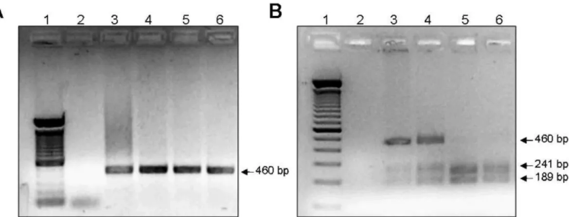
Figure 3 -JAK2V617F genotyping.(A) PCR amplification ofJAK2: lane 1: 100 bp ladder; lane 2: negative control; lanes 3 to 6 – 460-bp
amplicons obtained from the genomic DNA of a patient with PV (3), a CMML patient after disease progression (4) and two MDS patients (with RA) (5 and 6). (B) BsaXI digestion: lane 1: 100 bp ladder, lane 2: negative control; lanes 3 and 4: digestion pattern observed in a PV patient (3) and in the CMML patient positive for theJAK2V617F allele after disease progression (4); lanes 5 and 6:
digestion pattern observed in two MDS patients (with RA) with wild-typeJAK2alleles.
Figure 4 -FLT3-D835 genotyping.(A) PCR amplification ofFLT3: lane 1: 100 bp ladder; lane 2: negative control; lanes 4 to 5: 114-bp
amplicons obtained from the genomic DNA of patients with MDS (3-4) and AML (5). (B) Eco321 digestion: lane 1: 100 bp ladder, lane 2: negative control; lanes 3 and 4: digestion pattern observed in two MDS patients negative for theFLT3-D835 allele, lane 5: digestion
observed the presence of the JAK2V617F mutation in one patient with CMML after disease progression (case 1; Table 2). Figures 3 and 4 represent the RFLP analysis of theJAK2V617 andFLT3-D835 mutations, respectively.
DISCUSSION
Acute leukemia results from a combination of muta-tions and changes in protein function that lead to an increase in proliferation and defects in differentiation and apoptosis.24 Although FLT3 and NPM1 mutations have been described with great frequency in cases of AML,25,26 these mutations were not detected in the MDS patients included in this study. As the presence of these mutations was investigated at the time of diagnosis, screening during disease progression could be interesting. Pinheiro and colleagues27 have reported the acquisition of the FLT3-ITD mutation in 2 of 50 MDS patients included in their study one year after diagnosis. These patients later progressed toward AML, suggesting that the ac-quisition of this mutation may be related to leukemic transformation.
JAK2mutations were not found in the MDS patients in this study at diagnosis. Interestingly, theJAK2V617F mutation was identified in one CMML patient after disease progres-sion. Initially, this patient presented with fewer than 5% bone marrow blasts and lacked the JAK2 V617F mutation. We observed the presence of theJAK2V617F mutation during disease progression, with increased white blood cell (WBC) counts and bone marrow blasts (at diagnosis: 9000 WBC/L, 3% bone marrow blasts; at disease progression: 60000 WBC/ L, 10% bone marrow blasts).JAK2mutations occur in 10% of CMML cases and are associated with clinical and morpho-logical features.28 The JAK2 V617F mutation leads to constitutive activation of the JAK2/STAT3 pathway and aberrant signaling, resulting in growth factor independence, increased proliferation and differentiation failure.29In light of the frequency of these events during MDS progression, our results suggest that the acquisitionof JAK2mutations may be involved in disease progression and should be investigated in more cases of MDS evolution. This finding is in agreement with other authors.30,31A recent publication by Malcovatiet al.31reported 3 patients who evolved from RARS with normal platelet counts and wild-type JAK2 to RARS-T with JAK2 mutation at the time of transformation.
Mutations in exons 9 and 20 of the PI3K gene are frequently described in cancer.4,5,13 However, we did not observe the presence of these mutations in the MDS patients included in this study. Constitutive activation of PI3K occurs in AML and high-risk MDS patients at diagnosis32-34, and mutations in exons 9 and 20 result in constitutive activation of this protein.5,14-17 The presence of PI3K mutations in AML5justifies the evaluation of these muta-tions in a larger number of MDS patients, as they represent a possible factor involved in disease progression.
The presence or absence of these mutations has prog-nostic value in AML,21 and therefore, the investigation of similar mutations in other myeloid diseases such as MDS could be interesting in the context of developing targeted therapies. The PI3K/Akt pathway has already been targeted in acute leukemia, and specific PI3K inhibitors, such as LY294002, have been testedin vitro.35Other members of the PI3K signaling pathway have also been investigated as targets for leukemia treatment. Clinical studies with
rapamycin analogues, which inhibitor mTOR, are currently in phase II AML trials, alone or in combination with other chemotherapeutics.36 Furthermore, FLT3 inhibitors have shown therapeutic activity in AML patients with FLT3 mutations,37and selective JAK2 inhibitors have been tested in patients withJAK2mutations.38
In summary, our study has shown that mutations in the JAK2,FLT3,NPM1andPI3Kgenes are not common in patients with MDS at diagnosis and thatJAK2mutations may occur in MDS during disease progression. Further studies may be helpful to understand the involvement of genetic changes and the impact of these mutations in MDS progression and in different subgroups of patients with the disease.
ACKNOWLEDGMENTS
The authors would like to thank Raquel S. Foglio for English review. This work received financial support from the Conselho Nacional de Desenvolvimento Cientı´fico e Tecnolo´gico (CNPq) and the Fundac¸a˜o de Amparo a` Pesquisa do Estado de Sa˜o Paulo (FAPESP).
Contributions: Joa˜o Agostinho Machado-Neto contributed to the
selection of patients, carried out all experiments and participated in the writing of the manuscript; Fabiola Traina contributed to the selection of patients, clinical follow-up of the patients, analysis of the results and the writing of the manuscript; Mariana Lazarini provided technical assistance with the experiments and participated in the writing of the manuscript; Paula de Melo Campos contributed to the selection of patients and clinical follow-up of the patients; Katia Borgia Barbosa Pagnano contributed to the clinical follow-up of the patients and with the techniques to detect the FLT3-ITD andNPM1mutation; Irene Lorand-Metze was responsible for the morphological diagnosis of myelodysplastic syndrome in the patients included in this study; Fernando Ferreira Costa contributed to the analysis of the results; Sara T. Olalla Saad was the principal investigator.
REFERENCES
1. List AF, Vardiman J, Issa JP, DeWitte TM. Myelodysplastic syndromes. Hematology Am Soc Hematol Educ Program. 2004:297-317.
2. Parker JE, Mufti GJ. Excessive apoptosis in low risk myelodysplastic syndromes (MDS). Leuk Lymphoma. 2000;40:1-24, doi: 10.3109/ 10428190009054877.
3. Parker JE, Mufti GJ, Rasool F, Mijovic A, Devereux S, Pagliuca A. The role of apoptosis, proliferation, and the Bcl-2-related proteins in the myelodysplastic syndromes and acute myeloid leukemia secondary to MDS. Blood. 2000;96:3932-8.
4. Levine DA, Bogomolniy F, Yee CJ, Lash A, Barakat RR, Borgen PI, et al. Frequent mutation of the PIK3CA gene in ovarian and breast cancers. Clin Cancer Res. 2005;11:2875-8, doi: 10.1158/1078-0432.CCR-04-2142. 5. Karakas B, Bachman KE, Park BH. Mutation of the PIK3CA oncogene in
human cancers. Br J Cancer. 2006;94:455-459.
6. Baxter EJ, Scott LM, Campbell PJ, East C, Fourouclas N, Swanton S, et al. Acquired mutation of the tyrosine kinase JAK2 in human myeloproli-ferative disorders. Lancet. 2005;365:1054-61.
7. Lerario AM, Brito LP, Mariani BM, Fragoso MC, Machado MA, Teixeira R. A missense TCF1 mutation in a patient with mody-3 and liver adenomatosis. Clinics. 2009;65:1059-60, doi: 10.1590/S1807-59322010001000024.
8. Torres LC, de Lourdes Lopes Chauffaille M, Delboni TP, Okay TS, Carneiro-Sampaio M, Sugayama S. Rubinstein-taybi syndrome: a female patient with a de novo reciprocal translocation t(2; 16)(q36.3; p13.3) and dysgranulopoiesis. Clinics. 2009;65:107-9, doi: 10.1590/S1807-59322010000100016.
9. Bittencourt PL, Marin ML, Couto CA, Cancado EL, Carrilho FJ, Goldberg AC. Analysis of HFE and non-HFE gene mutations in Brazilian patients with hemochromatosis. Clinics. 2009;64:837-1.
10. Shaw RJ, Cantley LC. Ras, PI(3)K and mTOR signalling controls tumour cell growth. Nature. 2006;441:424-30, doi: 10.1038/nature04869. 11. Stroud RM, Wells JA. Mechanistic diversity of cytokine receptor
signaling across cell membranes. Sci STKE. 2004;2004(231):re7, doi: 10. 1126/stke.2312004re7.
12. Ihle JN, Kerr IM. Jaks and Stats in signaling by the cytokine receptor superfamily. Trends Genet. 1995;11:69-74, doi: 10.1016/S0168-9525(00)89000-9.
13. Muller CI, Miller CW, Hofmann WK, Gross ME, Walsh CS, Kawamata N, et al. Rare mutations of the PIK3CA gene in malignancies of the hematopoietic system as well as endometrium, ovary, prostate and
Mutations in Patients with Myelodysplastic Syndromes
Machado-Neto JA et al. CLINICS 2011;66(5):793-799
osteosarcomas, and discovery of a PIK3CA pseudogene. Leuk Res. 2007;31:27-32, doi: 10.1016/j.leukres.2006.04.011.
14. Qiu W, Schonleben F, Li X, Ho DJ, Close LG, Manolidis S, et al. PIK3CA mutations in head and neck squamous cell carcinoma. Clin Cancer Res. 2006;12:1441-6, doi: 10.1158/1078-0432.CCR-05-2173.
15. Hafner C, Lopez-Knowles E, Luis NM, Toll A, Baselga E, Fernandez-Casado A, et al. Oncogenic PIK3CA mutations occur in epidermal nevi and seborrheic keratoses with a characteristic mutation pattern. Proc Natl Acad Sci U S A. 2007;104:13450-4, doi: 10.1073/pnas.0705218104. 16. Zhao L, Vogt PK. Helical domain and kinase domain mutations in
p110alpha of phosphatidylinositol 3-kinase induce gain of function by different mechanisms. Proc Natl Acad Sci U S A. 2008;105:2652-7, doi: 10. 1073/pnas.0712169105.
17. Riener MO, Bawohl M, Clavien PA, Jochum W. Rare PIK3CA hotspot mutations in carcinomas of the biliary tract. Genes Chromosomes Cancer. 2008;47:363-7, doi: 10.1002/gcc.20540.
18. Tefferi A. Classification, diagnosis and management of myeloprolifera-tive disorders in the JAK2V617F era. Hematology Am Soc Hematol Educ Program. 2006:240-5.
19. Small D, Levenstein M, Kim E, Carow C, Amin S, Rockwell P, et al. STK-1, the human homolog of Flk-2/Flt-3, is selectively expressed in CD34+ human bone marrow cells and is involved in the proliferation of early progenitor/stem cells. Proc Natl Acad Sci U S A. 1994;91:459-63, doi: 10. 1073/pnas.91.2.459.
20. Cheng K, Grisendi S, Clohessy JG, Majid S, Bernardi R, Sportoletti P, et al. The leukemia-associated cytoplasmic nucleophosmin mutant is an oncogene with paradoxical functions: Arf inactivation and induction of cellular senescence. Oncogene. 2007;26:7391-400, doi: 10.1038/sj.onc. 1210549.
21. Gale RE, Green C, Allen C, Mead AJ, Burnett AK, Hills RK, et al. The impact of FLT3 internal tandem duplication mutant level, number, size, and interaction with NPM1 mutations in a large cohort of young adult patients with acute myeloid leukemia. Blood. 2008;111:2776:84. 22. Bennett JM, Catovsky D, Daniel MT, Flandrin G, Galton DA, Gralnick
HR, et al. Proposals for the classification of the myelodysplastic syndromes. Br J Haematol. 1982;51:189-99.
23. Vardiman JW, Thiele J, Arber DA, Brunning RD, Borowitz MJ, Porwit A, et al. The 2008 revision of the World Health Organization (WHO) classification of myeloid neoplasms and acute leukemia: rationale and important changes. Blood. 2009;114:937-51, doi: 10.1182/blood-2009-03-209262.
24. Steelman LS, Pohnert SC, Shelton JG, Franklin RA, Bertrand FE, McCubrey JA. JAK/STAT, Raf/MEK/ERK, PI3K/Akt and BCR-ABL in cell cycle progression and leukemogenesis. Leukemia. 2004;18:189-218, doi: 10.1038/sj.leu.2403241.
25. Lin P, Jones D, Medeiros LJ, Chen W, Vega-Vazquez F, Luthra R. Activating FLT3 mutations are detectable in chronic and blast phase of chronic myeloproliferative disorders other than chronic myeloid leukemia. Am J Clin Pathol. 2006;126:530-3.
26. Grundler R, Miething C, Thiede C, Peschel C, Duyster J. FLT3-ITD and tyrosine kinase domain mutants induce 2 distinct phenotypes in a murine bone marrow transplantation model. Blood. 2005;105:4792-9, doi: 10.1182/ blood-2004-11-4430.
27. Pinheiro RF, de Sa Moreira E, Silva MR, Alberto FL, Chauffaille Mde L. FLT3 internal tandem duplication during myelodysplastic syndrome follow-up: a marker of transformation to acute myeloid leukemia. Cancer Genet Cytogenet. 2008;183:89-93, doi: 10.1016/j.cancergencyto.2008.02.006. 28. Pich A, Riera L, Sismondi F, Godio L, Davico Bonino L, Marmont F, et al. JAK2V617F activating mutation is associated with the myeloproliferative type of chronic myelomonocytic leukaemia. J Clin Pathol. 2009;62:798-801, doi: 10.1136/jcp.2009.065904.
29. James C, Ugo V, Le Couedic JP, Staerk J, Delhommeau F, Lacout C, et al. A unique clonal JAK2 mutation leading to constitutive signalling causes polycythaemia vera. Nature. 2005;434:1144-8, doi: 10.1038/nature03546. 30. Hellstrom-Lindberg E. Significance of JAK2 and TET2 mutations in myelodysplastic syndromes. Blood Rev. 2010;24:83-90, doi: 10.1016/j. blre.2010.01.002.
31. Malcovati L, Della Porta MG, Pietra D, Boveri E, Pellagatti A, Galli A, et al. Molecular and clinical features of refractory anemia with ringed sideroblasts associated with marked thrombocytosis. Blood. 2009;114:3538-45, doi: 10.1182/blood-2009-05-222331.
32. Chapuis N, Tamburini J, Cornillet-Lefebvre P, Gillot L, Bardet V, Willems L, et al. Autocrine IGF-1/IGF-1R signaling is responsible for constitutive PI3K/Akt activation in acute myeloid leukemia: therapeutic value of neutralizing anti-IGF-1R antibody. Haematologica. 2010;95:415-23, doi: 10.3324/haematol.2009.010785.
33. Tamburini J, Elie C, Bardet V, Chapuis N, Park S, Broet P, et al. Constitutive phosphoinositide 3-kinase/Akt activation represents a favorable prognostic factor in de novo acute myelogenous leukemia patients. Blood. 2007;110:1025-8, doi: 10.1182/blood-2006-12-061283. 34. Nyakern M, Tazzari PL, Finelli C, Bosi C, Follo MY, Grafone T, et al.
Frequent elevation of Akt kinase phosphorylation in blood marrow and peripheral blood mononuclear cells from high-risk myelodysplastic syndrome patients. Leukemia. 2006;20:230-8, doi: 10.1038/sj.leu.2404057. 35. Billottet C, Grandage VL, Gale RE, Quattropani A, Rommel C, Vanhaesebroeck B, et al. A selective inhibitor of the p110delta isoform of PI 3-kinase inhibits AML cell proliferation and survival and increases the cytotoxic effects of VP16. Oncogene. 2006;25:6648-59, doi: 10.1038/sj. onc.1209670.
36. Park S, Chapuis N, Tamburini J, Bardet V, Cornillet-Lefebvre P, Willems L, et al. Role of the PI3K/AKT and mTOR signaling pathways in acute myeloid leukemia. Haematologica.95:819-28, doi: 10.3324/haematol. 2009.013797.
37. Weisberg E, Barrett R, Liu Q, Stone R, Gray N, Griffin JD. FLT3 inhibition and mechanisms of drug resistance in mutant FLT3-positive AML. Drug Resist Updat. 2009;12:81-9, doi: 10.1016/j.drup.2009.04.001.