Original Article
Artigo Original
Electrophysiological characterization of
hearing in individuals with Down syndrome
Caracterização eletrofisiológica da audição em
indivíduos com Síndrome de Down
Hellen Medeiros Kazan1
Alessandra Giannella Samelli1
Ivone Ferreira Neves-Lobo1
Fernanda Cristina Leite Magliaro1
Suelly Cecília Olivan Limongi1
Carla Gentile Matas1
Keywords
Down Syndrome Auditory Evoked Potentials Brainstem P300 Evoked Potential
Descritores
Síndrome de Down Potenciais Evocados Auditivos Tronco Encefálico Potencial Evocado P300
Correspondence address: Carla Gentile Matas Departamento de Fisioterapia, Fonoaudiologia e Terapia Ocupacional, Faculdade de Medicina, Universidade de São Paulo – USP
Rua Cipotânea, 51, Cidade
Universitária, São Paulo (SP), Brazil, CEP: 05360-160.
E-mail: [email protected]
Received: October 23, 2015
Study carried out at the Departamento de Fisioterapia, Fonoaudiologia e Terapia Ocupacional, Faculdade de Medicina, Universidade de São Paulo – USP - São Paulo (SP), Brazil.
1 Universidade de São Paulo – USP - São Paulo (SP), Brazil.
Financial support: nothing to declare.
Conlict of interests: nothing to declare.
ABSTRACT
Introduction: Few studies have performed Brainstem (BAEP) and P300 Auditory Evoked Potentials simultaneously to assess central auditory pathways in normal hearing individuals with Down syndrome (DS),
mainly because of the dificulty in applying these procedures to this population. Previous studies have suggested
that individuals with DS might present different patterns of response compared with those of individuals with
typical development; nevertheless, the identiication of these potentials would be crucial for the establishment
of an accurate audiological diagnosis. Purpose: To characterize BAEP and P300 in normal-hearing individuals with DS. Methods: BAEP and P300 were analyzed in 17 individuals with DS and in 21 individuals with typical development aged 7 to 15 years. The results were quantitatively and qualitatively analyzed using descriptive measures and hypothesis tests. Results: In the quantitative analysis, latency values were lower in the BAEP
for the DS group, with statistically signiicant difference for wave V and interpeaks III-V and I-V; there were no signiicant differences in the P300 latency values. In the qualitative analysis, there were a larger number of
individuals with early values for BAEP latencies and late latencies for P300 in the DS group; both comparisons
showed statistically signiicant differences. Conclusion: Children and adolescents with DS can present early responses to the components of BAEP, suggesting that their auditory pathway requires less time for the neural transmission of acoustic stimuli to the brainstem. Concerning P300, individuals with DS may present increased latencies, suggesting impairment in the central auditory pathway for the cortical processing of auditory information.
RESUMO
Introdução: Poucos estudos realizaram, concomitantemente, o potencial evocado auditivo de tronco encefálico (PEATE) e o P300 na Síndrome de Down (SD), em indivíduos audiologicamente normais, para a avaliação
da via auditiva central, principalmente pela diiculdade de realizar estes procedimentos nesta população.
Estudos anteriores sugeriram que indivíduos com SD podem apresentar padrões de respostas diferentes das
encontradas em indivíduos com desenvolvimento típico, sendo que a identiicação destes seria fundamental para o
estabelecimento de um diagnóstico audiológico preciso. Objetivo: Caracterizar o PEATE e o P300 em indivíduos com SD audiologicamente normais. Método: Foram analisados o PEATE e o P300 de 17 indivíduos com SD e 21 com desenvolvimento típico de sete a 15 anos. A análise foi quantitativa e qualitativa, utilizando medidas descritivas e os testes de hipótese. Resultados: Os valores de latência foram menores no PEATE para o grupo
SD, com diferença estatisticamente signiicante para a onda V, interpicos III-V e I-V; não foram encontradas diferenças signiicantes nos valores de latência do P300. Observou-se maior número de indivíduos com valores
precoces para as latências do PEATE e com latências atrasadas para o P300 no grupo SD; ambas as comparações
mostraram diferenças signiicantes. Conclusão: Crianças e adolescentes com SD podem apresentar respostas precoces para os componentes do PEATE, sugerindo que a via auditiva destes necessita de menor tempo para
a transmissão neural do estímulo acústico até o tronco encefálico. Quanto ao P300, indivíduos com SD podem
INTRODUCTION
Down’s syndrome (DS) was irst described by John Langdon Down in 1866, but it was characterized as a genetic disorder caused by the presence of the extra full or partial chromosome 21 by Lejeune in 1957(1,2).
It is the most common genetic occurrence, estimated at one in 800 or 1000 live births. Its diagnosis is conirmed by cytogenetic investigation considering a series of signs and symptoms, with the following most commonly found characteristics: intellectual impairment (100%), muscular hypotonia (99%), oblique palpebral issure (90%), increased vascularity (90%), microcephaly (85%), lat occiput (80%), and joint hypermobility (80%)(3).
Auditory impairment in individuals with DS has been widely reported in the speciic literature(4-6). Hearing loss - conductive,
sensorineural, or mixed - occurs in approximately two thirds of children with DS(7,8), with highest incidence for conductive
hearing losses (around 80%) owing to the presence of otitis caused by constant respiratory tract infections(4,9,10).
In addition, studies have shown that the cochlea of individuals with DS is anatomically smaller, but apparently the abnormalities of the inner ear are not frequent(11). However, some authors
have reported that, as of the second decade of life, individuals with DS may present progressive sensorineural hearing loss, similar to presbycusis(12).
Impairment of central areas of the brain can also be veriied in DS, characterized by lesions or dysfunctions. Some authors have attributed these alterations to failures in habituation mechanisms, central inhibition deicits in stimulus afferentation, or disturbances in the cognition mechanism, which may cause reduction of cerebral neuroelectric activities(13).
Accurate audiological assessment in children with DS is a challenge because, most of the time, it is performed through subjective tests, which depend on the child’s participation and collaboration. Accordingly, studies using objective tests such as auditory evoked potentials are extremely important in this population.
The Brainstem Auditory Evoked Potential (BAEP) evaluates the integrity of the auditory pathway until the brainstem and it has been one of the most effective tests to assist in the identiication of hearing loss by means of identiication of the electrophysiological threshold. A study using BAEP conducted with individuals with DS found reduced values of absolute latency for waves I, III, and V in children with or without hearing loss, including conductive hearing loss(14).
Nevertheless, other authors observed reduced latency values for waves I and III and the interpeak I-III only in children aged 18 months or less(15).
Thus it seems to be no consensus on this decrease in latency values. One of the justiications for these decreased values would be the smaller head circumference of the child with DS, reducing the distance of the transmission of the nervous stimulus(16); another would be the higher transmission velocity
because of early myelination for the age(15). Therefore, the aim
of the present study was to verify whether the reduced latency values previously described can also be observed in older children or adolescents.
The late, event-related, auditory evoked potential (P300 or Cognitive Potential) is generated when, among a series of frequent stimuli, an infrequent, random stimulus related to sensory and cognitive aspects in the processing of auditory information is detected. Some studies have described an increase in P3 or P300 wave latency values in adult individuals with DS(13,17). The justiication for this inding, suggested by some
authors, is that these late P300 latency values may indicate an early aging process in these individuals; this procedure is useful for the identiication and prevention of pre-senile dementia(17).
Because of the dificulty in applying the test, there are few studies involving P300 in children with DS. Considering the small number of studies on the theme, the current research intends to analyze the P300 from the perspective of objectively evaluating the processing of auditory information in individuals with DS without hearing loss.
Moreover, there are few studies that performed the BAEP and P300 on Down syndrome, in individuals with normal audiologic proile, to evaluate the central auditory pathway, mainly due to the dificulty in conducting these procedures in this population. Previous studies have suggested that individuals with DS may present patterns of responses different from those found in individuals with typical development, and their identiication would be critical for the establishment of an accurate auditory diagnosis.
Therefore, the objective of this study was to record and characterize the BAEP with clicks and the P300 in audiologically normal individuals with DS.
METHODS
The present study was approved by the Research Ethics Committee of the College of Medicine of the University of Sao Paulo - FMUSP under protocol number 138/11. The parents/guardians of all participants signed an Informed Consent Form prior to commencement of the study.
Sample
Study participants were divided into two groups:
Down syndrome group (DSG) - composed of 17 individuals (eight males and nine females) with Down syndrome aged seven to 15 years (mean age = 10.9±1.6 years). All individuals in this group were undergoing Speech-language Pathology treatment at the aforementioned institution; 10 presented a history of otitis media according to their parents, with number of disease episodes ranging from 2 to 5 (mean of 3.1 episodes).
with number of disease episodes ranging from 1 to 3 (mean of 2.42 episodes).
The following inclusion criteria were adopted for both groups: • Auditory thresholds lower than 15 dBHL for all frequencies
assessed(18);
• Type A tympanometry curve and presence of ipsilateral acoustic relexes;
• Absence of history of evident psychiatric and neurological diseases.
It is worth mentioning that, according to the inclusion criteria adopted at the time of the evaluation, the individuals did not present middle ear impairment.
Procedures
The following procedures were conducted:
a) Anamnesis: information on the medical and otological history was collected.
b) Meatoscopy: was performed, using a Heine manufactured otoscope, prior to the audiological evaluation to eliminate possible interferences in the examinations (presence of cerumen, foreign bodies, etc.).
c) Immittanciometry (tympanometry and measure of ipsilateral acoustic relexes at the frequencies of 500, 1000, 2000, and 4000 Hz): was performed using AT 235 (Interacoustic®)
equipment. All participants were submitted to this assessment prior to the recording of the BAEP; recording of the P300 was conducted on a day different from that of the BAEP; this procedure was repeated to ensure absence of middle ear impairment.
d) Pure-tone audiometry: was performed at the frequencies of 500, 1000, 2000, and 4000 Hz using supra-aural headphones in an acoustic cabin (Grason-Stadler®, GSI-61 audiometer). e) Electrophysiological hearing evaluation: these procedures were performed using portable, 2-channel equipment (Biologic®,
Traveller Express) with individuals sitting in a reclining armchair in an acoustically and electrically treated room. The acoustic stimuli were presented through a supra-aural headphone. The impedance values of the electrodes should be below 5 kOhm. The following parameters were used for each electrophysiological evaluation:
- BAEP (Brainstem Auditory Evoked Potential): electrodes were placed on the regions of the left (M1) and right (M2) mastoids and forehead (Fz) and the earth electrode was placed on the contralateral ear. Acoustic stimulus was generated through clicks with duration of 0.1 ms and presentation rate of 19.1 stimuli per second, totalizing 2000 stimuli in rareied polarity. Low-pass ilters of 1500 Hz and high-pass ilters of 100 Hz were used with analysis window of 10 ms. Acoustic stimulus intensity of 80 dB nHL was used to analyze auditory pathway integrity with assessment of one ear at a time. Absolute latency values of waves I, III and V
and interpeaks I-III, III-V and I-V were analyzed bilaterally according to the normal values of the equipment (Evoked Potential User Manual)(19).
In addition to the quantitative analysis, the results were analyzed qualitatively for each individual, considering the criterion of normality of the absolute latencies of waves I, III, and V plus two standard deviations. The results were classiied as follows:
- Normal: for absolute latencies of waves I, III and V within
the normality criteria plus two standard deviations, for each individual (considering both ears).
- Early: for absolute latencies of any of the waves (I, III, and V) below the established values plus two standard deviations, for each individual (considering at least one ear).
- Late: for absolute latencies of any of the waves (I, III, and V) above the established values plus two standard deviations, for each individual (considering at least one ear).
- P300: electrodes were placed on the regions of the left (M1) and right (M2) mastoids and vertex (Cz) and the earth electrode was placed on the forehead (Fz). Tone-burst acoustic stimulus was used at frequencies of 1000 and 1500 Hz, intensity of 75 dB nHL, and presentation rate of 1.1 stimuli per second, totalizing 300 stimuli. High-pass ilters of 1 Hz and low-pass ilters of 30 Hz were used with analysis window of 800 ms. Participants were asked to count aloud the rare stimuli (20% of the stimuli) that would appear randomly among the frequent stimuli (80% of stimuli) - oddball paradigm; assessment occurred in one ear at a time.
At the end of the evaluation, participants were instructed to report to the researcher the number of rare stimuli they heard. To certify that individuals would be able to discriminate the rare sounds from the frequent ones, they were instructed to raise their arms when they heard the irst rare stimulus. P300 was identiied as a wave of positive polarity with post-stimulus approximate latency of 300 ms, obtained by subtracting the tracing corresponding to the rare stimuli in the corresponding tracing of frequent stimuli. The latency values of the P300 wave were analyzed. The normality criteria proposed by McPherson(20) was
used according to the age range evaluated. It is worth noting that 15 of the 17 children in the DSG underwent P300 recording.
In addition to the quantitative analysis, the results were analyzed qualitatively for each individual with respect to the absolute value of the P300 wave. The results were classiied as follows:
- Normal: when the P300 wave latency was within the values established by McPherson(20) according to the age range.
- Late: when increased P300 wave latency was observed in at least one of the ears compared with the normality values established by McPherson(20), according to the age range.
Statistical analysis of the data
The study results were analyzed both quantitatively and qualitatively: for analysis of the quantitative data, mean and standard deviation were calculated for the latency values, whereas for analysis of the qualitative data, a study was performed on the occurrence of alterations in the BAEP and P300 and on the types of changes found per individual according to the normality criteria previously described.
ANOVA and Chi-square association tests were conducted for comparison between the data of the groups and ears at 5% signiicance level.
RESULTS
Quantitative analysis
Initially, the right and left ears were compared within each group. As no statistically signiicant difference was observed for any of the comparisons, the ears were grouped for the quantitative analyses between groups.
Table 1 shows the latencies of the waves for BAEP and P300 compared between the groups. Note that the latencies for all the comparisons are smaller for the DSG and there was
statistically signiicant difference for the wave V and interpeaks III-V and I-V.
Qualitative analysis
Considering waves I, III, and V of the PEATE for both ears, early latencies were observed for most of the individuals in the DSG, with 82.35% presenting early latency for the wave V. Regarding the control group (CG), all individuals presented normal results. There was statistically signiicant difference between groups for the distribution of results in the three categories (early, normal, and late) for waves I, III, and V (Table 2).
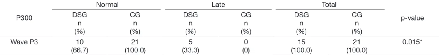
With regard to the P300, there was predominance of normal results for both groups (67% for DSG and 100% for CG). However, statistically signiicant difference was observed between groups for the distribution of results (normal and late) in the qualitative analysis (Table 3).
DISCUSSION
In the present study, no statistically signiicant difference between the ears was observed for the absolute and interpeak latency values of the waves in the BAEP and for the latency
Table 1. Mean and standard deviation (in ms) of the latency values for waves I, III, and V and for interpeaks I-III, III-V, and I-V of the BAEP and the
latency of the P300 for the Down syndrome (DSG) and control (CG) groups
BAEP / P300 Mean
DSG SD DSG Mean CG SD CG p-value
I 1.49 0.14 1.50 0.10 0.776
III 3.55 0.26 3.61 0.11 0.310
V 5.37 0.30 5.59 0.12 0.002*
I – III 2.05 0.25 2.11 0.11 0.303
III – V 1.82 0.20 1.98 0.07 0.001*
I – V 3.88 0.27 4.08 0.11 0.002*
P300 323 58.80 329.05 28.54 0.558
*p-value statistically significant
Table 2. Distribution of the occurrence of normal, early, and late results in the BAEP for both groups
BAEP
Early Normal Late Total
p-value DSG n (%) CG n (%) DSG n (%) CG n (%) DSG n (%) CG n (%) DSG n (%) CG n (%)
Wave I 9 (52.9) 0
(0.0) 5 (29.4) 21 (100.0) 3 (17.7) 0 (0.0) 17 (100.0) 21 (100.0) <0.0001*
Wave III 12
(70.6) 0 (0.0) 3 (17.6) 21 (100.0) 2 (11.8) 0 (0.0) 17 (100.0) 21 (100.0) <0.0001*
Wave V 14
(82.3) 0 (0.0) 2 (11.8) 21 (100.0) 1 (5.9) 0 (0.0) 17 (100.0) 21 (100.0) <0.0001* *p-value statistically significant
Table 3. Distribution of the occurrence of normal and late results in the P300 for the Down syndrome (n=15) and control (n=21) groups
P300
Normal Late Total
p-value DSG n (%) CG n (%) DSG n (%) CG n (%) DSG n (%) CG n (%)
Wave P3 10
(66.7) 21 (100.0) 5 (33.3) 0 (0) 15 (100.0) 21 (100.0) 0.015*
values in the P300 for both groups. Such indings corroborate those of previous studies in which no statistically signiicant differences were found between the ears for latency values of the BAEP and P300 in audiologically normal individuals and in individuals with Down syndrome(21,22). These results reinforce
the fact that, for individuals with DS, the reference values and analysis criteria for the BAEP and P300 with non-complex stimuli (clicks and tone burst) can be used equally for both ears, as it already occurs in clinical practice in individuals with typical development.
Regarding gender, the indings were not analyzed in relation to this variable, considering that previous studies have not found statistically signiicant differences between male and female genders for BAEP and P300 responses(13).
With respect to the electrophysiological evaluation performed using the BAEP, the DSG presented lower mean latency values, with statistically signiicant difference only for the absolute latency of the wave V and for interpeaks III-V and I-V, compared with those of the CG (Table 1). These indings are in agreement with studies in the literature that report a decrease in the absolute latency values of the wave V and the interpeak I-V in individuals with DS(14,23). It is noteworthy that, in the study by Squires et al. (14), decreased latency values were observed for all BAEP waves
in children with DS with or without hearing loss.
Our results also corroborate those of a previous study conducted with newborns with DS in which the researchers reported a decrease in the latency values of the wave V and interpeaks III-V and I-V of the BAEP compared with those of newborns of the CG(24).
In the qualitative analysis, according to Table 2, statistically signiicant differences were found between the groups in relation to the early classiication for the values of absolute latencies of waves I, III, and V. Thus it was possible to observe that 58.82% of the individuals with DS presented early latency for the wave I, 70.58% for the wave III, and 82.35% for the wave V. These results agree with those of other studies that found reduced absolute latency values for the BAEP waves in individuals with DS compared with individuals with typical development(15,25-27).
It was also possible to observe higher standard deviation values in the data of the DSC, thus demonstrating greater variability of responses in these individuals compared with those of the control group.
There is no agreement in the speciic literature regarding the justiication of early latency values in the BAEP in Down syndrome. Among the several hypotheses, authors use the DS phenotype as a justiication, considering the smaller skull circumference and the consequent shorter distance between the cochlea and the brainstem present in these individuals. Other theories refer to early progress of myelination in brainstem; changes in the cochlea and auditory pathway, or simpliication of pathway; greater conduction velocity of the nervous iber; and smaller brainstem(28).
It is worth noting that the early latency values of the wave V, that is, the region of the upper brainstem, identiied in the quantitative and qualitative analyses were more evident in the
present study. This inding was not in agreement with those of other studies conducted with children with DS. Many studies are in agreement as for the low values of the latencies of waves III and V and interpeaks I-III and III-V. It is believed that the age range involved in this study may have interfered with this outcome. Some authors have veriied early latency values in the BAEP only for children younger than 18 months, justiied by maturational aspects, that is, with maturational development, responses to the BAEP of children with DS tend to resemble those of children with typical development(15,25-27).
These early responses are extremely important for the proper analysis and interpretation of the BAEP in children with DS, mainly due to the middle ear changes frequently present in this population, which instead of causing late latency of the waves (a result often found in patients with conductive hearing loss), could lead to latency values within normal range.
It is important to mention that participants with DS of the present study showed a history of otitis, with mean number of disease episodes of 3.1, slightly higher than that of participants in the CG (mean number of disease episodes of 2.42). Thus the number of otitis episodes in childhood is a factor that must be considered in the analysis of results of auditory evoked potentials, because it may inluence the maturation of the central auditory pathways(29).
Therefore, it is important to emphasize that normative data from normal individuals should be used with caution in interpreting the results when the BAEP is used in the audiological assessment of individuals with DS.
Regarding the quantitative and qualitative analyses of the P300 (Tables 1 and 3), statistically signiicant difference was found between the groups only for the qualitative analysis (Table 3). This analysis shows that 33% of the individuals with DS were classiied as presenting increased latency in at least one of the ears. Data from the present survey disagree with those of other studies in which increased mean latency values of the P300 were found in individuals with DS, but agree to verify a larger number of individuals with altered results in the DSC compared with the CG. Some authors report that increased latency would be related to sensory and cognitive deicits in the processing of acoustic information(30).
of sounds, or even premature aging. Some authors report that P300 analysis is quite complex because it is an auditory evoked potential resulting from multiple cognitive factors(30). In the
present study, although no altered mean latency values were found in the P300, the number of individuals with increased latency values may be related to the cortical processing of auditory information(13), which is directly inluenced by external factors
such as the history of otitis episodes in childhood(29). It should be
emphasized that the processes of attention and auditory memory may also be impaired in this population, which may have directly inluenced the generation of this electrophysiological response.
Further studies on the maturational development of auditory evoked potentials in children with DS, especially with regard to cortical auditory evoked potentials, would be of extreme interest for the better understanding of this aspect in this population.
CONCLUSION
Children and adolescents with DS can present early responses to the components of Brainstem Auditory Evoked Potential (BAEP), suggesting that their auditory pathway requires less time for the neural transmission of acoustic stimuli to the brainstem.
Concerning P300, individuals with DS may present increased latencies, suggesting impairment in the central auditory pathway for the cortical processing of auditory information.
REFERENCES
1. Down JLH. Observations on an ethnic classification of idiots. London Hospital Clinical Lecture and Reports. 1866;3:259-62.
2. Lejeune J, Gauthier M, Turpin R. Les chromosomes humains en culture de tissus. C R Hebd Seances Acad Sci. 1959;248(4):602-3. PMid:13629913. 3. Sommer CA, Henrique-Silva F. Trisomy 21 and Down syndrome: a short
review. Braz J Biol. 2008;68(2):447-52. PMid:18660978. http://dx.doi. org/10.1590/S1519-69842008000200031.
4. Bull MJ. Health supervision for children with Down syndrome. Pediatrics. 2011;128(2):393-406. PMid:21788214. http://dx.doi.org/10.1542/peds.2011-1605.
5. Cheng W-W, Lau W-L, Ko C-H. Prevalence and parental awareness
of hearing loss in children with Down syndrome. Chin Med J (Engl).
2015;128(8):1091-5. PMid:25881605. http://dx.doi.org/10.4103/0366-6999.155105.
6. Austeng ME, Akre H, Falkenberg ES, Øverland B, Abdelnoor M, Kværner
KJ. Hearing level in children with Down syndrome at the age of eight. Res Dev Disabil. 2013;34(7):2251-6. PMid:23644229. http://dx.doi. org/10.1016/j.ridd.2013.04.006.
7. Saliba I, Sbeity S, El-Zir E, Yammine FG, Noun CT, Haddad A. Down syndrome: an electrophysiological and radiological profile. Laryngoscope. 2014;124(4):E141-7. PMid:24114773. http://dx.doi.org/10.1002/lary.24375. 8. Park AH, Wilson MA, Stevens PT, Harward R, Hohler N. Identification of hearing loss in pediatric patients with Down syndrome. Otolaryngol Head Neck Surg. 2012;146(1):135-40. PMid:21987652. http://dx.doi. org/10.1177/0194599811425156.
9. Rodman R, Pine HS. The otolaryngologist′s approach to the patient with Down syndrome. Otolaryngol Clin North Am. 2012;45(3):599-629, vii-viii. PMid:22588039. http://dx.doi.org/10.1016/j.otc.2012.03.010.
10. Chin CJ, Khami MM, Husein M. A general review of the otolaryngologic
manifestations of Down Syndrome. Int J Pediatr Otorhinolaryngol.
2014;78(6):899-904. PMid:24704318. http://dx.doi.org/10.1016/j. ijporl.2014.03.012.
11. Harada T, Sando I. Temporal bone histopathologic findings in Down’s syndrome. Arch Otolaryngol. 1981;107(2):96-103. PMid:6451213. http:// dx.doi.org/10.1001/archotol.1981.00790380026007.
12. Carrico B, Samelli AG, Matas CG, Magliaro FCL, Carvallo RMM, Limongi SCO, et al. Avaliação auditiva periférica em crianças com síndrome de Down. Audiol. Commun. Res. 2014;19(3):280-5. http://dx.doi.org/10.1590/ S2317-643120140003000012.
13. Cesar CPHAR, Caovilla HH, Munhoz MSL, Ganança MM. Potencial evocado auditivo tardio relacionado a eventos (P300) na Síndrome de
Down. Braz J Otorhinolaryngol. 2010;76(2):206-12. PMid:20549081. http://dx.doi.org/10.1590/S1808-86942010000200010.
14. Squires N, Ollo C, Jordan R. Auditory brain stem responses in the mentally retarded: audiometric correlates. Ear Hear. 1986;7(2):83-92. PMid:2938998. http://dx.doi.org/10.1097/00003446-198604000-00006.
15. Krecicki T, Zalesska-Krecicka M, Kubiak K, Gawron W. Brain auditory
evoked potentials in children with Down syndrome. Int J Pediatr
Otorhinolaryngol. 2005;69(5):615-20. PMid:15850683. http://dx.doi. org/10.1016/j.ijporl.2004.11.025.
16. Teipel SJ, Hampel H. Neuroanatomy of Down syndrome in vivo: a model of preclinical Alzheimer’s disease. Behav Genet. 2006;36(3):405-15. PMid:16485178. http://dx.doi.org/10.1007/s10519-006-9047-x. 17. Blackwood DH, St Clair DM, Muir WJ, Oliver CJ, Dickens P. The
development of Alzheimers disease in Downs syndrome assessed by
auditory event-related potentials. J Ment Defic Res. 1988;32(Pt 6):439-53.
PMid:2976841.
18. Northern JL, Downs MP. Hearing in children. 5. ed. Baltimore: Lippincott Williams and Wilkins; 2002.
19. Evoked PUM. Evoked Potential: Program Version 5.00. User manual.
1993. Bio-logic Systems Corp. Part n˚ 590-BLSUEP rev.1.
20. McPherson DL. Late potentials of the auditory system (evoked potentials). San Diego: Singular Publishing Group; 1996.
21. Soares IA, Menezes PL, Carnaúba ATL, Pereira LD. Standardization of brainstem auditory evoked potential using a new device. Pró-Fono Revista de Atualização Científica. 2010;22(4):421-6. PMid:21271093. http://dx.doi. org/10.1590/S0104-56872010000400010.
22. Arisi E, Forti S, Amadeo C, Fagnani E, Filipponi E, Iacona E, et al. A. Auditory late potentials in normal-hearing adult subjects with Down’s Syndrome. Otol Neurotol. 2012;33(7):1113-7. PMid:22892803. http:// dx.doi.org/10.1097/MAO.0b013e3182659d02.
23. Ferri R, Del Gracco S, Elia M, Musumeci SA, Stefanini MC. Age, sex and mental retardation related changes of brainstem auditory evoked potentials
in Down’s syndrome. Ital J Neurol Sci. 1995;16(6):377-83. PMid:8626215. http://dx.doi.org/10.1007/BF02229173.
24. Sato M, Suzuki S. Auditory brainstem responses in newborns with Down
25. Kakigi R, Kuroda Y. Brain-stem auditory evoked potentials in adults with Down’s syndrome. Electroencephalogr Clin Neurophysiol. 1992;84(3):293-5. PMid:1375889. http://dx.doi.org/10.1016/0168-5597(92)90011-Y. 26. Kaga K, Marsh R. Auditory brainstem responses in young children
with Down’s syndrome. Int J Pediatr Otolaryngol. 1986;11(1):29-38.
PMid:2940199. http://dx.doi.org/10.1016/S0165-5876(86)80025-8. 27. Kittler PM, Phan HTT, Gardner JM, Miroshnichenko I, Gordon A, Karmel
BZ. Auditory brainstem evoked responses in newborns with Down Syndrome.
Am J Intellect Dev Disabil. 2009;114(6):393-400. PMid:19792055. http:// dx.doi.org/10.1352/1944-7558-114.6.393.
28. Forti S, Amadeo C, Fagnani E, Filipponi E, Pignataro L, Cesarani A, et al. Auditory brainstem responses (ABR) in normal hearing adult subjects with Down’s syndrome. Brain Res. 2008;3:58-62. PMid:18703027. http://dx.doi. org/10.1016/j.brainres.2008.07.078.
29. Borges LR, Paschoal JR, Colella-Santos MF. (Central) Auditory Processing: the impact of otitis media. Clinics. 2013;68(7):954-9. PMid:23917659. http://dx.doi.org/10.6061/clinics/2013(07)11.
30. van Dinteren R, Arns M, Jongsma MLA, Kessels RPC. P300 development across the lifespan: a systematic review and meta-analysis. PLoS One. 2014;9(2):e87347. PMid:24551055. http://dx.doi.org/10.1371/journal. pone.0087347.
Author contributions