845
CLINICS 2008;63(6):845-8
LETTER TO THE EDITOR
I Head and Neck Surgery, CLIMEDCAP - São Paulo/SP, Brazil.
II Department of Pathology, Salomão & Zoppi Medicina Diagnóstica - São
Paulo/SP, Brazil.
III Faculdade de Medicina do ABC - Santo André/SP, Brazil.
IV Head and Neck Surgery, Hospital das Clínicas, Faculdade de Medicina,
Universidade de São Paulo - São Paulo/SP, Brazil. Email: [email protected]
Tel.: 55 11 5058.5738
UNUSUAL LOCATION OF A CERVICAL
PARAGANGLIOMA BETWEEN THE THYROID GLAND
AND THE COMMON CAROTID ARTERY: CASE
REPORT
doi: 10.1590/S1807-59322008000600024
Fábio Roberto Pinto,I Fábio de Aquino Capelli,I Sueli Aparecida Maeda,II Emílio Marcelo Pereira,II Marcela Benetti Scarpa,III Lenine Garcia BrandãoIV
INTRODUCTION
Paragangliomas are rare tumors derived from the extra-adrenal paraganglionic system, which is composed of cells from the neural crest that are associated with the autonomous nervous system.1,2 They have been observed at many sites in the head and neck area, including the carotid body, the jugular-tympanic region, and the vagus nerve.2,3 Unlike pheochromocytomas, head and neck paragangliomas are usually non-functioning tumors; in other words, they do not secrete catecholamines, and the primary symptom is a slow growing mass in the neck.2,4 Paragangliomas arising within the thyroid gland or the neighboring area are extremely rare, and are believed to derive from the inferior laryngeal paraganglia.5-9 The laryngeal paraganglia was first described in 1963 by Watzka,10 who showed the presence of this particular tissue in the upper and anterior third of ventricular folds. One year later, Kleinsasser11 reported the presence of paraganglia in the subglottic larynx and named it the inferior laryngeal glomus; this author also labeled Watzka’s discovery as the superior laryngeal glomus. In anatomical studies of the human larynges, Zak12 and Lawson13 demonstrated that the position of the inferior laryngeal paraganglia can vary from patient to patient. They found paraganglionic tissue to lie between the inferior horn of the thyroid cartilage and the cricoid cartilage, to lie between the cricoid and
the first tracheal ring, or to be in intimate contact with the medial aspect of the capsule of the thyroid gland. Aberrant positions, such as in front of the cricoid cartilage and over the cricothyroid membrane, have also been described.11-13 The association between the inferior laryngeal paraganglia and the inferior laryngeal nerve and its branches has been well document.11-14
The objective of this paper is to report the diagnosis and treatment of a paraganglioma located in the anterior visceral cervical space, between the thyroid gland and the common carotid artery, which during surgery showed no relationship with the recurrent laryngeal nerve and was easily dissected from the thyroid capsule. The origin of the reported tumor, which cannot be explained by the previous anatomical descriptions of the inferior laryngeal paraganglia, is discussed.
CASE DESCRIPTION
846
CLINICS 2008;63(6):845-8 Unusual location of a cervical paraganglioma
Pinto FR et al.
or other operative or post-operative complications were observed. The histopathological examination as well as a new immunohistochemical test with a panel of antibodies confirmed the diagnosis of paraganglioma (Table 1, Figures 4 and 5). No tumor recurrence or metastasis was detected six months after surgery, and the patient continues to be observed in regular follow-up exams.
DISCUSSION
Paragangliomas may theoretically arise at any site where normal paraganglionic tissue exists. In the head and neck, the carotid body is the most frequently described location of such tumors, but several other sites have been described in the literature.2,3 Uncommon sites include the laryngeal,14-17 tracheal,18 and thyroid paragangliomas.5-9,19-31 The laryngeal paraganglia has been well described in anatomical studies performed by German researchers,10,11 and more recently by Zak12 and Lawson.13 The latter studies reported that the inferior laryngeal paraganglionic tissue may extend to the
Figure 1 - Neck CT scan showing a well-defined and highly contrast
en-hanced tumor between the right common carotid artery (CCA) and the right thyroid lobe (RTL)
Figure 2 - Intra-operative view of the paraganglioma (PG), which is being
dissected from the right thyroid lobe (RTL)
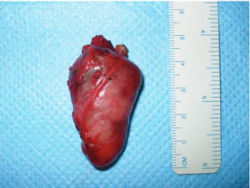
Figure 3 - Surgical specimen after resection
Cytological analysis using Giemsa stain showed numerous cells with round or oval nuclei, finely granular chromatin, and scanty cytoplasm. The cells were grouped in single layers around branched capillaries. An immunohistochemical study was performed on the cell block: it tested positive for chromogranin, synaptophysin, and vimentin; it tested negative for thyroglobulin, calcitonin, parathyroid hormone, and cytokeratins. These results suggested a diagnosis of paraganglioma. Urinary tests for catecholamines were normal.
The patient was then submitted to a cervicotomy through a traditional thyroidectomy incision. During surgery, a reddish tumor was easily dissected from the postero-lateral aspect of the right thyroid lobe, which was preserved (Figures 2 and 3). The recurrent laryngeal nerve was identified and preserved using standard procedures. No relationship was observed between the tumor and this nerve or any of its branches. No excessive bleeding
Table 1 - Immunohistochemical panel of antibodies of the present case
ANTIGEN RESULT
Vimentin (monoclonal antibody V9) positive
Chromogranin positive
Synaptophysin positive
CD56 positive
S-100 Protein positive in sustentacular cells
Thyroglobulin negative
Cytokeratins (monoclonal antibodies AE1/AE3)
negative
847
CLINICS 2008;63(6):845-8 Unusual location of a cervical paraganglioma
Pinto FR et al.
medial aspect of the thyroid capsule and may be in contact with the trachea. This finding explained most cases of thyroid paragangliomas.5-9,19-29,31
The first remarkable observation in the present case was the asymptomatic anterior neck swelling reported by the patient. This complaint may suggest a benign goiter. However, the immobility of the mass during deglutition caught our attention as a possible indicator of a non-thyroidal disease. The imaging tests, particularly the CT scan, demonstrated clearly that the tumor was not inside the thyroid gland. The fine needle aspiration biopsy together with an immunohistochemical panel of antibodies was essential to figure out the diagnosis. Positive results for chromogranin, synaptophysin, and vimentin, coupled with negative results for thyroglobulin, calcitonin, and cytokeratins were strongly suggestive of a paraganglioma, as described in the literature.3,8,14,16,22-31 Among the disorders most
easily mistaken for cervical paragangliomas, particularly those arising within the larynx or the thyroid, are the neuroendocrine carcinomas, such as atypical carcinoids and medullary thyroid carcinoma.14,16,19,22-29,31 As postulated by Martinez-Madrigal et al.,3 the anti-cytokeratin antibody is the single most useful marker for differentiating between paraganglioma and neuroendocrine carcinomas. Both atypical carcinoids and medullary thyroid carcinoma often exhibit diffuse positivity for cytokeratins. Another disorder that may be confused with paragangliomas is hyalinizing trabecular adenoma, a benign thyroid neoplasm that shows a prominent nesting pattern resembling a paraganglioma.24,25,29 This rare entity is usually negative for chromogranin and positive for thyroglobulin and cytokeratins.
During surgery in the present case, the tumor was easily dissected from the thyroid gland and the common carotid artery. No relationship was observed between the tumor and the recurrent laryngeal nerve. Although the patient’s gender (female) and age (40s and 50s), as well as the laterality of the tumor (right side predominance), favored the diagnosis of an inferior laryngeal paraganglioma,9,14-16 the site where this tumor developed could not be explained by previous anatomical descriptions of where the inferior laryngeal paraganglia was located. The lesion was too far from the carotid body to be considered a displaced carotid body tumor. To our knowledge, only one prior report has described a similar localization for a cervical paraganglioma.30 Those authors described a 3.5-cm paraganglioma located immediately posterior to the left thyroid lobe, but they could not dissect it from the gland, which was therefore partially removed. Other previous papers have reported paragangliomas located within the thyroid,5,7-9,21-29,31 between the gland and the trachea,15 and even over the thyroid cartilage.6,20 All of these locations may be explained by anatomical studies documenting the distribution of cervical paraganglionic tissue.
The histopathological and immunohistochemical findings of the specimen confirmed the diagnosis of paraganglioma in the present case. The classic organoid Zellballen pattern, composed of nests of chief cells surrounded by sustentacular cells, was observed, consistent with most previous papers.3,5,7,8,14-19,22-27,29-31 The diagnosis was further supported by diffuse positivity for chromogranin and synaptophysin in the chief cells, a focal positivity for S-100 protein in the sustentacular cells, and negativity for cytokeratins. Despite the absence of histopathological signs of aggressiveness, malignance cannot be excluded based only on morphological criteria. The diagnosis of a malignant paraganglioma is based on the detection of regional or distant metastasis and is not related to the histology of the primary tumor. Hence, all patients submitted to a paraganglioma resection must be
Figure 4 - Classic nesting pattern (Zellballen) of the paraganglioma of the
present case Clusters of tumors cells are surrounded by capillaries in an organoid pattern (H & E, 100x)
Figure 5 - Immunohistochemical findings. Diffuse positivity for
848
CLINICS 2008;63(6):845-8 Unusual location of a cervical paraganglioma
Pinto FR et al.
observed in regular follow-up exams to rule out recurrence or metastasis. However, according to the literature, the ratio of proven cervical malignant paragangliomas is low: it ranges from 2% to 10% for carotid and vagal body tumors, to less than 2% for laryngeal paragangliomas.16,19
In conclusion, we have described here an atypical location of a cervical paraganglioma. This tumor probably originated
from an ectopic inferior laryngeal paraganglia or perhaps from a novel paraganglionic tissue not yet described. This description supports the idea that the anatomical distribution of human paraganglia is incompletely understood. We expect that, with the development and spread of immunohistochemical techniques, new and unusual sites where paragangliomas can arise will be described in the future.
1. Tischler AS, Kimura N, Mcnicol AM. Pathology of pheochromocytoma and extra-adrenal paraganglioma. Ann N Y Acad Sci. 2006;1073:557-70. 2. Paris J, Facon F, Thomassin JM, Zanaret M. Cervical paragangliomas: neurovascular surgical risk and therapeutic management. Eur Arch Otorhinolaryngol. 2006;263:860-5.
3. Martinez-Madrigal F, Bosq J, Micheau C, Nivet P, Luboinski B. Paragangliomas of the head and neck: immunohistochemical analysis of 16 cases in comparison with neuroendocrine carcinomas. Pathol Res Pract. 1991;187:814-23.
4. Plouin PF, Gimenez-Roqueplo AP. Initial work-up and long-term follow-up in patients with phaeochromocytomas and paragangliomas. Best Pract Res Clin Endocrinol Metab. 2006;20:421-34.
5. Haegert DG, Wang NS, Farrer PA, Seemayer TA, Thelmo W. Non-chromaffin paragangliomatosis manifesting as a cold thyroid nodule. AJCP. 1974;61:561-70.
6. Kay S, Montague JW, Dodd RW. Nonchromaffin paraganglioma (chemodectoma) of thyroid region. Cancer. 1975;36:582-5.
7. Mitsudo SM, Grajower MM, Balbi H, Silver C. Malignant paraganglioma of the thyroid gland. Arch Pathol Lab Med. 1987;111:378-80. 8. LaGuette J, Matias-Guiu X, Rosai J. Thyroid paraganglioma: a
clinicopathologic and immunohistochemical study of three cases. Am J Surg Pathol. 1997;21:748-53.
9. Aribas OK, Kanat F, Avunduk MC. Inferior laryngeal paraganglioma presenting as plunging goiter. Eur J Cardiothorac Surg. 2004;25:655-7. 10. Watzka M. Über die paraganglien in der plica ventricularis des
menschlichen kehlkopfes. Dtsch Med Forsch. 1963;1:19-20. 11. Kleinsasser O. Das glomus laryngicum inferior - Ein bisher unbekanntes,
nichtchromaffines Paraganglion vom Bau der sogenannten: Carotisdruse im menschlichen Kehlkopf. Arch Ohren Nasen und Kehlkopfheilk. 1964;184:214-24.
12. Zak FG, Lawson W. Glomic (paraganglionic) tissue of the larynx and capsule of the thyroid gland. Mt Sinai J Med. 1972;39:82-90. 13. Lawson W, Zak FG. The glomus bodies (“paraganglia”) of the human
larynx. Laryngoscope. 1974;84:98-111.
14. Barnes L. Paraganglioma of the larynx. A critical review of the literature. ORL J Otorhinolaryngol Relat Spec. 1991;53:220-34.
15. Brandwein M, Levi G, Som P, Urken ML. Paraganglioma of the inferior laryngeal paraganglia. Arch Otolaryngol Head Neck Surg. 1992;118:994-6.
16. Ferlito A, Barnes L, Wenig BM. Identification, classification, treatment, and prognosis of laryngeal paraganglioma: review of the literature and eight new cases. Ann Otol Rhinol Laryngol. 1994;103:525-36.
REFERENCES
17. Peterson KL, Fu YS, Calcaterra T. Subglottic paraganglioma. Head Neck. 1997;19:54-6.
18. Liew SH, Leong ASY, Tang HMK. Tracheal paraganglioma: a case report with review of the literature. Cancer. 1981;47:1387-93. 19. Hinojar AG, Prieto JR, Muñoz E, Hinojar AA. Relapsing paraganglioma
of the inferior laryngeal paraganglion: case report and review of the literature. Head Neck. 2002;24:95-102.
20. Banner B, Morecki R, Eviatar A. Chemodectoma in the mid-thyroid region. J Otolaryngol. 1979;8:271-3.
21. Cayot F, Bastien H, Justrabo E, Mottot C, Cuisenier J, Bruchon Y, Cabanne F. Paragangliomes multiples du cou avec localization thyroïdienne. Cancer papillaire thyroïdien et adenoma parathyroïdien associés. Sem Hop. 1982;58:2004-7.
22. de Vries EJ, Watson CG. Paraganglioma of the thyroid. Head Neck. 1989;11;462-5.
23. Brownlee RE, Shockley WW. Thyroid paraganglioma. Ann Otol Rhinol Laryngol. 1992;101:293-9.
24. Hughes JH, El-Mofty S, Sessions D, Liapis H. Primary intrathyroidal paraganglioma with metachronous carotid body tumor: report of a case and review of the literature. Pathol Res Pract. 1997;193:791-6. 25. Bizollon MH, Darreye G, Berger N. Paragangliome thyroïdien: à propos
d’un cas. Ann Pathol. 1997;17:416-8.
26. Kronz JD, Argani P, Udelsman R, Silverberg L, Westra WH. Paraganglioma of the thyroid: two cases that clarify and expand the clinical spectrum. Head Neck. 2000;22:621-5.
27. Vodovnik A. Fine needle aspiration cytology of primary thyroid paraganglioma. Report of a case with cytologic, histologic and immunohistochemical features and differential diagnostic considerations. Acta Cytol. 2002;46:1133-7.
28. Zantour B, Guilhaume B, Tissier F, Louvel A, Jeunemaitre X, Gimenez-Roqueplo AP, Bertagna X. A thyroid nodule revealing a paraganglioma in a patient with a new germline mutation in the succinate dehydrogenase B gene. Eur J Endocrinol. 2004;151:433-8.
29. Corrado S, Montanini V, De Gaetani C, Borghi F, Papi G. Primary paraganglioma of the thyroid gland. J Endocrinol Invest. 2004;27:788-92.
30. Schmit GD, Gorman B, van Heerden JA, Gharib H. Inferior laryngeal paraganglioma mimicking a primary thyroid tumor. Endocr Pract. 2006;12:432-5.