CLINICAL SCIENCE
Markers of autoimmune liver diseases in
postmenopausal women with osteoporosis
Umit Secil Demirdal,IIhsan Hakkı Ciftci,IIVural KavuncuI
IDepartment of Physical Medicine and Rehabilitation, School of Medicine, Afyon Kocatepe University, Afyonkarahisar, Turkey. IIDepartment of
Microbiology, School of Medicine, Afyon Kocatepe University, Afyonkarahisar, Turkey.
INTRODUCTION: Osteoporosis is a common complication of chronic liver diseases. However, there is limited information about autoimmune liver diseases as a factor of secondary osteoporosis. Therefore, we aimed to investigate the autoantibodies of autoimmune liver diseases in patients with osteoporosis.
METHODS: One hundred fifty female patients with postmenopausal osteoporosis were included. Bone mineral density was measured by dual energy X-ray absorptiometry. We analysized autoantibodies including antinuclear antibodies, liver membrane antibodies, liver/kidney microsomal autoantibodies1, liver-specific protein, anti-smooth muscle antibodies, and anti-mitochondrial antibodies by indirect immunofluorescence. Serum was assayed for the levels of aminotransferases.
RESULTS:The mean age of the patients was 63,13¡8,6 years. The mean values of L1-L4 T-scores and femur total T-scores were -3,08¡0,58 and -1,53¡0,81, respectively. Among the 150 patients with osteoporosis, 14 (9.3%) were antinuclear antibodies, four (2.7%) were liver membrane antibodies, three (2.0%) were anti-liver/kidney microsomal autoantibodies1, and two (1.3%) were liver-specific protein positive. None of the patients had anti-mitochondrial antibodies or smooth muscle antibodies positivity. The mean values of levels of aminotransferases were within normal range.
CONCLUSIONS:The presence of liver membrane antibodies, liver-specific protein, and anti-liver/kidney microsomal autoantibodies1 has permitted us to see that there may be some suspicious clues of autoimmune liver diseases in patients with osteoporosis as a secondary risk factor. On the other hand, there is a need for comprehensive studies with a larger sample size and studies designed to compare the results with a normal population to understand the clinical importance of our findings.
KEYWORDS: Osteoporosis; Autoimmune; Liver disease; liver autoantibodies; Aminotransferases.
Demirdal US, Ciftci IH, Kavuncu V. Markers of autoimmune liver diseases in postmenopausal women with osteoporosis. Clinics. 2010;65(10):971-974.
Received for publication onMay 2, 2010;First review completed onMay 25, 2010;Accepted for publication onJuly 13, 2010 E-mail: [email protected]
Tel.:+90 272 246 33 33-3012
INTRODUCTION
Osteoporosis (OP) is usually defined as a skeletal disease characterized by low bone mass and microarchitectural deterioration of bone tissue, leading to enhanced bone fragility and increase in fracture risk.1It affects up to one in two women and one in five men over the age of 50.2It is a worldwide disease seen in all racial groups and in both males and females.3In addition to being a common disease, OP becomes a serious health issue due to the increase in morbidity, mortality, and financial burden related with osteoporotic fractures.4-6
Osteoporosis can be classified as primary and secondary OP according to the underlying causes. Secondary OPmay be described as the low mineral density in which an
underlying cause or factor can be defined other than those attributable to the postmenopausal state or aging.7Primary
OPrefers to OP when a secondary cause cannot be found.8 There are a number of secondary causes of osteoporosis such as hypogonadism or hyperparathyroidism, which are treatable, and renal failure, which should be considered more seriously. Chronic liver diseases also play an important role among secondary factors of OP.9,10
Bone disease is a major complication of chronic liver disease. OP is more commonly seen in patients with liver diseases as compared to the normal population, showing variable prevalence according to the patient selection and diagnostic criteria.11,12 OP is a common complication of chronic liver diseases such as cholestatic disorders, alcoholic liver diseases, posthepatitic cirrhosis, and autoimmune liver diseases (ALD).13Types of ALD are autoimmune hepatitis (AH), primary biliary cirrhosis (PBC), and sclerosing cholangitis. ALD affects women at any age with a wide range of clinical presentations. In some cases, the disease is incidentally diagnosed during routine laboratory tests, while it shows a fulminate course in some others.14 Copyrightß2010CLINICS– This is an Open Access article distributed under
the terms of the Creative Commons Attribution Non-Commercial License (http:// creativecommons.org/licenses/by-nc/3.0/) which permits unrestricted non-commercial use, distribution, and reproduction in any medium, provided the original work is properly cited.
CLINICS 2010;65(10):971-974 DOI:10.1590/S1807-59322010001000008
One of the main characteristics of the disease is the presence of circulating autoantibodies. It has been sug-gested that liver membrane antibodies (LMA) and liver specific protein (LSP) are associated with ALD.15,16Defined antibodies are antinuclear antibodies (ANA), anti-smooth muscle antibodies (SMA), and anti-liver/kidney microso-mal autoantibodies1 (anti-LKM1) in addition to perinuclear antineutrophil cytoplasmic antibodies (pANCA), antibodies to liver-cytosol type 1 (anti-LC-1), autoantibodies to soluble liver antigen/liver pancreas antigen (SLA/LP), anti-bodies to the asialoglycoprotein receptor (anti-ASGPR), and anti-mitochondrial antibodies (AMA).14,17
The prevalence of OP is higher in patients with PBC, and osteoporotic fractures are more prevalent in patients with AH treated with glucocorticoids.18However, it is not clear whether or not chronic liver diseases – especially ALD – are related to the secondary OP. There are a few studies investigating liver diseases as a secondary cause of OP.8,19 Therefore, we aimed to assess the circulating autoantibodies related to ALD in patients with OP.
MATERIALS AND METHODS
The study was conducted at Afyon Kocatepe University Hospital following the approval of the Ethical Committee for Medical Research of the university. Postmenopausal women age 50 or older with OP were included in the study. Two hundred and eighty-seven postmenopausal women were evaluated. Bone mineral density (BMD) was measured by dual energy X-ray absorptiometry (DXA, HOLOGIC Q DR 4500 W) both at the lumbar spine (anteroposterior projection of L1–L4), and the proximal femur (total score) if the patients were 50 years or older. Osteoporosis is defined as T score below -2.5 at any site according to WHO guidelines.1 Smoking, alcohol use, medication (especially drugs with high risk of liver toxicity), malignancy, history of gastrointestinal system (thyroid gland, kidney, and liver diseases), rheumatologic conditions, and nutrition were questioned in detail. Patients with secondary risk factors and anatomic deformity in lumbar and femoral region that affects BMD were excluded. In total, 287 postmenopausal women were evaluated; of these, 150 presented the criteria for participation in this study.
Venous blood samples were drawn in the morning after an overnight fast. Serum levels of aminotransferases (aspartate aminotransferase-AST, alanine aminotransferase-ALT), cal-cium (Ca), phosphate (PO4), alkaline phosphatase (ALP), parathyroid hormone (PTH), 25-(OH) vitamin D, osteocalcin (OC), and serum C-telopeptide cross-linked collagen type I (CTX) were measured on the same day. Serum PTH was measured by immunoradiometric assay (IRMA, USA). Serum CTX was measured by electrochemiluminescence immunoassay (ECLIA, Germany). Serum osteocalcin was measured with an RIA (radioimmunoassay) technique (DiaSorin, Saluggia, Italy). The level of 25-(OH) vitamin D was measured by radioimmunoassay (RIA) (Nichols Institute Diagnostics, USA). The levels of Ca, PO4, and ALP were measured by colorimetric method. Serum was assayed using an autoanalyzer to measure the levels of aminotransferases. All blood tests were performed in a single laboratory using reference ranges of this laboratory’s data.
Serum samples were analyzed for autoantibodies by indirect immunofluorescence (IIF) on a substrate kit (Euroimmun, Germany) that included
fluorescein-conju-gated goat antibodies to human immunoglobulin G (IgG). IIF patterns were read at serum dilutions of 1:100 for ANA, SMA, AMA, and anti-LKM1 positivity on a Zeiss Axioskop (Carl Zeiss, Jena, Germany) by the same experienced microbiologist. LMA and LSP were tested by the same method with minimal titers for positivity being 1:80. The levels of aminotransferases were re-examined when auto-antibody positivity was determined.
The analyses were carried out using SPSS version 13.5. Descriptive analysis was carried out and data were expressed as percentages (%), means, and standard devia-tions.
RESULTS
Two hundred and eighty-seven postmenopausal women were evaluated and, of these, 150 patients were found to be eligible to include in this study. The mean age of the patients was 63,13¡8,6 years. The mean values of L1-L4
T-scores and femur total T-scores, were -3,08¡0,58 and
-1,53¡0,81, respectively. Upon evaluation of the question-naires, none of the patients had signs or symptoms of liver diseases.
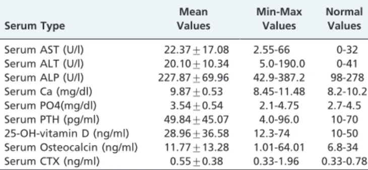
The mean values of the enzymes reflecting liver functions and markers of bone metabolism are shown in Table 1. The mean values for all the laboratory tests were within the normal range.
Among the 150 patients with OP, 14 (9.3%) were ANA, four (2.7%) were LMA for 1:80 titers, three (2.0%) were anti-LKM1, and two (1.3%) were LSP positive. None of the patients had AMA or SMA positivity. Serological para-meters are summarized in Table 2.
Only one patient was found to have positivity for two and more autoantibodies (LMA and anti-LKM1). In laboratory examination of the patient, all biochemical tests were within the normal range.
Table 1-Values of serum aminotransferases and bone
metabolism markers.
Serum Type
Mean Values
Min-Max Values
Normal Values
Serum AST (U/l) 22.37¡17.08 2.55-66 0-32
Serum ALT (U/l) 20.10¡10.34 5.0-190.0 0-41
Serum ALP (U/l) 227.87¡69.96 42.9-387.2 98-278
Serum Ca (mg/dl) 9.87¡0.53 8.45-11.48 8.2-10.2
Serum PO4(mg/dl) 3.54¡0.54 2.1-4.75 2.7-4.5
Serum PTH (pg/ml) 49.84¡45.07 4.0-96.0 10-70
25-OH-vitamin D (ng/ml) 28.96¡36.58 12.3-74 10-50 Serum Osteocalcin (ng/ml) 11.77¡13.28 1.01-64.01 6.8-34
Serum CTX (ng/ml) 0.55¡0.38 0.33-1.96 0.33-0.78
Table 2 -Liver autoantibodies positivity (%) in patients with osteoporosis
Positivity Autoantibodies n (150) %
ANA 14 9.3
LMA 4 2.7
Anti-LKM1 3 2.0
LSP 2 1.3
SMA 0 0
AMA 0 0
Lıver autoantıbodıes ın patients with osteoporosis
Demirdal US et al. CLINICS 2010;65(10):971-974
Two patients were found to have positive for both ANA and higher ALT levels, whereas three patients had higher ALT levels without autoantibodies positivity. AST and ALT levels were within normal range in patients with the other autoantibodies positivity.
DISCUSSION
In this study, we investigated the autoantibodies of ALD in patients with OP. Among the 150 patients with OP, 14 (9.3%) were ANA, four (2.7%) were LMA, three (2.0%) were anti-LKM1, and two (1.3%) were LSP positive. None of the patients had AMA or SMA positivity. The levels of amino-transferases of the patients with autoantibodies positivity were within normal range.
Assessment of patients with OP is usually focused on the measurement of bone mineral density and investigation of bone metabolism markers. However, factors that affect the bone structure are various, and some should be kept in mind even if they have no place in routine laboratory evaluation. Dimutrescu et al. denote that 32% to 37% of women with low BMD have a history of other diseases or medications known to contribute to OP.20 Among women with OP, between 30% and 60% have a secondary cause.9 Liver diseases are one of the secondary causes investigated in a limited number of studies. In one study, 272 patients with OP, 96% of whom were women, were examined by laboratory evaluation for liver diseases in addition to endocrinologic disorders such as osteomalacia or hyperpar-athyroidism and hematologic diseases such as anemia. They mentioned that no unknown liver diseases were found in their study cohort.19 Melton et al. investigated secondary
causes, including cirrhosis, in women and men with OP and osteopenia. Cirrhosis was not found in study cases, but one patient in the control group was found to have cirrhosis.21 Odabasi et al. found that three patients had chronic liver diseases among 947 postmenopausal women with OP when examined for secondary causes.8 Among the studies mentioned above, ALD or definite autoantibodies of ALD were not investigated. It is obvious that the diagnosis of ALD is not totally dependent on the presence of auto-antibodies; however, autoantibody positivity is the clue to the disease. In the view of this, the present study is a rare example of the studies investigating liver diseases in patients with OP.
Among the 150 patients with OP, 14 (9.3%) were positive for ANA at 1:100 titers. ANAs were the first autoantibodies observed in AH and are still the most sensitive marker of the disease. On the other hand, ANA positivity is not specific for AH because positivity may occur in patients with other diseases and even in healthy subjects.22,23The prevalence of ANA positivity is variable in the healthy population. In spite of the variability, the prevalence is age and sex dependant. Elderly over 60 years and females have relatively high frequencies of ANA. It is estimated that 10% to 15% of healthy people over the age of 65 are ANA positive, and the titers are usually# 1:160.23,24Consistent with the literature, all our cohort were women, the mean age of the patients was 63,13¡8,6 years, and 9.3% of the patients
were found ANA positivity for the 1:100 titers. Thereby, the age and sex of the cohort may be the causes of the highest prevalence of autoantibodies positivity for ANA. Tan et al. describe that there is no cut-off value that can reliably distinguish between normal and diseased populations.
However, it is considered that some healthy individuals have low-titer ANA.23-25In the present study, it is accepted that 1:100 titers is enough for ANA positivity; the maximum titers of ANA was not studied. Therefore, this cut-off value was not enough to distinguish the pathologic value of ANA positivity of the cohort. The abnormal levels of liver function tests with ANA positivity may be useful for the association with AH. Only two patients were positive for ANA with ALT levels higher than upper limits. These patients had no history for signs or symptoms of any liver diseases. Therefore, clinical presentation of AH for the patients was not considered.
Previous studies mention the diagnostic importance of LSP and LMA in ALD.15,26,27In our study population, four
(2.7%) patients were found to have positive for LMA and two (1.3%) for LSP. The biochemical assessments of the patients were within the normal range. The findings may be coincidental, but AH should be considered for patients when the elevated serum levels of aminotransferases are determined during the follow-up period. LMA were also found in some cases of alcoholic liver diseases,15but one of the exclusion criteria of the study was alcohol use.
Anti-LKM1 typically occurs in the absence of SMA and ANA in patients with AH.28In accordance with this data,
two patients were found to have with anti-LKM1 positivity in the absence of SMA and ANA positivity. In Europe, anti-LKM1 are found mainly in pediatric patients with AH and are demonstrated in only 20% of adults with the disease.28 The result that 2% of women without history of liver disease were found to positive for anti-LKM1 suggests that we follow up the patients after a period of time.
The detection of AMA is nearly diagnostic of PBC, even in the absence of symptoms. AMA positivity with normal liver tests may be incidental and reflects a normal 0.5% incidence in the general population.29,30 Among the patients of the
present study, AMA positivity was not detected. A possible explanation for the apparent difference between the result presented in this study and the number considered for the normal population (0.5%) may be the sample size. SMA facilitates diagnosis of AH and discriminates subtypes of the disease.22 No patients were found to have SMA positivity in the present study.
Jamal et al. investigated the clinical utility of laboratory testing including serum aminotransferases to assess if an underlying medical condition is contributing to bone loss. They report that the prevalence of abnormal liver function tests in women with OP compared to women without was not different.31 Among the subjects included the present study, the mean levels of AST and ALT were within the normal range. Analyzing aminotransferases may be helpful to determine the abnormal laboratory tests findings condi-tional upon a diagnosis of liver diseases. On the other hand, this analysis has not permitted us to obtain any additional information because two patients were found to have positive for both ANA and higher ALT levels, whereas three patients had higher ALT levels without any auto-antibodies positivity.
Our study has several limitations. First, the clinical importance of our findings seems debatable. The clinical importance can be discussed if it is possible to compare the prevalence of autoantibodies detected in the study popula-tion with the prevalence in age-matched postmenopausal women without OP. Unfortunately, the number of volun-teers without OP was not enough to constitute a control CLINICS 2010;65(10):971-974 Lıver autoantıbodıes ın patients with osteoporosis Demirdal US et al.
group for this study. In addition, there were no similar studies to compare our findings. Notwithstanding, the percentage of patients with positive autoantibodies may be significant. On the other hand, these findings could also attributed to normal variables or could be considered as coincidental. Studies with larger sample sizes may help us to elucidate the clinical importance of our findings. The second limitation of the study is adapting our findings to clinical practice. Our results may not be generalized, and similar studies are required to determine whether labora-tory testing should include autoantibodies as a marker of secondary causes of OP. Finally, the sample size is relatively small – the population of the study was derived from a single academic center– whereas OP is a widespread disease. Furthermore, our exclusion of subjects ineligible to participate in the study may have caused a smaller sample size than hoped.
CONCLUSIONS
This study assessed the presence of definite autoantibo-dies related with ALD in patients with OP. Regardless of the most frequently detected antibody-ANA in the present study, the presence of LMA, LSP, and anti-LKM1 has permitted us to see there may be some suspicious clues of ALD in patients with OP as a secondary risk factor. On the other hand, the clinical importance of the detected auto-antibodies is unclear. There is a need for comprehensive studies with a larger sample size and a design to compare the results with normal population to understand if the findings are normal variability or coincidental.
REFERENCES
1. Assessment of fracture risk and its application to screening for postmenopausal osteoporosis. 1994. Geneva: WHO Study Group. Technical Report 843.
2. Keen R. Osteoporosis: Strategies for prevention and management. Best Pract Res Clin Rheumatol. 2007; 21:109–22, doi: 10.1016/j.berh.2006.10. 004.
3. Wilkins CH. Osteoporosis screening and risk management. Clin Interv Aging. 2007;2:389–94.
4. Fink HA, Ensrud KE, Nelson DB, Kerani RP, Schreiner PJ, Zhao Y, et al. Disability after clinical fracture in postmenopausal women with low bone density: The fracture intervention trial (FIT). Osteoporos Int. 2003;14:69–76, doi: 10.1007/s00198-002-1314-y.
5. Johnell O, Kanis JA, Oden A, Sernbo I, Redlund-Johnell I, Petterson C, et al. Mortality after osteoporotic fractures. Osteoporos Int. 2004;15:38– 42, doi: 10.1007/s00198-003-1490-4.
6. Ethgen O, Tellier V, Sedrine WB, De Maeseneer J, Gosset C, Reginster JY. Health-related quality of life and cost of ambulatory care in osteoporosis: How may such outcome measures be valuable information to health decision-makers and payers? Bone. 2003; 32:718–24, doi: 10.1016/S8756-3282(03)00089-9.
7. Satomi E, Sitta Mdo C, Machado AN, Garcez Leme LE. Identification and treatment of osteoporosis among elderly patients with hip fractures. Clinics. 2009;64:1201-4, doi: 10.1590/S1807-59322009001200010. 8. Odabası E, Turan M, Tekbas F, Kutlu M. Evaluation of secondary causes
that may lead to bone loss in women with osteoporosis: A retrospective study. Arch Gynecol Obstet. 2009;279:863–7, doi: 10.1007/s00404-008-0846-6.
9. Lash RW, Nicholson JM, Velez L,Van Harrison R, McCort J. Diagnosis and management of osteoporosis. Prim Care. 2009;36:181-98, doi: 10. 1016/j.pop.2008.10.009.
10. Sikon AL, Thacker HL, Carey J, Deal C, Licata AA. Secondary osteoporosis: Are we recognizing it? J Womens Health (Larchmt). 2006;15:1174–83, doi: 10.1089/jwh.2006.15.1174.
11. Leslie WD, Bernstein CN, Leboff MS; , American Gastroenterological Association Clinical Practice Commitee. AGA technical review on osteoporosis in hepatic disorders. Gastroenterology 2003;125:941–66, doi: 10.1016/S0016-5085(03)01062-X.
12. Wariaghli G, Mounach A, Achemlal L, Benbaghdadi I, Aouragh A, Bezza A, et al. Osteoporosis in chronic liver disease: A case-control study. Rheumatol Int. 2009. [Epub ahead of print]
13. Mounach A, Ouzzif Z, Wariaghli G, Achemlal L, Benbaghdadi I, Aouragh A, et al. Primary biliary cirrhosis and osteoporosis: A case-control study. J Bone Miner Metab. 2008;26:379–84, doi: 10.1007/s00774-007-0833-1.
14. Manns MP, Vogel A. Autoimmune hepatitis, from mechanisms to therapy. Hepatology 2006;43:132-44, doi: 10.1002/hep.21059.
15. Hopf U, Jahn HU, Mo¨ller B, Stemerowicz R, Wittenbrink C, Klein R, et al. Liver membrane antibodies (LMA) recognize a 26-kD protein on the hepatoceluler surface. Clin Exp Imunol. 1990;79:54–61, doi: 10.1111/j. 1365-2249.1990.tb05126.x.
16. McFarlane IG, McFarlane BM, Major GN, Tolley P, Williams R. Identification of the hepatic asialo-glycoprotein receptor (hepatic lectin) as a component of liver specific membrane lipoprotein (LSP). Clin Exp Immunol. 1984;55:347–54.
17. Teufel A, Galle PR, Kanzler S. Update on autoimmune hepatitis. World J Gastroenterol. 2009;15:1035–41.
18. Pare´s A, Guan˜abens N. Treatment of bone disorders in liver disease. J Hepatol. 2006;45:445–53.
19. Barzel US. Recommended testing in patients with low bone density. J Clin Endocrinol Metab. 2003;88:1404–5, doi: 10.1210/jc.2002-021660. 20. Dumitrescu B, van Helden S, ten Broeke R, Nieuwenhuijzen-Kruseman
A, Wyers C, Udrea G, et al. Evaluation of patients with a recent clinical fracture and osteoporosis, a multidisciplinary approach. BMC Musculoskelet Disord. 2008;9:109, doi: 10.1186/1471-2474-9-109. 21. Melton LJ III, Atkinson EJ, Khosla S, O’Fallon WM, Riggs BL. Secondary
osteoporosis and the risk of vertebral deformities in women. Bone. 1999;24:49–55, doi: 10.1016/S8756-3282(98)00150-1.
22. Invernizzi P, Lleo A, Podda M. Interpreting serological tests in diagnosing autoimmune liver diseases. Semin Liver Dis. 2007;27:161– 72, doi: 10.1055/s-2007-979469.
23. Muro Y. Antinuclear antibodies. Autoimmunity 2005;38:3–9, doi: 10. 1080/08916930400024612.
24. Lyons R, Narain S, Nichols C, Satoh M, Reeves WH. Effective use of autoantibody tests in the diagnosis of systemic autoimmune disease. Ann NY Acad Sci. 2005;1050:217–28, doi: 10.1196/annals.1313.023. 25. Wiik AS. Anti-nuclear autoantibodies: Clinical utility for diagnosis,
prognosis, monitoring, and planning of treatment strategy in systemic immunoinflammatory diseases. Scand J Rheumatol. 2005;34: 260–8. 26. Chattopadhyay D, Sen MR, Aryya NC. Immunohistopathological
reactions for liver-specific membrane lipo-protein in experimental autoimmune hepatitis. Indian J Pathol Microbiol. 1999;42:291–7. 27. Meyer zum Bu¨schenfelde KH, Manns M, Hu¨tteroth TH, Hopf U, Arnold
W. LM-Ag and LSP—two different target antigens involved in the immunopathogenesis of chronic active hepatitis? Clin Exp Immunol. 1979;37:205–12.
28. Czaja AJ, Norman GL. Autoantibodies in the diagnosis and management of liver disease. J Clin Gastroenterol. 2003;37:315–29, doi: 10.1097/ 00004836-200310000-00011.
29. Muratori L, Granito A, Muratori P, Pappas G, Bianchi FB. Anti-mitochondrial antibodies and other antibodies in primary biliary cirrhosis: Diagnostic and prognostic value. Clin Liver Dis. 2008;12:261–76, doi: 10. 1016/j.cld.2008.02.009.
30. Kumagi T, Onji M. Presentation and diagnosis of primary biliary cirrhosis in the 21st century. Clin Liver Dis. 2008;12:243–59, doi: 10.1016/ j.cld.2008.02.014.
31. Jamal SA, Leiter RE, Bavoumi AM, Bauer DC, Cummings SR. Clinical utility of laboratory testing in women with osteoporosis. Osteoporosis Int. 2005;16:534–40, doi: 10.1007/s00198-004-1718-y.
Lıver autoantıbodıes ın patients with osteoporosis
Demirdal US et al. CLINICS 2010;65(10):971-974