ISSN 1557-4555
© 2011 A.E. Hefnawy et al., This open access article is distributed under a Creative Commons Attribution (CC-BY) 3.0 license
Corresponding Author: Abd Elghany Hefnawy, Department of Internal Medicine, Faculty of Veterinary Medicine, Benha University, 13736 Moshtohor, Egypt Tel: +20132460640 Fax: +20132463074
88
Effect of Gestation and Maternal Copper on the Fetal
Fluids and Tissues Copper Concentrations in Sheep
1
Abd Elghany Hefnawy,
2Jorge Tortora-Perez,
3Saad Mohamed Shousha
and
4Seham Youssef AbuKora
1
Department of Internal Medicine, Faculty of Veterinary Medicine,
Benha University, 13736 Moshtohor, Egypt
2
Faculty Of High Studies, Cuautitlan UNAM, Cuautitlan,
Teoloyucan Cuautitlan Izcalli, 54700 Mexico
3
Department of Physiology,
4
Department of Pharmacology,
Faculty of Veterinary Medicine, Benha University,
13736 Moshtohor, Egypt
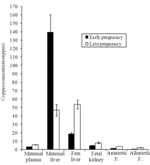
Abstract: Problem statement: Samples of allantoic, amniotic fluid, fetal liver, kidney, maternal plasma and liver were collected from 30 ewes and classified into either early or late gestation and copper concentrations were measured. Approach: The Cu concentrations in the maternal plasma, allantoic, amniotic fluid, fetal liver and kidney increased significantly (p<0.01) during late gestation while maternal liver Cu decreased significantly (p<0.01). Results: Significant positive relationships were recorded between age of the fetus and Cu concentrations in the allantoic and amniotic fluid (r = 0.71-0.83, p<0.001), fetal liver (r = 0.80, p<0.001), kidney (r = 0.59, p<0.01) and maternal plasma (r = 0.75, p<0.001). Significant (p<0.01) positive relationships were also recorded between the Cu concentrations in the amniotic, allantoic fluid and maternal plasma with fetal liver Cu concentrations (r = 0.36-0.73), the maternal plasma and liver Cu concentrations were significantly negative correlated (r = -42, p<0.05). Conclusion: A significant negative correlation was recorded between the Cu concentrations in the maternal liver and fetal age (r = -0.74, p<0.01). Strong fetal-maternal relationships in Cu concentration were evident throughout the gestational period and dams seem to sacrifice Cu levels in order to maintain that in the fetus. Cu concentrations in the amniotic and allantoic fluids could be used as a possible indicator of the Cu status of the fetus throughout gestation.
Key words: Maternalcopper, fetal fluids,amniotic fluid, fetal liver, maternal plasma, Cu concentration
INTRODUCTION
Pregnancy is a period of rapid growth and cell differentiation for both the mother and fetus. Consequently, it is a period when both are vulnerable to changes in dietary supply, especially of those nutrients that are marginal under normal circumstances (Gambling and McArdle, 2004). Each fetus is completely dependent on its dam via the placenta for its supply of essential trace elements (Abdelrahman and Kincaid, 1993). Copper is often one of the most limiting trace elements for the fetus and neonate for normal development. Deficiency of this element impairs fetal growth and can cause death (Mertz and Underwood, 1987). Calves normally are born with liver
clearly understood (Solaiman et al., 2001). Hepatic concentrations of trace elements are commonly used to estimate trace element storage pools because dietary intake is rarely available and nutrient interactions affect availability or retention (Hill and Matrone, 1970; Mertz, 1988).
Collection of fetal tissues from local slaughter houses may enable endemic deficiencies of minerals to be determined. However, if fetal tissues are to be used to assess the nutritional status of the dam, the effect of gestational age on the concentrations of minerals in fetal tissues must be known. Therefore, the present study was conducted to estimate and correlate maternal liver, plasma, fetal liver, fetal kidney and amniotic fluid and allantoic fluid copper concentrations and studying the effect of maternal copper on the fetal copper status through gestation in sheep.
MATERIAL AND METHODS
Samples were taken from 30 pregnant singleton Pelibuey sheep of 3-4 years old and their corresponding fetuses, at the time of slaughter. Maternal samples included blood and liver, while the fetal samples included amniotic fluid, allantoic fluid, liver and kidney. All samples were used for Cu determination and investigation the effect of the fetal age and maternal Cu on the Cu concentrations in the fetal fluids and tissue throughout the gestation. Samples were classified according to the estimated age of the fetus (Lyngest, 1971). The early stage of gestation was defined as before day 90 when fetal length was less than 20 cm which included 10 animals at this stage; while the late stage of gestation was defined as after day 90 which included 20 animals at this stage. Ewes have been kept on an adequate Cu diet.
Preparation and analysis of the samples: Maternal blood samples were centrifuged (2000 G; 15 min) to obtain plasma (-20°) for later analyses. Amniotic and allantoic fluid, maternal liver, fetal liver and kidney were frozen (-20°) for Cu determinations.
The plasma, amniotic and allantoic fluid samples were processed by mixing 1 mL of each sample with 10 ml of deionized water, 5 mL of concentrated nitric acid and 2 mL of hydrogen peroxide (30%) (J.T. Baker, Phillipsburg, N. J.)-keeping the solution at room temperature for 30 min in sealed Teflon vessels (Lyngest, 1971). Subsequently, the samples were placed in a microwave digester (Mars 5 CEM Corporation USA) with an increasing temperature slope of 5 min to reach 120°C and it was held in this
temperature for 2 min. The temperature was then increased to 170°C within 5 min and maintained for 2 min with a maximum pressure of 350 psi (Ortman and Pehrson, 1997). The samples were allowed to cool for 5 min in an oven and then left to obtain room temperature (for 1 h). The samples were then transferred to 50 mL volumetric flasks and filled to the top with 7M HCl and left overnight (4°C) to be analyzed the following day. Cu concentrations were determined with the aid of atomic absorption spectrophotometer (Varian, model Spectra AA-800).
Tissue samples of 0.5+0.1 g were digested using a microwave oven using 10 mL of nitric acid and 2 mL of double distilled water in a Teflon vessel (Rowntree et al., 2004; Shaw et al., 2002). Samples were then allowed to be digested for approximately 1h at room temperature. Vessels were then placed in a microwave digester (Mars 5 CEM Corporation USA), in order to gradually increase the pressure to 210 psi with the maximal vessel temperature being 190°C. The vessels were maintained at 210 psi for 10 min, allowed to cool for 10 min and then ventilated. Thereafter 2 mL of hydrogen peroxide (30%) (J.T. Baker, Phillipsburg, N.J) Was added to each tissue sample. The digested samples were transferred to volumetric flasks and brought to a uniform volume of 25 mL with 7M HCl and then stored until analyses. Cu concentrations were determined with the aid of atomic absorption spectrophotometer (Varian, model Spectra AA-800).
Statistical analysis: Means, Pearson correlation coefficient, Analysis Of Variance (ANOVA) by general linear model and regression analyses were performed using the Statistical Analysis System software (SAS, 1985).
RESULTS
90 Fig. 1: Copper concentration (ppm) in maternal plasma,
liver, fetal liver, kidney, amniotic fluid and allantoic fluid in the early and late stage of pregnancy in sheep
Fig. 2: The relationships between maternal liver Cu and fetal liver Cu concentrations with the fetal age in sheep
There was significant (r = -0.50, p<0.01) negative relationship between the fetal and maternal liver Cu concentrations and maternal Cu tended to be significantly higher than fetal Cu in early gestation (p<0.001) while there was no significant changes between maternal and fetal liver Cu concentrations in late gestation. There were significant positive relationships between maternal plasma Cu concentrations with amniotic fluid (r = 0.42, p<0.01),
Table 1: The relationships between Cu concentrations in the fetal liver, kidney, amniotic fluid, allantoic fluid, maternal liver, plasma and age of the fetus in sheep
Age of Maternal Maternal Fetal the fetus plasma Cu liver Cu liver Cu Amniotic fluid Cu 0.83** 0.42* -0.63** 0.73** Allantoic fluid Cu 0.71** 0.48** -0.55** 0.56**
Fetal liver Cu 0.80** 0.36* -0.5** -
Fetal kidney Cu 0.59** NS -0.43* -
Maternal liver Cu -0.74** -0.42* - -0.5**
Maternal plasma Cu 0.75** - -0.42* 0.36*
allantoic fluid (r = 0.48, p<0.01) and fetal liver (r = 0.36, p<0.01) Cu concentrations, while the relationship between maternal plasma and maternal liver Cu concentrations was significantly negative (r = -0.42, p<0.01). There were significant negative relationships between maternal liver Cu concentrations with amniotic fluid (r = -0.63, p<0.001) and allantoic fluid (r = -0.55, p<0.01), fetal liver (r = -0.50, p<0.01) and kidney (r = - 0.43, p<0.01) Cu concentrations. There were significant positive relationships between fetal liver Cu concentration with amniotic fluid (r = 0.73, p<0.001) and allantoic fluid (r = 0.56, p<0.01) Cu concentrations Table 1.
DISCUSSION
Maternal liver Cu was negatively correlated with fetal age, this results agree with the results obtained by (Gonneratne and Christensen, 1989b). While (Graham
In humans, fetal Cu concentrations reportedly increased or remained stable through gestation (Casey and Robinson, 1978; Widdowson et al., 1972). Ovine maternal and fetal liver Cu were negatively correlated in this and previous reports (Gonneratne and Christensen, 1989a). Presence of significant negative relationship between age of the fetus and maternal liver Cu concentration as well as the relationships between maternal liver and amniotic and allantoic fluid Cu concentrations were significantly negative, while the relationship between age of the fetus and maternal plasma, fetal liver, amniotic fluid, allantoic fluid and fetal kidney Cu concentrations were significantly positive may indicate that, the dam and fetus depend on the maternal liver Cu contents during gestation and it can be used as an indicator of the Cu status through gestation and fetuses have a capacity to sequester maternal Cu, even when the dam is Cu deficient (Graham et al., 1994). Parkinson et al. (1981) found that amniotic fluid copper concentration gradually increased during pregnancy.
CONCLUSION
From this study we can concluded that, there is a strong relationship between the fetus and dam concerning Cu metabolism through gestation. The dams seem to sacrifice their Cu level in order to maintain the fetal Cu disposition. Cu concentrations in the amniotic and allantoic fluids play a role in the metabolism and utilization of Cu through gestation and may be used as an indicator of Cu status in the fetus throughout gestation.
REFERENCES
Abdelrahman, M.M. and R.L. Kincaid, 1993. Deposition of copper, manganese, zinc, and selenium in bovine fetal tissue at different stages of gestation. J. Dairy Sci., 76: 3588-3593. DOI: 10.3168/jds.S0022-0302(93)77698-5
Casey, C.E. and M.F. Robinson, 1978. Copper, manganese, zinc, nickel, cadmium and lead in human foetal tissues. Birch J. Nutr., 39: 639-646. PMID: 638131
Gambling, L. and H.J. McArdle, 2004. Iron, copper and fetal development. Proc. Nutr. Soc. 63: 553-562. DOI: 10.1079/PNS2004385
Gonneratne, S.R. and D.A. Christensen, 1989b. A survey of maternal and fetal tissue zinc, iron, manganese and selenium concentrations in bovine. Canadian J. Anim. Sci., 69: 151-159. DOI: 10.4141/cjas89-018
Gonneratne, S.R. and D.A. Christensen. 1989a. A survey of maternal copper status and fetal tissue copper concentrations in Saskatchewan bovine. Canadian J. Anim. Sci., 69: 141-150. DOI: 10.4141/cjas89-017
Graham, T.W., M.C. Thurmond, F.C. Mohr, C.A. Holmberg and M.L. Anderson et al., 1994. Relationships between maternal and fetal liver copper, iron, manganese, and zinc concentrations and fetal development in California Holstein dairy cows. J. Veter. Diagnosis Invest., 6: 77-87. DOI: 10.1177/104063879400600114 PMID: 8011786 Hidiroglou, M. and J.E. Knipfel, 1981. Maternal-fetal
relationships of copper, manganese, and sulfur in ruminants. A review. A review. J. Dairy Sci., 64: 1637-1647. DOI: 10.3168/jds.S0022-0302(81)82741-5
Hill, C.H. and G. Matrone, 1970. Chemical parameters in the study of in vivo and in vitro interactions of transition elements. Feed Proc., 29: 1474-1481. PMID: 5459894
Keen, C.L., J.Y. Uriu-Hare, S.N. Hawk, M.A. Jankowski and G.P. Daston et al., 1998. Effect of copper deficiency on prenatal development and pregnancy outcome. Am. J. Clin. Nutr., l 67: 1003-1011.
Lyngest, O. 1971. Studies on reproduction in the goat. VII. Pregnancy and the development of the foetus and the foetal accessories of the goat. Acta Vet. Scand., 12: 185-201. PMID: 5106994
Mertz, W. and E.J. Underwood, 1987. Trace Elements in Human and Animal Nutrition. 4th Edn., Academic Press, New York, ISBN: 0124912516, pp: 480.
Mertz, W., 1988. Trace Elements in Human and Animal Nutrition. 5th Edn., Academic Press, San Diego, CA., ISBN-10: 0124912516, pp: 480.
Ortman, K. and B. Pehrson, 1997. Selenite and selenium yeast as feed supplements for dairy cows. Zentralbl Veterinarmed A., 44: 373-380. PMID: 9342929
Parkinson, C.E., J.C.Y. Tan, P.J. Lewis and M.J. Bennett. 1981. Amniotic fluid zinc and copper and neural tube defects. J. Obstetric Gynecol., 1: 207-212. Richards, M.P., 1999. Zinc, copper, and iron metabolism during porcine fetal development. Biol. Trace Element Res., 69: 27-44. DOI: 10.1007/BF02783913 PMID: 10383097
92 SAS, 1985. SAS user's guide: Statistics. 5th Edn., SAS
Institute, Cary, NC., ISBN: 0917382668, pp: 956. Shaw, D.T., D.W. Rozeboom, G.M. Hill, A.M. Booren
and J.E. Link, 2002. Impact of vitamin and mineral supplement withdrawal and wheat middling inclusion on finishing pig growth performance, fecal mineral concentration, carcass characteristics, and the nutrient content and oxidative stability of pork. J. Anim. Sci., 80: 2920-2930. PMID: 12462260
Solaiman, S.G., M.A. Maloney, M.A. Qureshi, G. Davis and G. D’Andrea, 2001. Effects of high copper supplements on performance, health, plasma copper and enzymes in goats. Small Ruminant Res., 41: 127-139. DOI: 10.1016/S0921-4488(01)00213-9
Widdowson, E.M., H. Chan, G.E. Harrison and R.D.G. Milner. 1972. Accumulation of Cu, Zn, Mn, Cr and Co in the human liver before birth. Biol. Neonate, 20: 360-367. DOI: 10.1159/000240478
Widdowson, E.M., J. Dauncey and J.C.L. Shaw, 1974. Trace elements in foetal and early postnatal development. Proc. Nutr. Soc., 33: 275-284. DOI: 10.1079/PNS19740050
Williams, R.B. and I. Bremner, 1976. Copper and zinc deposition in the foetal lamb. Proc. Nutr. Soc., 35: 86-88.PMID: 972901
