from
E. coli
into Human Cells
Ana Blanco-Toribio1, Serge Muyldermans2,3, Gad Frankel4, Luis A´ ngel Ferna´ndez1*
1Department of Microbial Biotechnology, Centro Nacional de Biotecnologı´a, Consejo Superior de Investigaciones Cientı´ficas (CSIC), Campus Cantoblanco Universidad Auto´noma de Madrid (UAM), Madrid, Spain,2Laboratory of Cellular and Molecular Immunology, Vrije Universiteit Brussel, Brussels, Belgium,3Department of Molecular and Cellular Interactions, Vrije Universiteit Brussel, Brussels, Belgium,4Centre for Molecular Microbiology and Infection, Division of Cell and Molecular Biology, Imperial College London, London, United Kingdom
Abstract
Intracellular proteins have a great potential as targets for therapeutic antibodies (Abs) but the plasma membrane prevents access to these antigens. Ab fragments and IgGs are selected and engineered inE. coliand this microorganism may be also an ideal vector for their intracellular delivery. In this work we demonstrate that single-domain Ab (sdAbs) can be engineered to be injected into human cells byE. colibacteria carrying molecular syringes assembled by a type III protein secretion system (T3SS). The injected sdAbs accumulate in the cytoplasm of HeLa cells at levels ca. 105–106molecules per cell and their functionality is shown by the isolation of sdAb-antigen complexes. Injection of sdAbs does not require bacterial invasion or the transfer of genetic material. These results are proof-of-principle for the capacity ofE. colibacteria to directly deliver intracellular sdAbs (intrabodies) into human cells for analytical and therapeutic purposes.
Citation:Blanco-Toribio A, Muyldermans S, Frankel G, Ferna´ndez LA´ (2010) Direct Injection of Functional Single-Domain Antibodies fromE. coliinto Human Cells. PLoS ONE 5(12): e15227. doi:10.1371/journal.pone.0015227
Editor:Sebastian D. Fugmann, National Institute on Aging, United States of America
ReceivedAugust 6, 2010;AcceptedNovember 1, 2010;PublishedDecember 8, 2010
Copyright:ß2010 Blanco-Toribio et al. This is an open-access article distributed under the terms of the Creative Commons Attribution License, which permits unrestricted use, distribution, and reproduction in any medium, provided the original author and source are credited.
Funding:This work was supported by grants to LAF from the Spanish Ministry of Science and Innovation (MICINN) (BIO2008-05201). ABT is a holder of a PhD fellowship from the FPI Program of the Spanish MICINN (http://www.micinn.es/portal/site/MICINN/), Autonomous Community of Madrid (S-BIO-236-2006) (http:// www.madrimasd.org/), and the VI Framework Program from the European Union (FP6-LSHB-CT-2005-512061 NoE ‘‘EuroPathogenomics’’) (http://www.noe-epg. uni-wuerzburg.de/). GF is supported by the Wellcome Trust (http://www.wellcome.ac.uk/). This work was partially supported by a Royal Society-CSIC joint grant to GF and LAF (2004GB0017). The funders had no role in study design, data collection and analysis, decision to publish, or preparation of the manuscript.
Competing Interests:The authors have declared that no competing interests exist.
* E-mail: [email protected]
Introduction
The ability to express antibody (Ab) fragments inEscherichia coli has an enormous biotechnological and therapeutic potential [1]. The smallest Ab fragments (,12–15 kDa) are the so-called single-domain antibodies (sdAbs), which are composed of a single variable (V) immunoglobulin (Ig) domain [2,3]. The sdAbs are generated by engineering conventional Igs (e.g. human or murine) [4] or obtained from natural heavy-chain-only Igs expressed by certain animals like camelids [5]. The sdAbs from camelid heavy-chain-only Igs are known as VHH domains or Nanobodies. Importantly, the absence of a paired V domain in VHHs does not hinder their affinity for their cognate antigens, which is in the same
range of conventional Abs with paired VH/VL domains
(KD,1028–10210M). Targets for therapeutic Abs are extracellu-lar including cytokines, matrix proteins, and extracelluextracellu-lar domains of membrane receptors [6]. Intracellular proteins (e.g. components of cell signaling cascades) are excellent therapeutic targets but plasma membrane prevents the access of Abs to them. Nonethe-less, Ab fragments against different antigens have been expressed intracellularly (intrabodies) as inhibitors of proteins involved, for instance, in carcinogenesis and viral replication [7,8]. Intrabody expression requires transfer of the encoding gene into the cell, either using transfection with naked DNA, liposomes, or infection with recombinant viral vectors, which raises concerns given its possible integration into the host cell genome. Therefore direct transfer of antibody polypeptides into target cells constitute an
attractive alternative. Since E. coli is employed for selection, engineering and production of IgGs and Ab fragments [9,10] this microorganism is an excellent candidate for delivery of intrabo-dies. Preferably, the delivery system should avoid the use of invasiveE. colistrains that release their cell content after lysis in the phagosome [11]. Interestingly, intestinal pathogenicE. colistrains, such as the enteropathogenicE. coli(EPEC) O127:H6 [12] and enterohaemorragic E. coli (EHEC) O157:H7 [13], remain extracellular while using a type III protein secretion system (T3SS) to inject specific bacterial proteins, referred to as ‘‘effectors’’, into mammalian cells [14,15].
Effectors and translocators, the substrates of T3SSs, contain a non-cleavable N-terminal translocation signal usually comprising the first,15–30 residues [20,22]. N-terminal fusions of effectors with viral antigens and certain enzymes have been secreted through T3SSs for the generation of live vaccines [23,24] or as translocation reporters [25,26]. The aim of this study was to determine whether non-invasiveE. colibacteria carrying a T3SS can be used to translocate Ab fragments into human cells.
Results
Secretion of functional sdAbs intoE. coliculture media
The N-terminal 20 amino acids of the effector EspF, which are fully conserved in EPEC strain E2389/69 and EHEC strain EDL933stx(Table 1), were selected to drive the T3 secretion of the sdAb fragments. We chose VHHs as sdAb fragments due to their favorable biophysical properties and ability to function as potent enzyme inhibitors [5,27]. Two characterized VHHs, named Vamy and Vgfp, recognizing amylase (Amy) and the green fluorescent protein (GFP) respectively, were employed as models [28,29]. We used the IPTG-inducible bacterial expresion vector pSA10 (Table 2) to express EspF20 T3 signal (T3s) fused to Vamy (T3sVamy) or Vgfp (T3sVgfp) (Fig. S1). The VHHs were tagged with His and E-tag epitopes at their C-termini to allow metal-affinity purification and detection with monoclonal antibodies (mAbs). Although T3SS are cell-contact dependent, secretion can be triggered in vitro under by growing bacteria under certain growth conditions [20]. In EPEC this is achieved by growth in DMEM 5% CO2at 37uC. Thus, we used this growth conditions to
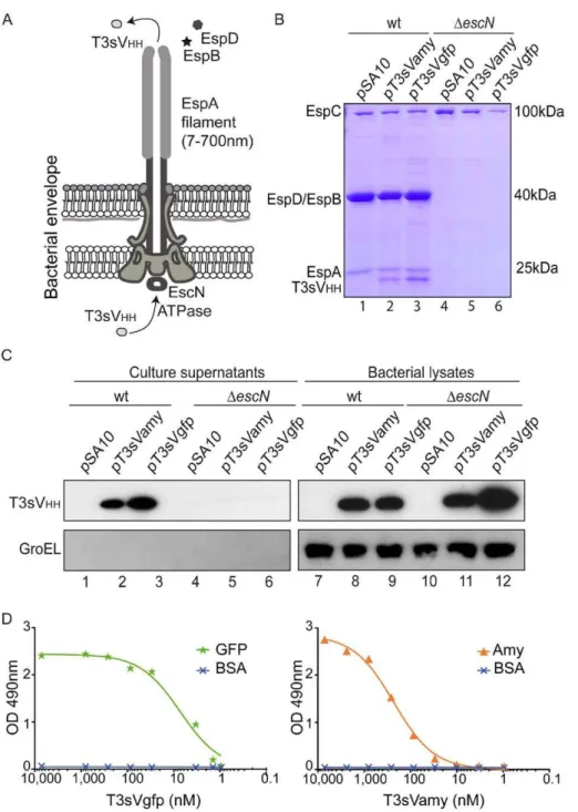
analyze whether T3sVHHs are secreted by the T3SS of EPEC (Fig. 1). EPEC wild-type (wt) strain and DescN strain (Table 1), which lacks the T3SS ATPase EscN [30], transformed with pSA10 (empty vector), pT3sVamy, or pT3sVgfp, were grown in DMEM and induced with IPTG for 3 h. Induction of T3sVHHfusions did not affect the growth of EPEC strains, which reached the same final optical density (OD600 nm,1.2) as cultures with the empty vector. Coomassie staining of proteins secreted from wt EPEC revealed discrete bands corresponding to the T3SS substrates (e.g. EspA, EspB, EspD) and protein bands of ca. 21–23 kDa, corresponding to the expected size of T3sVHHs, when bacteria carried pT3sVamy or pT3sVgfp (Fig. 1B; lanes 1–3). No secreted T3sVHHs were seen from wt EPEC carrying the empty vector. Neither protein bands corresponding to the T3SS substrates nor the T3sVHHs were present in the supernatant of DescN strain (Fig. 1B; lanes 4–6). A band corresponding to the autotransporter EspC [31], which is secreted by the Sec-pathway, was present in the supernatants of both the wt andDescNstrains.
We next evaluated expression and secretion of the T3sVHHs by Western blot (WB). The anti-E-tag mAb detected the T3sVHHs (ca. 21–23 kDa) in the culture supernatants of wt EPEC but not in the
DescN strain containing pT3sVamy and pT3sVgfp (Fig. 1C; top panel; lanes 1–6). In contrast, T3sVHHs were detected in the bacterial lysates of both the wt andDescNstrains with these plasmids (Fig. 1C; lanes 7–12). The membranes were also probed with anti-GroEL mAb (Fig. 1C; bottom panels) to control for non-specific release of cytoplasmic EPEC proteins. The result showed no reactivity with anti-GroEL in culture supernatants (lanes 1–6) whereas strong signals were detected in bacterial lysates (lanes 7–12). Secretion of the
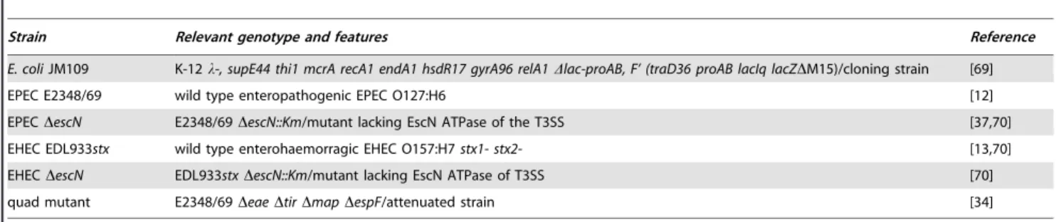
Table 1.E. colistrains employed in this study.
Strain Relevant genotype and features Reference
E. coliJM109 K-12l-, supE44 thi1 mcrA recA1 endA1 hsdR17 gyrA96 relA1Dlac-proAB, F’ (traD36 proAB lacIq lacZDM15)/cloning strain [69]
EPEC E2348/69 wild type enteropathogenic EPEC O127:H6 [12]
EPECDescN E2348/69DescN::Km/mutant lacking EscN ATPase of the T3SS [37,70]
EHEC EDL933stx wild type enterohaemorragic EHEC O157:H7stx1- stx2- [13,70]
EHECDescN EDL933stxDescN::Km/mutant lacking EscN ATPase of T3SS [70]
quad mutant E2348/69DeaeDtirDmapDespF/attenuated strain [34]
doi:10.1371/journal.pone.0015227.t001
Table 2.Plasmids employed in this study.
Name Relevant features and application Reference
pSA10 Apr, pUC-ori,Ptacpromoter, lacIq
/expression vector [72]
pT3sVamy pSA10 derivative/expression of T3sVamy This work
pT3sVgfp pSA10 derivative/expression of T3sVgfp This work
pDsVgfp pSA10 derivative/expression ofDsVgfp; lacks T3 signal (EspF20) This work
pCX340 Tcr, pBR-ori;
Ptrcpromoter, ‘blaM(TEM-1)/ vector forb-lactamase fusions [26]
pT3s-Bla pCX340 derivative/expression of T3s-Bla fusion This work
pT3sVgfp-Bla pCX340 derivative/expression of T3sVgfp-Bla fusion This work
pT3sVamy-Bla pCX340 derivative/expression of T3sVamy-Bla fusion This work
pEGFP-N1 Kmr, pUC/pSV40-ori, P
CMVpromoter, enhanced GFP Clontech
pCS2+MT Apr, pUC-ori, P
CMVpromoter, 6xmyc-tag, vector [74]
T3sVHHs to culture supernatants was also observed in EHEC wt strain but not in the EHECDescNstrain (Fig. S2; Table 1). As an additional proof of the T3-dependent secretion, a derivative of pT3sVgfp was constructed in which the T3s was deleted (pDsVgfp). WB of induced wt EPEC and EHEC carrying pDsVgfp resulted in intracellular accumulation ofDsVgfp but not in its secretion (Fig. S3).
To determine whether secreted T3sVHHs remained as soluble proteins, and to rule out their aggregation or association with outer membrane vesicles (OMVs), the culture supernatants from induced EPEC were ultracentrifuged at 100,000 g. The presence of T3sVHHs in the soluble and pellet fractions was analyzed by WB (Fig. S4) showing that$95% of T3sVHHs are found as soluble proteins. Figure 1. Secretion of sdAbs to the extracellular medium with T3SS of EPEC.(A) Schematic representation of the T3SS-complex encoded by EPEC, labeling the essential ATPase EscN, the extracellular EspA filament and the secreted EspB and EspD translocators. The secretion of T3sNbs from the cytoplasm of the bacteria to the extracellular medium is indicated. (B) Coomassie staining of TCA-precipitated proteins found in the extracellular media of cultures of wt EPEC orDescNstrains carrying plasmids pSA10, pT3sVamy, or pT3sVgfp, as indicated. Cultures were grown at 37uC in DMEM and induced with 0.1 mM IPTG for 4 h. The protein bands of EspA, EspB, EspD, and that of the Sec-dependent autotransporter EspC, are labeled. Size in kDa of protein standards for SDS-PAGE are shown on the right. (C) Western blot analysis of the proteins found in extracellular media (Culture supernantants; lanes 1–6) and cells (bacterial lysates; lanes 7–12) from the same cultures as in (B). WB developed with mAbs anti-E-tag (top panels) or anti-GroEL (bottom panels) to control the absence of cytoplasmic proteins in the extracellular media. (D) Binding activity of the secreted sdAbs. ELISA with His-tag purified T3sVgfp (left) and T3sVamy (right), at the indicated concentrations (nM), against their cognate antigens (GFP or Amy) and BSA (negative control). Bound T3sVHHs developed with anti-E-tag mAb-POD and their Optical Density (O.D.) determined at 490 nm.
Taking advantage of the His-tag present at their C-termini, both T3sVHHs were purified (Fig. S5) with standard yields between 0.5– 1 mg/L of culture supernatant. Binding activities of the purified T3sVHHs were tested by ELISA using antigens Amy, GFP or BSA (as negative control). Bound T3sVHHs were developed with anti-E-tag mAb demonstrating the specific antigen-binding activity of the T3sVHHs over a range of concentrations (Fig. 1D).
Translocation of sdAbs into human cells
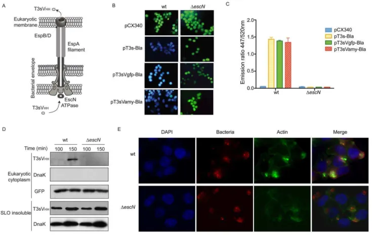
We tested whether T3sVHHs can be injected into human cells with EPEC strain (Fig. 2A). Toward this end, vector pCX340 (Table 2), which encodes the TEM-1 b-lactamase (Bla) reporter devoid of its natural N-terminal Sec-dependent signal peptide [26], was used to generate T3s-Bla, T3sVamy-Bla, and T3sVgfp-Bla fusions (Fig. S6A). Plasmids pT3s-T3sVgfp-Bla and pCX340 were used as positive and negative controls of translocation, respectively. Cultured HeLa cells, infected with wt andDescNEPEC carrying these Bla-expressing plasmids, were incubated with the nonfluo-rescent esterified CCF2/AM substrate. Upon passive entry into
the eukaryotic cell, CCF2/AM is transformed by eukaryotic esterases to the fluorescent substrate of Bla CCF2, which is mostly contained inside the eukaryotic cell [26]. Injection of Bla into the cytoplasm of the eukaryotic cells leads to hydrolysis of CCF2 changing its fluorescence emission from 520 nm (green) to 447 nm (blue), which could be detected under the fluorescence microscope or quantified in a fluorimeter. Examination by fluorescence microscopy revealed a clear shift to blue fluorescence of HeLa cells infected with wt EPEC carrying pT3s-Bla, pT3sVamy-Bla, or pT3sVgfp-Bla (Fig. 2B). In contrast, green fluorescence was observed in HeLa cells infected either with wt EPEC/pCX340 or with any of the DescN strains (Fig. 2B), demonstrating that the hydrolysis of CCF2 is only due to the Bla translocated into the cytosol of the HeLa cells. The fluorescence intensity of infected cells was also quantified in a fluorimeter and is shown as the ratio between blue emission fluorescence (447 nm) and green emission fluorescence (520 nm) (Fig. 2C). Expression of Bla fusions in wt andDescNEPEC was confirmed by WB with anti-Bla antibodies (Fig. S6B). Thus, these results demonstrate that EPEC carrying a
Figure 2. Translocation of sdAbs into HeLa cells using T3SS of EPEC.(A) Schematic representation of T3SS-complex from EPEC with EspB/D pore complex assembled in the mammalian cell plasma membrane. Injection of T3sNbs from the cytoplasm of the bacteria to the cytoplasm of the mammalian cell is shown. (B) Fluorescence microscopy images of cultured HeLa cells infected with EPEC wt (left column) andDescN(right column) strains, harbouring pCX340, pT3s-Bla, pT3sVamy-Bla or pT3sVgfp-Bla, as indicated, and incubated with CCF2/AM. Hydrolysis of CCF2 by translocated Bla changes the fluorescence emission of the cytosol of HeLa cells from 520 nm (green) to 447 nm (blue). (C) Quantification of the activity of translocated Bla by measuring the ratio of fluorescence at 447/520 nm in HeLa cells infected with the indicated bacteria. (D) Western blot of protein extracts after SLO treatment of HeLa cell cultures infected for the indicated time (min) with wt EPEC andDescNstrains harboring pT3sVgfp. Eukaryotic cytoplasm extracts (top panels) were developed with anti-E-tag mAb to detect T3sVHHs, anti-DnaK mAb to control the absence of bacterial
contamination, and anti-GFP mAb to test the efficacy of SLO pore formation in all samples and as a loading control. SLO-insoluble protein extracts (bottom panels), corresponding to EPEC bacteria and HeLa cell debris, were developed with anti-E-tag mAb, to show the expression level of T3sNb in bacteria, and with anti-DnaK mAb to control similar attachment of both strains to HeLa cells. (E) Immunofluorescence microscopy images of Hela cells infected with EPEC wt andDescNstrains to demonstrate similar adhesion and microcolony formation of both strains in HeLa cells. Bacteria are labeled with anti-O127 serum (red), F-actin labeled with conjugated phalloidin (green), and DNA and cell nuclei labeled with DAPI (blue). Actin accumulation is only observed underneath wt EPEC.
functional T3SS are able to inject T3sVHHs-Bla fusions into human cells.
Next, we investigated whether T3sVHHs could be detected in the cytoplasm of infected HeLa cells. Toward this end we employed a fractionation method of the infected cells based on Streptolysin-O (SLO), a pore-forming cytolysin from Streptococcus pyogenesthat selectively bind to cholesterol groups in the eukaryotic plasma membrane [32,33]. HeLa cells were infected for 90 or 150 min with EPEC expressing T3sVHHs in the presence of IPTG, placed on ice and the monolayer washed with PBS. Following SLO treatment the cytoplasmic content of HeLa cells was collected (‘‘Eukaryotic cytoplasm’’ extract). Ghost HeLa cells and bound bacteria were lysed in a SDS-containing buffer (‘‘SLO-insoluble’’ extract). Protein extracts were subjected to WB revealing a time-dependent accumulation of T3sVgfp in the cytoplasm of HeLa cells infected with wt EPEC but not with EPEC DescN(T3sVHH top panel, Fig. 2D). No sign of bacterial contamination was detected in the eukaryotic cytoplasmic extracts using a mAb directed against DnaK (Fig. 2D). Efficiency of the SLO-treatment in the different samples was controlled by WB developed with anti-GFP mAb (Fig. 2D). In addition, WB of the ‘‘SLO-insoluble’’ extracts demonstrated similar expression levels of T3sVgfp in wt EPEC andDescN strains (T3sVHH bottom panel, Fig. 2D). The comparable signal from the ‘‘SLO-insoluble’’ extracts probed with anti-DnaK indicated similar cell attachment levels of wt EPEC and DescN (Fig. 2D). This was confirmed by fluorescence microscopy of the infected cultures (Fig. 2E), in which microcolonies of wt EPEC andDescN bacteria (red) were stained adhered to HeLa cells at similar levels, although actin pedestals are only observed in cells infected by wt EPEC (Fig. 2E, staining of F-actin in green).
Since wt EPEC also secretes T3sVgfp to the extracellular medium, we wanted to exclude the possibility that the T3sVgfp molecules detected in the cytoplasmic extracts entered the cells indirectly from the extracellular medium through the pores formed in the plasma membrane by the EspBD translocation pore or during the SLO-treatment (despite removal of the extracellular medium before addition of SLO). For this, HeLa cells were infected with wt EPEC harboring empty pSA10 (expressing a functional T3SS but not T3sVgfp) or EPEC DescN/pT3sVgfp (lacking a functional T3SS and thus unable to inject T3sVgfp). After 120 min of infection, the culture media was replaced by conditional, bacteria-free, medium obtained from an induced wt EPEC/pT3sVgfp culture (containing secreted T3sVgfp). After a further 30 min incubation, infected cells were fractionated with SLO into ‘‘eukaryotic cytoplasm’’ and ‘‘SLO-insoluble’’ extracts, as above. WB showed that T3sVgfp was not found in the ‘‘eukaryotic cytoplasm’’ (Fig. S7), hence ruling out the possibility that T3sVgfp molecules were entering HeLa cells from the extracellular medium. Taken together, the experiments with Bla reporter and biochemical fractions demonstrated that wt EPEC are able to directly inject T3sVHHs into human cells.
We also investigated whether the presence of their cognate antigen influence the stability of T3sVHHs. Toward this end HeLa cells, transfected with pEGFP-N1 or with the empty vector pCS2+MT (Table 2), were infected with wt EPEC and EPEC
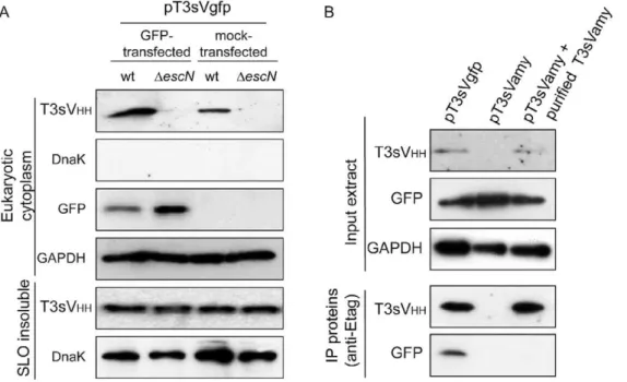
DescN harboring pT3sVgfp. ‘‘Eukaryotic cytoplasm’’ and ‘‘SLO-insoluble’’ extracts were analyzed by WB revealing that the amount of T3sVgfp found in the cytoplasm of HeLa cells was, 3-fold higher when GFP was expressed (Fig. 3A, T3sVHH panel). Detection of the cytoplasmic enzyme glyceraldehyde 3-phosphate dehydrogenase (GAPDH) was used as a loading control of the ‘‘eukaryotic cytoplasm’’ extracts (Fig. 3A, GAPDH panel). WB with anti-DnaK demonstrated the absence of bacterial
contami-nation in the ‘‘eukaryotic cytoplasm’’ extracts and similar signals in the ‘‘SLO-insoluble’’ extracts (Fig. 3A, DnaK panels). Therefore, higher levels of injected T3sVHHs are found when HeLa cells express the relevant antigen. We estimated that after 150 min infection in the presence of IPTG, an average of ,76105 molecules of T3sVgfp per cell are found in pEGFPN1-transfected cells. This estimation was done by densitometry of the WB signals of T3sVgfp in ‘‘eukaryotic cytoplasm’’ extracts from three independent infections with the signals generated with known protein concentrations of purified T3sVgfp used as standard curve (Fig. S8).
Antigen binding activity of injected sdAbs
To investigate formation of intracellular antigen-VHH complex-es, ‘‘eukaryotic cytoplasm’’ extracts from pEGFP-N1-transfected cells, infected with wt EPEC carrying pT3sVgfp or pT3sVamy, were immunoprecipitated (IP) with anti-E-tag mAb bound to protein G-beads. WB of the input extracts and IP proteins (Fig. 3B) revealed that GFP was specifically co-IP from the cytoplasmic extracts of cells infected with EPEC/pT3sVgfp demonstrating the formation of intracellular GFP-T3sVgfp complexes. Since T3sVamy does not accumulate at detectable levels in the cytoplasm of HeLa cells (likely caused by the absence of its antigen in the cytoplasm; see Discussion), purified T3sVamy was added to the input extract of cells infected with EPEC/pT3sVamy to provide a control of the specific co-IP of GFP with T3sVgfp (Fig. 3B). Similar amounts of GFP and GAPDH proteins were detected in the input extracts (Fig. 3B). These data provide direct evidence that the injected T3sVHHs have the capacity to bind their specific antigen in the cytoplasm of human cells.
Translocation of sdAbs by attenuated EPEC
The EPEC wt strain is a pathogen causing strong cytopathic effects due to the injection of its natural repertoire of T3 effectors [14,18]. Therefore most biotechnological applications will require using attenuated strains deficient in the major or all of the T3 effectors. To obtain a proof-of-principle of the injection of VHHs with attenuated bacteria, we employed quadruple (‘‘quad’’) mutant strain of EPEC that assembles functional injectisomes but carries four deletions in genes encoding the adhesin intimin and the effectors Tir, EspF and Map [34]. Bla translocation assays in HeLa cells infected with the ‘‘quad’’ mutant or with EPEC
Figure 4. Injection of sdAbs by attenuated bacterial strain.(A) Quantification of the Bla activity by measuring the ratio of fluorescence at 447/ 520 nm in HeLa cells infected with quad mutant andDescNstrains, carrying the indicated plasmids. (B) Western blot of ‘‘eukaryotic cytoplasm’’ (top panels) and ‘‘SLO-insoluble’’ (bottom panels) extracts from infected HeLa cell cultures with wt EPEC, quad mutant, andDescNstrains harboring pT3sVgfp. WB developed as in Figs 2 and 3. (C) Immunofluorescence microscopy images of Hela cells infected with the quad mutant andDescN
strains, to demonstrate the absence of actin pedestals and similar adhesion and microcolony formation by the attenuated quad mutant. Bacteria are labeled with anti-O127 serum (red), F-actin labeled with conjugated phalloidin (green), and DNA and cell nuclei labeled with DAPI (blue). doi:10.1371/journal.pone.0015227.g004
Figure 3. Antigen binding recognition by translocated sdAbs.(A) Antigen expression (GFP) increases the level of T3sVgfp detected in the cytoplasm of HeLa cells. Western blot of ‘‘eukaryotic cytoplasm’’ (top panels) and ‘‘SLO-insoluble’’ (bottom panels) protein extracts from infected HeLa cell cultures transfected with the indicated plasmids: pEGFPN1 (GFP) or control pCS2+MT (mock). Infections were carried out for 150 min with EPEC wt andDescNstrains harboring pT3sVgfp. The level of T3sVgfp in the cytoplasmic extracts of HeLa cells was developed with anti-E-tag mAb (T3sVHHtop panel). Absence of bacterial contamination in eukaryotic cytoplasm protein extracts was controlled with anti-DnaK mAb (DnaK top
panel). Transfection and expression of GFP was controlled with and anti-GFP mAb (GFP panel). Levels of human cytoplasmic GAPDH were determined with anti-GAPDH mAb to control equal SLO pore formation in all samples and as a loading control. SLO-insoluble protein extracts (bottom panels) were developed with anti-E-tag mAb to show the level of T3sVgfp in bacteria and with anti-DnaK mAb to control bacterial attachment. (B) Immunoprecipitation of T3sVgfp:GFP complexes from the cytoplasm of infected HeLa cells. Western blot of input eukaryotic cytoplasm protein extracts and immunoprecipitated proteins with anti-E-tag mAb bound to protein G-Sepharose resin. Input extracts were obtained by SLO treatment from pEGFP-N1-transfected HeLa cell cultures infected with wt EPEC strain carrying pT3sVgfp or pT3sVamy. Purified T3sVamy was added to one aliquot of input extract from cells infected with EPEC/pT3sVamy (lane 3) to reach a level similar to the translocated T3sVgfp (lane 1). Input extracts were developed with anti-E-tag, anti-GFP, and anti-GAPDH mAbs (top panels). Immunoprecipitated (IP) proteins were developed with anti-E-tag and anti-GFP mAbs (bottom panels).
Discussion
Expression of Ab fragments in the cytoplasm of human cells allows targeting of intracellular proteins that participate in disease and infection processes [7]. In this work we have shown that non-invasive E. coli bacteria carrying a functional T3SS are able to secrete and translocate to the cytoplasm of human cells sdAb fragments with full capacity to bind their cognate antigens. Single-domain VHHs appeared specially suited for this application given their potential as enzyme inhibitors, monomeric nature, stability, and size (2–3 nm diameter) [5] that fit in the protein channels of T3 needles and EspA filament [19,35]. We have shown that T3-secreted VHHs present in the extracellular medium do not enter in the cytoplasm of HeLa cells via EspBD pores, and that translocation only occurs directly from bacteria to the mammalian cell. In addition, we obtained evidence of the formation of antigen-VHH complexes in the cytoplasm of the infected cells and quantified that,76105VHH molecules accumulated per cell in the presence of antigen. Interestingly, we found that expression of the antigen in the cytoplasm of the mammalian cell increases the level of VHHs in the cytoplasm, which suggests that formation of antigen-VHH complex could stabilize the intracellular VHH. Although the actual reason of this stabilization is unclear, it is possible that the higher molecular weight of the antigen-VHH complex might reduce its susceptibility to proteolyic degradationin vivo. In this regard, detection of translocated T3sVHH-Bla in cells that do not express their cognate antigens could also be explained by their higher molecular weight of these fusions.
To establish this proof-of-principle we took advantage of non-invasive intestinal pathogenicE. coli strains that harbor a T3SS, such as EPEC and EHEC [12,13]. Although in vivoinfection by these strains is restricted to humans and certain animals (e.g. cattle),in vitro they can infect many mammalian cell lines from distinct cell types and species, including human and murine epithelial cells, fibroblasts, and macrophages among others [36– 38]. Interestingly, the mouse pathogen Citrobacter rodentium also carries a LEE pathogenicity island encoding a T3SS almost identical to that found in EPEC and EHEC strains [39].
Applications of intrabody delivery by bacterial injection require the use non-pathogenic bacterial strains. In this study we employed an attenuated strain lacking the adhesin Intimin and three major T3-effectors (EspF, Map, Tir) of EPEC. In the absence of intimin and Tir EPEC looses its intimate adhesion and actin pedestal formation capabilities, while the lack of EspF and Map diminishes major cytopathic effects due to disruption of the mitochondria and activation of Rho GTPases [14,34]. A study with human volunteers who ingested an EPEC null mutant in the intimin gene (eae) demonstrated a strong attenuation of this strain [40]. The EPEC strain E2348/69 used in this study encodes 21 effectors of the T3SS [12]. Accordingly, it would be possible to engineer an attenuated E. coli strain lacking all these effectors following a genome minimization approach [41]. Such bacterial strain will be extremely useful for antibody injection into mammalian cells. Another appealing strategy for bacterial injection of sdAbs is the use of commensalE. colistrains endowed with a functional T3SS. It has been reported that E. coli K-12 strain carrying the completeLEEfrom EPEC on a cosmid is able to induce actin polymerization in human cells in vitro [42]. However, theLEEis expressed weakly inE. coli K-12 [43] and, therefore, additional engineering is needed for efficient injection fromE. coliK-12.
The use of extracellular bacteria with a T3SS as vectors for delivery of proteins (including intrabodies) into mammalian cells differs from other approaches that need the transfer of the
protein-encoding gene by viral infection or transfection [8]. InvasiveE. coli expressing the Invasin (Inv) fromYersinia and the Listeriolysin O (LLO) from Listeria have been employed previously to deliver proteins, DNA, and interfering shRNA into mammalian cells [11]. Upon cell invasion, invasiveE. colicells (Inv+LLO+) are lysed in the phagosomes releasing their total cellular content. In contrast to this situation, non-invasive E. colicells carrying a T3SS remain extracellular and could inject specifically the desired sdAb. In addition, extracellular E. coli bacteria are more sensitive to antibiotic treatment (e.g. gentamycin) facilitating their elimination fromin vitrocultures as well as from whole animalsin vivo.
The use of live bacteria has a great potential for delivery of therapeutic proteins in vivo, in specific organs or tissues of the animal, where they can produce a continuous supply of the polypeptide. For instance, probiotic strains of lactic acid bacteria have been usedin vivofor the extracellular secretion of cytokines, enzymes, antibody fragments, etc. against infectious and inflam-matory diseases [44–46]. Also mucosal and systemic infections with live attenuated invasive bacterial strains (e.g. Salmonella, Listeria) have been employed for intracellular delivery of antigens for vaccination and cytotoxins for tumor therapy [47–54]. Interestingly, probiotic E. coli strains are currently used as therapeutic agents in humans. For instance, colonization of the gastrointestinal tract byE. coli Nissle 1917 [55] is used to treat some inflammatory bowel diseases such as Crohn’s disease [56,57]. In addition, deliberate colonization of the urinary bladder with probiotic E. coli strain ABU83972, isolated from an asymptomatic bacteriuria patient [58], is being used to treat recurrent urinary tract infections by uropathogenicE. colistrains [59,60]. Therefore, an attractive possibility is to engineer probiotic E. colistrains to carry a functional T3SS to deliver intracellularly therapeutic sdAbs targeting proteins involved in diseases such as inflammation and cancer in the gastrointestinal and urinary tracts. Systemic infections to treat other organs and solid tumors are not excluded [61,62]. Importantly, sdAbs interfering the function of relevant intracellular targets involved in cell proliferation (e.g. Ras), apoptosis (e.g. Caspase-3), cell migration (e.g. Gelsolin), and HIV-replication proteins (e.g. Rev) have already been selected [63–66]. The levels of intracellular sdAbs reported here with theE. coli T3SS (105–106 molecules per cell) seem appropriated to modulate the activity of regulatory and cell-signaling proteins, which often have intracellular levels below 106molecules per cell [67,68] and, in addition, could trigger a downstream signaling cascade that would amplify the initial effect of the intrabody.
In conclusion, we believe that injection of sdAbs into mammalian cells using non-pathogenic bacterial strains carrying a T3SS is a promising technology forin vitroandin vivointrabody applications that target host cell functions and signaling pathways.
Materials and Methods
Bacterial strains, growth, induction and infection conditions
capped Falcon tube (Beckton Dickinson) and incubation continued at 37uC with shaking until OD600,0.4. At this point, 0.1 mM isopropyl-1-thio-b-D-galactoside (IPTG) was added for 4 h. For infection experiments, overnight LB cultures (as above) were used to inoculate 15 ml of DMEM (initial OD600,0.1) and the cultures were incubated under static conditions at 37uC with 5% CO2for 2 h, as a pre-activation step. Bacteria from these cultures were used for infection of HeLa cell cultures (,105cells/well in 24-well tissue culture plates; Falcon) at a multiplicity of infection (MOI) 300:1 (bacterial CFUs: HeLa cells) and 0.1 mM IPTG was added. Infection continued at 37uC with 5% CO2for the indicated time (90–150 min) and were stopped on ice.
Plasmids and DNA constructs
Plasmids employed in this study are summarized in Table 2. Standard methods of DNA manipulation were used [71]. All DNA constructs were sequenced using an automated DNA sequencer (Perkin Elmer). Oligonucleotides were synthesized by Sigma Genosys (Table 3). Plasmid pSA10 is a vector that containslacIq repressor and a multiple cloning site under the control of Ptac promoter [72]. Plasmid pT3sVamy contains a DNA fragment of 549 bp, cloned at the EcoRI site of pSA10 under the Ptac promoter, which encodes the VHH anti-amylase (Vamy) fused to the T3-signal EspF20at its 59-end and a six-histidine (His) tag and the 12-amino acid epitope E-tag (GAPVPYPDPLEP) at its 39-end. This DNA fragment was obtained byEcoRI digestion of a 560 bp DNA product generated by homology-driven PCR, fusing two PCR subfragments with a final amplification of the fused product with oligonucleotides R1-Xb-SD-espF y RI-stop-E (Table 3). Subfragment 1 (119 bp), containing EspF20signal, was amplified from genomic DNA from EDL933stx with primers R1-Xb-SD-espF and SfiI-R1-Xb-SD-espF. Subfragment 2 (517 bp), corresponding to Vamy with His and E-tag epitopes, was amplified using plasmid pEHLYA4SDVamy as a template and oligonucleotides SfiIVamy and RI-stop-E as primers. Plasmid pEHLYA4SDVamy is a derivative of pEHLYA2SDVamy [28] with the His and the E-tag epitope at the 39end of Vamy. Plasmid pT3sVgfp was obtained by SfiI andNotI digestion of pT3sVamy, substituting Vamy coding sequence by Vgfp. The DNA encoding Vgfp was obtained by PCR using plasmid pcAbGFP4 [73] as template and oligonucle-otides Vhh-sfiI2 and Vhh-NotI2 as primers. The amplified fragment was digested with SfiI and NotI and the resulting
358 bp DNA molecule was cloned in the backbone of vector pT3sVamy (,4.3 kb) digested with the same enzymes. Plasmid pDsVgfp was constructed by amplication of a DNA segment, encoding Vgfp-His-E-tag devoid of the T3-signal, from pT3sVgfp template with oligonucleotidesDsign-EcoRI and RI-stop-E. The ,0.5 kb PCR product was digested EcoRI and inserted in the same site of vector pSA10 under the control ofPtacpromoter.
Plasmids pT3s-Bla and pT3sVamy-Bla are derivatives of pCX340 [26], a vector employed to make fusions with the TEM
b-lactamase lacking its Sec-dependent-signal peptide (‘blaM). To construct pT3s-Bla, first an 83 bp DNA segment encoding the T3-signal of EspF was amplified from genomic DNA from EDL933stx with the oligonucleotides NdeI-espF and EcoRI-espF. Next, this DNA segment was fused, by homology-driven PCR, with a 1.2 kb DNA fragment encoding (‘blaM), that had been amplified from pCX340 with oligonucleotides EcoRI-TEM and BamHI-tetra. The resulting 1.3 kb fragment, amplified with oligonucleotides NdeI-espF and BamHI-tetra, was digested with NdeI and BamHI and ligated with backbone fragment of pCX340 digested with the same enzymes. To construct pT3sVamy-Bla, a DNA fragment encoding T3sVamy was amplified from plasmid pT3sVamy with oligonucleotides NdeI-espFVamy and EcoRIVamy-espF, digested withNdeI andEcoRI and ligated in the same sites of pCX340.
In vitrocell culture and plasmid transfection
The human epithelial cell line HeLa clone HtTA1 was grown as monolayer in DMEM, supplemented with 10% fetal bovine serum (FBS) and 2 mM glutamine, at 37uC with 5% CO2. For transfection, HeLa cells were seed in tissue 24-well culture plates (,105cells/well), grown for 20 h at 37uC with 5% CO2. Plasmid pEGFPN1 (Clontech) or pCS2+MT [74] was added (0.6mg DNA/well) to the cultures in calcium phosphate [75]. After 22 h incubation, the medium was removed, wells were washed three times with PBS, filled with 1 ml of complete medium and incubated for 1 h at 37uC with 5% CO2. This medium was replaced by serum-free medium before cell cultures were infected as described above.
SDS-PAGE and Western blot analysis
Sodium Dodecy Sulfate–Polyacrylamide gel electrophoresis (SDS-PAGE) and Western blot was performed following standard methods [71] using the Miniprotean III system (Bio-Rad). Proteins
Table 3.Oligonucleotides used in this study.
Name Sequence (59-39)
RI-Xb-SD-espF CCGGAATTCTCTAGAAAGAGGCATAAATTATGCTTAATGGAATTAGTA
SfiI-espF CTGCACCTGAGCCATGGCCGGCTGGGCCGCTGCGATACCTACAAGCTGCCGCCCTA
SfiIVamy CTTGTAGGTATCGCAGCGGCCCAGCCGGCCATGGCTCAGGTGCAGCTG
RI-stop-E CCGGAATTCTCATTAGGCCGGTTCCAGCGGATCCGGATACGGCAC
Vhh-SfiI2 GTCCTCGCAACTGCGGCCCAGCCGGCCATGGCTCAGGTGCAGCTGGTGGA
Vhh-NotI2 GGACTAGTGCGGCCGCTGAGGAGACGGTGACCTGGGT
NdeI-espF CCGGATCCATATGCTTAATGGAATTAGTAACGCTGCTTCT
EcoRI-espF GGTGCGAATTCGCTGCGATACCTACAAGCTGCCGCCCTA
EcoRI-TEM GCGGCAGCTTGTAGGTATCGCAGCGAATTCGCACCCAGAAACGCTGGTGA
BamHI-tetra ATGCGTCCGGCGTAGAGGATCCACAGGACGGGT
NdeI-espFVamy GGGAATTCCATATGCTTAATGGAATTAGTAACGCTGCT
EcoRIVamy-espF CCGGAATTCGCGGCCGGTTCCAGCGGATCCGGATA
separated by SDS-PAGE (in 10 or 12% gels) were stained with Coomassie Blue R-250 (Bio-Rad) or transferred to polyvinylidene difluoride membrane (PVDF, Immobilon-P, Millipore) as de-scribed previously [76]. Antibodies employed for Western blot were: anti-E-tag mAb conjugated to peroxidase (POD) (1:5000; GE Amersham Biosciences); anti-E-tag mAb (Phadia; 0.5 mg/ml), anti-GroEL mAb-POD (1:5000; Sigma); anti-b-lactamase mAb (1:1000; QED Bioscience); GFP mAb (1:1000; Roche); anti-GAPDH (1:2000; Santa Cruz Biotechnology); anti-DnaK (1:5000; Stressgen). Unconjugated mAbs were detected with anti-mouse IgG-POD (1:5000; Sigma) as secondary antibody. Membranes were developed by chemiluminiscence using Immun-Star Wes-ternC kit (Bio-Rad) and exposed to X-ray films (Konica) and to a ChemiDoc XRS+system for quantification (Bio-Rad).
Analysis of secreted and cellular proteins inE. colistrains
Whole-cell protein extracts from inducedE. coli cultures were obtained from cells harvested by centrifugation (30006g, 5 min)
from 1 ml aliquot of liquid cultures (OD600,1.2), resuspended in 100ml of phosphate-buffered saline (PBS), and mixed with the same volume of 2X SDS-PAGE sample buffer. Samples were boiled for 10 min, briefly sonicated (5 sec; Labsonic B Braun), and centrifuged (140006g, 5 min) to remove insoluble material before loading on SDS-PAGE. For analysis of proteins secreted in the culture medium, culture supernatants obtained after centrifugation (30006g, 15 min) were filtered utilizing a 0.22-mm sterile low-protein binding PVDF filter unit (Millex GV, Millipore) and the serine-protease inhibitor phenyl-methyl-sulphonyl-fluoride (PMSF) was added to 1 mM final concentration. The proteins in the filtered-culture supernatants were mixed with 1/5th of the volume of SDS-PAGE sample buffer (5X) for WB or were precipitated with trichloroacetic acid (TCA 20% w/v; Merck) for Coomassie staining. After centrifugation (140006g, 15 min),
TCA-precipitat-ed protein pellets were rinsTCA-precipitat-ed with cold acetone (220uC) and resuspended in SDS-PAGE sample buffer (1/10th of the precipitated volume). Purification of His-tagged T3-secreted sdAbs with Talon resin (Clontech) is described below. To test the solubility of the T3-secreted sdAbs, the filtered-culture super-nantants (see above) were centrifuged at 100.0006g in a Beckman TL-100 ultracentrifuge for 1 h at 4uC. The supernatant (Soluble) and the pellet (insoluble) fractions obtained after this centrifugation were adjusted to the same final volume in SDS-PAGE sample buffer, boiled and analyzed by WB.
Purification of T3-secreted sdAbs
Cultures of E. coli EPEC with the indicated plasmids (pT3sVamy or pT3sVgfp) were grown at 37uC with shaking in 200 ml of DMEM with ampicillin (using a capped bottle). IPTG was added at 0.1 mM final concentration when the OD600 reached ,0.4 and incubation continued for 4 h. Culture supernatants were filtered and PMSF was added as described above. Next, supernatants were equilibrated to PBS 1X and incubated overnight at 4uC with 2 ml of Cobalt-containing chromatography resin (Talon, Clontech) for binding of His-tagged sdAbs. Resin was packed in a column, washed 4 times with 10 ml of PBS containing 5 mM imidazole, and eluted in 1 ml aliquots with PBS containing 100 mM imidazole. Eluted fractions were stored at 4uC.
Enzyme-linked immunosorbent assays (ELISA)
ELISA conditions were based on those described previously [76]. Briefly, 96-well immunoplates (Maxisorp, Nunc) were coated for 2 h at room temperature with purified antigens (10mg/ml) in PBS. Antigens employed: alpha-amylase (Amy; Sigma), the green
fluorescent protein (GFP; Upstate), bovine serum albumin (BSA, Roche). Plates were washed with PBS and blocked in PBS buffer containing 3% (w/v) non-fat milk, before incubation with purified T3-secreted sdAbs or filtered-culture supernatants obtained after IPTG induction (at the indicated concentration or dilution in the same buffer). After PBS-wash, bound E-tagged sdAbs were revealed with anti-E-tag mAb-POD (1:2000) and developed with o-phenylenediamine (OPD, Sigma) and H2O2(Sigma). The OD at 490 nm of the plates was determined in a microplate reader (Bio-Rad).
b-lactamase translocation assay
We followed the method described by [26]. Briefly, the indicated bacterial strains were used to infect HeLa cells grown in vitroin 8-well Falcon culture slides (Beckton Dickinson), IPTG was added for induction and incubation continued for 90 min. The medium was removed and cells were washed three times with Hank’s balance salt solution (HBSS). Next, 200ml of HBSS and 40ml of the b-lactamase substrate CCF2/AM mix (K1024, Invitrogen) were added. Cells were incubated for additional 90 min at room temperature in the dark, washed three times with HBSS and analyzed by fluorescence microscopy (Nikon Eclipse E600, excitation UV light 330–380 nm). For quantitative analysis of Bla translocation, HeLa cells were seeded in a 96-well opaque plate (Nunc) at aprox. 85% confluence (,26104cells/well). After
16 h incubation at 37uC with 5% CO2, infection were done with pre-activated EPEC strains in serum-free DMEM and the cultures were further incubated for 30 min before addition of IPTG, and 60 min after this addition. Infections were washed three times with HBSS, and 200ml/well of HBSS were added plus 20ml of CCF2/ AM substrate mix. Samples were incubated for 90 min in the dark, washed three times with HBSS and finally 100ml/well of HBSS were added. Plates were read in a FLUOstar Optima Microplate Fluorometer with a filter set 450/520 nm.
Streptolysin-O (SLO) fractionation of infected cell cultures
Conditions of SLO treatment were based on those described by [77] with some modifications. Briefly, 24-well plates containing infected HeLa cell cultures (as described above) were placed on ice and washed three times with 1 ml/well of freshly prepared cold SLO-buffer (150 mM sucrose, 25 mM Hepes pH 7.4, 150 mM K-acetate, 2.5 mM MgCl2, 4 mM EGTA, 2 mM DTT). Next, 175ml/well of SLO-buffer containing 10mg/ml of Streptolysin O (purchase from Prof. Sucharit Bhakdi’s laboratory, Institute of Medical Microbiology and Hygiene, Hochhaus am Augustusplatz, Mainz, Germany) were added followed by 15 min of incubation on ice to allow binding of SLO to cells. After this incubation, unbound SLO was removed by washing three times with 1 ml/ well of cold SLO buffer and 175ml/well of this buffer was added. Plates were incubated at 37uC for 15 min for SLO-pore formation to allow release of the cytosolic content of HeLa cells (which was confirmed by observation in an inverted light microscope; Carl Zeiss). Extracellular media containing released cytoplasmic proteins were collected from plates. Protease inhibitors were added (Complete EDTA-free Protease Inhibitors Cocktail, Roche) to these extracts and centrifuged at 45006g (15 min, 4uC) in order
Immunoprecipitation assays
Eukaryotic cytoplasm protein extracts (350ml obtained as described above from two infected tissue culture wells) were incubated with 40ml of anti-E-tag mAb bound to protein G-Sepharose resin. Anti-E-tag mAb (1 mg, Phadia) was previously crosslinked to protein G-Sepharose resin (1 ml, Sigma) with dimethyl pimelimidate dihydrochloride (DMP; Sigma). After 16 h incubation at 4uC in an orbital shaker, the resin was collected by centrifugation (8006g, 1 min) and washed three times with 1.5 ml of 200 mM sodium phosphate buffer (pH 8.2). Bound proteins were eluted from resin by incubation with 60ml of 0.1 M glycine pH 2.8 (10 min at room temperature) followed by centrifugation (8006g, 1 min). Supernatants were collected and 30ml of 200 mM sodium phosphate buffer (pH 8.2) was added for pH neutralization, referred to as immunoprecipitated (IP) proteins. Routinely, 12ml of these IP proteins were mixed with 3ml of 5X SDS-PAGE sample buffer, boiled and gel-loaded for Western blot analysis.
Immunofluorescence microscopy
Infected HeLa cell cultures, grown on coverslips in 24-well plates, were washed three times with 1 ml/well of PBS, fixed with 3% (w/v) paraformaldehyde (in PBS) for 20 min at room temperature, and washed again with PBS three times. Cells were permeabilized by incubation with PBS containing 0.1% (v/v) of Triton X-100 (Sigma) for 5 min. To label EPEC, a rabbit polyclonal anti-O127 serum was diluted 1:200 in PBS with 10% donkey serum (Jackson Immunoresearch) and incubated 1 h at room temperature. Coverslips were washed three times with PBS and goat anti-rabbit IgG-Texas Red conjugated secondary antibody (1:500 in PBS with 10% donkey serum; Molecular Probes) was added along with Oregon-Green conjugated phalloi-din (1:100; Invitrogen) and DAPI (1:1000; Sigma) to label F-actin and DNA, respectively. Coverslips were washed three times with PBS after incubation and 4ml of mounting medium (DAKO) was added. Coverslips were analyzed by conventional epifluorescence microscopy using a Zeiss Axio imager microscope.
Supporting Information
Figure S1 Scheme of plasmid vectors used for
expres-sion of T3sVHHfusions. The sequence encoding the first 20
amino acids of EspF effector (T3s) is fused to the corresponding VHH (Vamy or Vgfp in pT3sVamy or pT3sVgfp, respectively). Epitope tags (His and E-tag) at the C-termini of fusions and unique restriction sites SfiI, NcoI and NotI flanking VHH domain are indicated. Gene constructs are under the control of the IPTG-inducible Ptac promoter. The presence of lacIq repressor, transcriptional terminators (T1,T2) from 5S ribosomal RNA gene, ampicillin-resistance (ampr) gene, and origin of replication (ori) are also shown.
(TIF)
Figure S2 Secretion of T3sVHHs by EHEC. (a) Proteins
found in extracellular media (Culture supernantants; lanes 1–6) and cells (Bacterial lysates; lanes 7–12) from cultures of wild type EHEC or DescN mutant strains carrying plasmids pSA10, pT3sVamy, or pT3sVgfp, as indicated, analyzed by Western blot with mAb anti-Etag (top panels), to detect T3sVHHfusions, or with anti-GroEL (bottom panels) to control the absence of bacterial lysis. Cultures were grown at 37uC in DMEM and induced with 0.1 mM IPTG for 4 h.(b)SDS-PAGE and Coomassie staining of proteins found in the extracellular media of cultures of EHEC and
DescNmutant strains carrying the indicated plasmids and induced
as in (a). The protein bands of T3sVHHfusions, T3SS- effectors EspA, EspB, EspD, and that of the Sec-dependent autotransporter EspP, are labelled. Size in kDa of protein standards for SDS-PAGE is shown on the left.
(TIF)
Figure S3 Requirement of the T3-signal for secretion of
VHHs. Western blot with anti-E-tag mAb-POD of proteins found
in extracellular media (Culture supernatants) and cells (Bacterial lysates) from cultures of wild type EPEC (top panels) and EHEC (bottom panels) carrying plasmids pT3sVgfp or pDsVgfp (lacking the T3-signal) as indicated. Cultures were grown at 37uC in DMEM and induced with 0.1 mM IPTG for 4 h.
(TIF)
Figure S4 Solubility of the T3-secreted VHHs. Extracellular
media from induced cultures of EPEC carrying plasmids pT3sVgfp or pT3sVamy, as indicated, were ultracentrifuged (100.0006g, 1 h) and proteins present in the resulting supernatants
(S) and pellet (P) fractions were analyzed by Western blot with anti-E-tag mAb-POD.
(TIF)
Figure S5 Purified T3sVHHs from culture supernatants.
Coomassie stained SDS-polyacrylamide gel of metal affinity purified His-tagged T3sVgfp and T3sVamy from extracellular media of EPEC strains harbouring pT3sVgfp or pT3sVamy. Size in kDa of protein standards for SDS-PAGE is shown on the left. (TIF)
Figure S6 Expression ofb-lactamase fusions in EPEC.
A)Scheme ofb-lactamase (Bla) gene fusions in plasmids pCX340, pT3s-Bla, pT3sVgfp-Bla and pT3sVamy-Bla. The position of Ptac promoter, T3 signal and VHHsequence, are indicated.B)Western blot developed with anti-b-lactamase mAb of whole cells protein extracts from induced EPEC wild type andDescNstrains carrying the indicated Bla plasmid. Size in kDa of protein standards for SDS-PAGE is shown on the right.
(TIF)
Figure S7 T3sVHHs do not enter into HeLa cells from
extracellular media. Western blot of ‘‘eukaryotic cytoplasm’’ (top panels) and ‘‘SLO-insoluble’’ (bottom panels) protein extracts from infected HeLa cell cultures with EPEC wt/pSA10 and EPEC
DescN/pT3sVgfp in which their extracellular media were replaced by medium containing T3sVgfp from induced EPEC wt/ pT3sVgfp. Western blot of ‘‘eukaryotic cytoplasm’’ extracts are developed with anti-E-tag to detect T3sVHH, anti-DnaK to control the absence of bacterial contamination and anti-GFP mAb to test the efficacy of SLO pore formation. Western blot of ‘‘SLO insoluble’’ extracts are developed with anti-E-tag to show the expression of T3sVHHin bacteria and anti-DnaK mAb to control attachment of both strains to HeLa cells.
(TIF)
Figure S8 Quantification of T3sVHHs molecules
Acknowledgments
We would like to thank Prof. Brendan Kenny (University of Newcastle, UK) for his kind gift of the quad mutant strain, and Prof. Jorge Blanco (Universidad de Santiago de Compostela, Spain) for his kind gift of anti-O127 serum. We thank Dr. Junkal Garmendia (IDAB, CSIC) and Dr. Gustavo Bodelo´n (CNB, CSIC) for helpful comments and discussions. The help of Drs. Cristina Risco and Alberto Fraile (CNB, CSIC) for establishing the SLO treatment conditions is greatly appreciated.
Author Contributions
Conceived and designed the experiments: ABT LAF. Performed the experiments: ABT. Analyzed the data: ABT SM GF LAF. Contributed reagents/materials/analysis tools: ABT SM GF LAF. Wrote the paper: ABT LAF.
References
1. Nelson AL, Reichert JM (2009) Development trends for therapeutic antibody fragments. Nat Biotech 27: 331–337.
2. Wesolowski J, Alzogaray V, Reyelt J, Unger M, Juarez K, et al. (2009) Single domain antibodies: promising experimental and therapeutic tools in infection and immunity. Med Microbiol Immunol 198: 157–174.
3. Holliger P, Hudson PJ (2005) Engineered antibody fragments and the rise of single domains. Nat Biotechnol 23: 1126–1136.
4. Holt LJ, Herring C, Jespers LS, Woolven BP, Tomlinson IM (2003) Domain antibodies: proteins for therapy. Trends Biotechnol 21: 484–490.
5. Muyldermans S, Baral TN, Retamozzo VC, De Baetselier P, De Genst E, et al. (2009) Camelid immunoglobulins and nanobody technology. Vet Immunol Immunopathol 128: 178–183.
6. Adams GP, Weiner LM (2005) Monoclonal antibody therapy of cancer. Nat Biotechnol 23: 1147–1157.
7. Lo AS, Zhu Q, Marasco WA (2008) Intracellular antibodies (intrabodies) and their therapeutic potential. Handb Exp Pharmacol. pp 343–373.
8. Kontermann RE (2004) Intrabodies as therapeutic agents. Methods 34: 163–170.
9. Ferna´ndez LA (2004) Prokaryotic expression of antibodies and affibodies. Curr Opin Biotechnol 15: 364–373.
10. Mazor Y, Van Blarcom T, Mabry R, Iverson BL, Georgiou G (2007) Isolation of engineered, full-length antibodies from libraries expressed inEscherichia coli. Nat Biotechnol 25: 563–565.
11. Critchley RJ, Jezzard S, Radford KJ, Goussard S, Lemoine NR, et al. (2004) Potential therapeutic applications of recombinant, invasiveE. coli. Gene Ther 11: 1224–1233.
12. Iguchi A, Thomson NR, Ogura Y, Saunders D, Ooka T, et al. (2009) Complete Genome Sequence and Comparative Genome Analysis of Enteropathogenic Escherichia coliO127:H6 Strain E2348/69. J Bacteriol 191: 347–354. 13. Perna NT, Plunkett G, 3rd, Burland V, Mau B, Glasner JD, et al. (2001)
Genome sequence of enterohaemorrhagicEscherichia coliO157:H7. Nature 409: 529–533.
14. Croxen MA, Finlay BB (2010) Molecular mechanisms of Escherichia coli pathogenicity. Nat Rev Micro 8: 26–38.
15. Kaper JB, Nataro JP, Mobley HL (2004) PathogenicEscherichia coli. Nature Reviews Microbiology 2: 123–139.
16. Knutton S, Lloyd DR, McNeish AS (1987) Adhesion of enteropathogenic Escherichia colito human intestinal enterocytes and cultured human intestinal mucosa. Infect Immun 55: 69–77.
17. McDaniel TK, Jarvis KG, Donnenberg MS, Kaper JB (1995) A genetic locus of enterocyte effacement conserved among diverse enterobacterial pathogens. Proc Natl Acad Sci U S A 92: 1664–1668.
18. Garmendia J, Frankel G, Crepin VF (2005) Enteropathogenic and enterohe-morrhagicEscherichia coliinfections: translocation, translocation, translocation. Infect Immun 73: 2573–2585.
19. Cornelis GR (2006) The type III secretion injectisome. Nat Rev Microbiol 4: 811–825.
20. Galan JE, Wolf-Watz H (2006) Protein delivery into eukaryotic cells by type III secretion machines. Nature 444: 567–573.
21. Knutton S, Rosenshine I, Pallen MJ, Nisan I, Neves BC, et al. (1998) A novel EspA-associated surface organelle of enteropathogenicEscherichia coliinvolved in protein translocation into epithelial cells. Embo J 17: 2166–2176.
22. Munera D, Crepin VF, Marches O, Frankel G (2010) The N-terminal type III secretion signal of enteropathogenicE. colitranslocator proteins. J Bacteriol 192: 3534–3539.
23. Ru¨ssmann H, Shams H, Poblete F, Fu Y, Galan JE, et al. (1998) Delivery of epitopes by theSalmonellatype III secretion system for vaccine development. Science 281: 565–568.
24. Ru¨ssmann H, Gerdemann U, Igwe EI, Panthel K, Heesemann J, et al. (2003) AttenuatedYersinia pseudotuberculosiscarrier vaccine for simultaneous antigen-specific CD4 and CD8 T-cell induction. Infect Immun 71: 3463–3472. 25. Sory MP, Boland A, Lambermont I, Cornelis GR (1995) Identification of the
YopE and YopH domains required for secretion and internalization into the cytosol of macrophages, using the cyaA gene fusion approach. Proc Natl Acad Sci U S A 92: 11998–12002.
26. Charpentier X, Oswald E (2004) Identification of the secretion and translocation domain of the enteropathogenic and enterohemorrhagicEscherichia colieffector Cif, using TEM-1 beta-lactamase as a new fluorescence-based reporter. J Bacteriol 186: 5486–5495.
27. Lauwereys M, Arbabi Ghahroudi M, Desmyter A, Kinne J, Holzer W, et al. (1998) Potent enzyme inhibitors derived from dromedary heavy-chain antibodies. Embo J 17: 3512–3520.
28. Fraile S, Munoz A, de Lorenzo V, Fernandez LA (2004) Secretion of proteins with dimerization capacity by the haemolysin type I transport system of Escherichia coli. Mol Microbiol 53: 1109–1121.
29. Rothbauer U, Zolghadr K, Muyldermans S, Schepers A, Cardoso MC, et al. (2008) A versatile nanotrap for biochemical and functional studies with fluorescent fusion proteins. Mol Cell Proteomics 7: 282–289.
30. Zarivach R, Vuckovic M, Deng W, Finlay BB, Strynadka NC (2007) Structural analysis of a prototypical ATPase from the type III secretion system. Nat Struct Mol Biol 14: 131–137.
31. Stein M, Kenny B, Stein MA, Finlay BB (1996) Characterization of EspC, a 110-kilodalton protein secreted by enteropathogenic Escherichia coli which is homologous to members of the immunoglobulin A protease-like family of secreted proteins. J Bacteriol 178: 6546–6554.
32. Heuck AP, Tweten RK, Johnson AE (2003) Assembly and topography of the prepore complex in cholesterol-dependent cytolysins. J Biol Chem 278: 31218–31225.
33. Palmer M, Harris R, Freytag C, Kehoe M, Tranum-Jensen J, et al. (1998) Assembly mechanism of the oligomeric streptolysin O pore: the early membrane lesion is lined by a free edge of the lipid membrane and is extended gradually during oligomerization. EMBO J 17: 1598–1605.
34. Dean P, Maresca M, Schuller S, Phillips AD, Kenny B (2006) Potent diarrheagenic mechanism mediated by the cooperative action of three enteropathogenic Escherichia coli-injected effector proteins. Proc Natl Acad Sci U S A 103: 1876–1881.
35. Daniell SJ, Kocsis E, Morris E, Knutton S, Booy FP, et al. (2003) 3D structure of EspA filaments from enteropathogenic Escherichia coli. Mol Microbiol 49: 301–308.
36. Marches O, Covarelli V, Dahan S, Cougoule C, Bhatta P, et al. (2008) EspJ of enteropathogenic and enterohaemorrhagic Escherichia coli inhibits opsono-phagocytosis. Cell Microbiol 10: 1104–1115.
37. Whale AD, Hernandes RT, Ooka T, Beutin L, Schuller S, et al. (2007) TccP2-mediated subversion of actin dynamics by EPEC 2 - a distinct evolutionary lineage of enteropathogenic Escherichia coli. Microbiology 153: 1743– 1755.
38. Wu Y, Lau B, Smith S, Troyan K, Barnett Foster DE (2004) Enteropathogenic Escherichia coliInfection Triggers Host Phospholipid Metabolism Perturbations. Infect Immun 72: 6764–6772.
39. Petty NK, Bulgin R, Crepin VF, Cerdeno-Tarraga AM, Schroeder GN, et al. (2010) TheCitrobacter rodentiumGenome Sequence Reveals Convergent Evolution with Human PathogenicEscherichia coli. J Bacteriol 192: 525–538.
40. Donnenberg MS, Tacket CO, James SP, Losonsky G, Nataro JP, et al. (1993) Role of the eaeA gene in experimental enteropathogenicEscherichia coliinfection. J Clin Invest 92: 1412–1417.
41. Posfai G, Plunkett G, 3rd, Feher T, Frisch D, Keil GM, et al. (2006) Emergent properties of reduced-genomeEscherichia coli. Science 312: 1044–1046. 42. McDaniel TK, Kaper JB (1997) A cloned pathogenicity island from
enteropathogenicEscherichia coliconfers the attaching and effacing phenotype onE. coliK-12. Mol Microbiol 23: 399–407.
43. Brinkley C, Burland V, Keller R, Rose DJ, Boutin AT, et al. (2006) Nucleotide sequence analysis of the enteropathogenic Escherichia coli adherence factor plasmid pMAR7. Infect Immun 74: 5408–5413.
44. Steidler L, Hans W, Schotte L, Neirynck S, Obermeier F, et al. (2000) Treatment of murine colitis byLactococcus lactissecreting interleukin-10. Science 289: 1352–1355.
45. Kru¨ger C, Hu Y, Pan Q, Marcotte H, Hultberg A, et al. (2002) In situ delivery of passive immunity by lactobacilli producing single-chain antibodies. Nat Biotechnol 20: 702–706.
46. Wells JM, Mercenier A (2008) Mucosal delivery of therapeutic and prophylactic molecules using lactic acid bacteria. Nat Rev Micro 6: 349–362.
47. Lewis GK (2007) Live-attenuatedSalmonellaas a prototype vaccine vector for passenger immunogens in humans: are we there yet? Expert Rev Vaccines 6: 431–440.
48. Stritzker J, Pilgrim S, Szalay AA, Goebel W (2008) Prodrug converting enzyme gene delivery by L. monocytogenes. BMC Cancer 8: 94.
50. Nishikawa H, Sato E, Briones G, Chen LM, Matsuo M, et al. (2006) In vivo antigen delivery by a Salmonella typhimurium type III secretion system for therapeutic cancer vaccines. J Clin Invest 116: 1946–1954.
51. Cheng CM, Lu YL, Chuang KH, Hung WC, Shiea J, et al. (2008) Tumor-targeting prodrug-activating bacteria for cancer therapy. Cancer Gene Ther 15: 393–401.
52. Friedlos F, Lehouritis P, Ogilvie L, Hedley D, Davies L, et al. (2008) Attenuated Salmonellatargets prodrug activating enzyme carboxypeptidase G2 to mouse melanoma and human breast and colon carcinomas for effective suicide gene therapy. Clin Cancer Res 14: 4259–4266.
53. Fu W, Lan H, Liang S, Gao T, Ren D (2008) Suicide gene/prodrug therapy usingSalmonella-mediated delivery ofEscherichia colipurine nucleoside phosphor-ylase gene and 6-methoxypurine 29-deoxyriboside in murine mammary carcinoma 4T1 model. Cancer Sci 99: 1172–1179.
54. Zhu X, Zhou P, Cai J, Yang G, Liang S, et al. (2010) Tumor antigen delivered bySalmonellaIII secretion protein fused with heat shock protein 70 induces protection and eradication against murine melanoma. Cancer Sci.
55. Grozdanov L, Raasch C, Schulze J, Sonnenborn U, Gottschalk G, et al. (2004) Analysis of the genome structure of the nonpathogenic probioticEscherichia coli strain Nissle 1917. J Bacteriol 186: 5432–5441.
56. Henker J, Muller S, Laass MW, Schreiner A, Schulze J (2008) Probiotic Escherichia coli Nissle 1917 (EcN) for successful remission maintenance of ulcerative colitis in children and adolescents: an open-label pilot study. Z Gastroenterol 46: 874–875.
57. Schultz M (2008) Clinical use ofE. coliNissle 1917 in inflammatory bowel disease. Inflamm Bowel Dis 14: 1012–1018.
58. Andersson P, Engberg I, Lidin-Janson G, Lincoln K, Hull R, et al. (1991) Persistence of Escherichia coli bacteriuria is not determined by bacterial adherence. Infect Immun 59: 2915–2921.
59. Sunden F, Hakansson L, Ljunggren E, Wullt B (2006) Bacterial interference–is deliberate colonization withEscherichia coli83972 an alternative treatment for patients with recurrent urinary tract infection? Int J Antimicrob Agents 28 Suppl 1: S26–29.
60. Sunden F, Hakansson L, Ljunggren E, Wullt B (2010)Escherichia coli83972 bacteriuria protects against recurrent lower urinary tract infections in patients with incomplete bladder emptying. J Urol 184: 179–185.
61. Stritzker J, Weibel S, Hill PJ, Oelschlaeger TA, Goebel W, et al. (2007) Tumor-specific colonization, tissue distribution, and gene induction by probiotic Escherichia coliNissle 1917 in live mice. Int J Med Microbiol 297: 151–162. 62. Forbes NS (2010) Engineering the perfect (bacterial) cancer therapy. Nat Rev
Cancer.
63. Tanaka T, Williams RL, Rabbitts TH (2007) Tumour prevention by a single antibody domain targeting the interaction of signal transduction proteins with RAS. EMBO J 26: 3250–3259.
64. McGonigal K, Tanha J, Palazov E, Li S, Gueorguieva-Owens D, et al. (2009) Isolation and functional characterization of single domain antibody modulators of Caspase-3 and apoptosis. Appl Biochem Biotechnol 157: 226–236. 65. Van den Abbeele A, De Clercq S, De Ganck A, De Corte V, Van Loo B, et al.
(2010) A llama-derived gelsolin single-domain antibody blocks gelsolin-G-actin interaction. Cell Mol Life Sci 67: 1519–1535.
66. Vercruysse T, Pardon E, Vanstreels E, Steyaert J, Daelemans D (2010) An Intrabody Based on a Llama Single-domain Antibody Targeting the N-terminal Œ6-Helical Multimerization Domain of HIV-1 Rev Prevents Viral Production. Journal of Biological Chemistry 285: 21768–21780.
67. Scheele JS, Rhee JM, Boss GR (1995) Determination of absolute amounts of GDP and GTP bound to Ras in mammalian cells: comparison of parental and Ras-overproducing NIH 3T3 fibroblasts. Proceedings of the National Academy of Sciences of the United States of America 92: 1097–1100.
68. Svingen PA, Loegering D, Rodriquez J, Meng XW, Mesner PW, et al. (2004) Components of the Cell Death Machine and Drug Sensitivity of the National Cancer Institute Cell Line Panel. Clinical Cancer Research 10: 6807–6820. 69. Yanish-Perron C, Viera J, Messing J (1985) Improved M13 Phage cloning
vectors and hosts strains: nucleotide sequences of the M13mp18 and pU19 vectors. Gene 33: 103–119.
70. Garmendia J, Phillips AD, Carlier MF, Chong Y, Schuller S, et al. (2004) TccP is an enterohaemorrhagicEscherichia coliO157:H7 type III effector protein that couples Tir to the actin-cytoskeleton. Cell Microbiol 6: 1167–1183. 71. Ausubel FM, Brent R, Kingston RE, Moore DD, Seidman JG, et al. (2002)
Short Protocols in Molecular Biology. New York: John Wiley & Sons, Inc.. 72. Schlosser-Silverman E, Elgrably-Weiss M, Rosenshine I, Kohen R, Altuvia S
(2000) Characterization ofEscherichia coliDNA lesions generated within J774 macrophages. J Bacteriol 182: 5225–5230.
73. Rothbauer U, Zolghadr K, Tillib S, Nowak D, Schermelleh L, et al. (2006) Targeting and tracing antigens in live cells with fluorescent nanobodies. Nat Methods 3: 887–889.
74. Turner DL, Weintraub H (1994) Expression of achaete-scute homolog 3 in Xenopus embryos converts ectodermal cells to a neural fate. Genes Dev 8: 1434–1447.
75. Jordan M, Schallhorn A, Wurm FM (1996) Transfecting mammalian cells: optimization of critical parameters affecting calcium-phosphate precipitate formation. Nucleic Acids Res 24: 596–601.
76. Jurado P, Ritz D, Beckwith J, de Lorenzo V, Ferna´ndez LA (2002) Production of functional single-chain Fv antibodies in the cytoplasm ofEscherichia coli. J Mol Biol 320: 1–10.