The 3
’
-J
Region of the TCR
Locus Bears
Gene Regulatory Activity in Thymic and
Peripheral T Cells
Martina Kučerová-Levisohn1,2, Stefan Knirr1, Rosa I. Mejia1, Benjamin D. Ortiz1,2*
1Department of Biological Sciences, City University of New York, Hunter College, New York, New York, United States of America,2Doctoral Program in Biology, City University of New York, Graduate Center, New York, New York, United States of America
Abstract
Much progress has been made in understanding the importantcis-mediated controls on mouse TCRαgene function, including identification of the Eαenhancer and TCRαlocus control region (LCR). Nevertheless, previous data have suggested that othercis-regulatory elements may reside in the locus outside of the Eα/LCR. Based on prior findings, we hypothesized the existence of gene regulatory elements in a 3.9-kb region 5’of the Cα exons. Using DNase hypersensitivity assays and TCRαBAC reporter transgenes in mice, we detected gene regulatory activity within this 3.9-kb region. This region is active in both thymic and peripheral T cells, and selectively affects upstream, but not downstream, gene expression. Together, these data indicate the existence of a novelcis-acting regulatory complex that contributes to TCRαtransgene expressionin vivo. The active chromatin sites we discovered within this region would remain in the locus after TCRαgene rearrangement, and thus may contribute to endogenous TCRαgene activity, particularly in peripheral T cells, where the Eαelement has been found to be inactive.
Introduction
The functional rearrangement and expression of the TCRαgene during T cell development results in cell surfaceαß TCR complex emergence. These processes are tightly regulated incis at the TCRαgene locus [1,2]. DNase I hypersensitive sites (HS) located 3’of the Cαconstant region exons comprise a locus control region (LCR) that supports a great deal of TCRα tran-scriptional regulatory characteristics [3,4]. In particular, the region of HS1 (which contains the Eαtranscriptional enhancer [5]) and the HS1 prime (HS1’) element [6] is critical for normal TCRαgene rearrangement [7]. This region also very strongly increases transcription levels in T cellsin vivo[6,7], likely via epigenetic regulation of chromatin states [8,9].
While the importance of the HS1/HS1’DNA region to the function of the TCRαlocus is indisputable, limited TCRαgene recombination is observed in its absence. This is accompanied by TCRαtranscription levels adequate to support maintenance of a peripheral T cell population
OPEN ACCESS
Citation:Kučerová-Levisohn M, Knirr S, Mejia RI, Ortiz BD (2015) The 3’-JαRegion of the TCRαLocus
Bears Gene Regulatory Activity in Thymic and Peripheral T Cells. PLoS ONE 10(7): e0132856. doi:10.1371/journal.pone.0132856
Editor:Tae-Young Roh, Pohang University of Science and Technology (POSTECH), REPUBLIC OF KOREA
Received:April 19, 2015
Accepted:June 18, 2015
Published:July 15, 2015
Copyright:© 2015 Kučerová-Levisohn et al. This is an open access article distributed under the terms of theCreative Commons Attribution License, which permits unrestricted use, distribution, and reproduction in any medium, provided the original author and source are credited.
Data Availability Statement:All relevant data are within the paper.
with normalαß TCR levels [7]. It has been speculated that the remaining elements of the TCRα
LCR might explain this HS1/HS1’region-independent TCRαgene activity [7]. Nevertheless, heterologous reporter transgenes linked to the TCRαLCR, while mimicking the kinetics/levels of TCRαgene expression in thymocytes [10], are transcribed at lower than expected levels in peripheral T cells [4]. This phenomenon is congruent with prior data indicating that while the Eα/HS1 element is active in thymocytes, its deletion from reporter transgenes had no impact on transgene mRNA levels in peripheral lymphoid organs [6]. Using completely different experi-mental models, a very recent report similarly concluded that, by multiple criteria, the Eα ele-ment is inactive in peripheral T cells [11]. Together these reports strongly suggest the presence of additional TCRαgene regulatory elements in the locus outside of the LCR capable of main-taining transcription of the TCRαgene in peripheral T cells.
In contrast to heterologous, TCRαLCR-driven reporter genes, transgenic mice bearing cog-nate TCRαtransgenes linked to the full TCRαLCR display normal levels of transgenicαß TCR in peripheral T cells [12]. A major difference between the TCRαtransgenes and the heterolo-gous TCRαLCR reporter constructs previously analyzed is that the former include TCRαlocus DNA sequences upstream of the LCR up to a SacI restriction site located near Jα3 [13]. The vast majority of this DNA region would remain present in the endogenous locus following functional TCRαgene rearrangement. We hypothesized that transcriptional control elements might be present in this DNA region between the Jα3-proximal SacI site and the LCR. In the present study, we examined this region for indications of gene regulatory activity.
We report the presence of an array of DNase I hypersensitive sites (HS) in a region of the mouse TCRαlocus that ranges from the Jα2 segment to the Cα1 exon. We previously described a TCRαlocus derived bacterial artificial chromosome (BAC) construct containing two reporter genes [14]. One, Vαpromoter-driven reporter lies upstream of the HS cluster in the orientation and position of the TCRαgene. The second gene reports the activity of the Dad 1 promoter that lies downstream of both the HS cluster and TCRαLCR. Deletion from this construct of a 3.9-kb region of TCRαlocus DNA, that includes the Jα3-proximal SacI site and the identified HS clusters, impairs upstream, but not downstream reporter gene activity in transgenic mice. The deleted region is active in both thymocytes and peripheral T cells. The HS cluster discov-ered here lies in a region of the locus that would remain in all functionally rearranged TCRα
alleles. Therefore, this novel regulatory region may play a role in endogenous TCRαgene activ-ity. It may be especially important to maintaining TCRαmRNA levels in peripheral T cells.
Materials and Methods
Ethics Statement
Transgenic animal studies presented in this work have been reviewed and approved by the Hunter College Institutional Animal Care and Use Committee (protocol # BO 10/17-01). Ani-mals are euthanized by carbon dioxide inhalation in conformance with American Veterinary Medical Association recommendations.
TCR
α
/Dad1 bacterial artificial chromosome (BAC) dual-reporter
constructs
The wild type dual-reporter BAC construct used in this study has been previously described [14]. The mutant BAC was engineered to delete a 3.9-kb region spanning from 38-bp 5’of a SacI site (located between Jα4 and Jα3) to 9-bp 3’of an EcoRV site within the Cαconstant region exon 1. BAC modifications utilized Red/ET recombination technology (Gene Brid-ges) [15].
collection and analysis, decision to publish, or preparation of the manuscript.
Transgenic mice
Wild type (76.2-kb) and mutant (72.3-kb) dual-reporter BAC fragments were released from the parent BAC pBACe3.6 vector backbone using NotI and FseI. Transgenic founders bearing intact integrants were identified by Southern blot and PCR screening. Founders were then out-crossed to C57BL/6 mice (Taconic) to establish transgenic mouse lines. The relative transgene copy numbers among the individual mouse lines were determined by multiple Southern blot experiments as described previously [16]. Four independent wild type (line 36, 42, 62 and 71) and four independent mutant reporter BAC transgenic mouse lines (line 4, 18, 25 and 30) are analyzed in this study.
DNase I Hypersensitivity Assay
These experiments were carried out as previously described [17] using nuclei of MACS (Milte-nyi) purified spleen T cells from C57BL/6 mice that were subjected to titrating amounts of DNase I (Worthington). NdeI restriction enzyme digestion was used to generate the 6.7-kb parent fragment of the TCRαlocus examined. A 669-bp probe was generated by PCR using the parent BAC clone as a template and the following primers: forward: 5’-atggctgagggaaaggtctacg-3’and reverse: 5’-agaaaagtctctgggaactggtgtc-3’. The probe was labeled with [α-32P] dCTP and/ or [α-32P] dATP using the RadPrime DNA Labeling System (Life Technologies).
Flow cytometric analyses
1x10⁶splenocytes were pretreated in 100μL of FACS staining medium (RPMI 1640 supple-mented with 3% FBS and 10mM HEPES buffer) for 10 min at 4°C with 1μg of rat anti-CD16/ 32 (Clone 2.4G2, Life Technologies) to block Fc receptors. Afterwards, 0.2-.5μg of the Abs (from BD Biosciences) were added and incubated for 20 min at 4°C, followed by three washes with FACS staining medium. The Ab clones used were mouse anti-human CD2 (clone S5.2), mouse anti-rat CD2 (clone OX-34), and hamster anti-mouse TCRß chain (clone H57-597). Samples were acquired using a FACSCalibur device (BD Biosciences) and the collected data was analyzed with FlowJo software (Tree Star).
RNA Analysis
Results
A region of active chromatin in the 3
’
-J
α
region
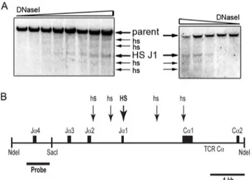
We used DNase I hypersensitivity assays to examine the chromatin state of a 6.7-kb NdeI frag-ment of the TCRαlocus containing the Jα1 to Jα4 segments and Cαexons 1 and 2 (Fig 1). Of the four Jαsegments present in this genomic fragment, only Jα2 supports functional TCRα
protein production [19]. To examine this region for HS in a way that minimizes the potential complication of Vα-Jαrecombination-mediated production of multiple parent restriction frag-ments, we selected a probe that would only detect alleles at which Vα-Jαrecombination occurred to a Jαsegment upstream of Jα3. To minimize the presence of non-functional Jα4 rearranged TCRαalleles in the DNase-treated genomic DNA samples, these assays were car-ried out using nuclei from isolated peripheral T cells.
A cluster of five HS was detected (Fig 1). The most prominent of these HS is proximal to the Jα1 segment (HS-J1), which is located near the center of the HS array. The other four HS are less prominent. In multiple, independent experiments, these four HS display variable intensity in comparison to each other. Two of these HS approximately localize to DNA in between the Jα2 and Jα1 segments. A third HS is found in the Jα1-Cα1 intron, while the fourth localizes to the Cα1 exon. Since Jα1 is a non-functional pseudo-Jαsegment [19], the region containing the HS array detected would remain present in all functionally rearranged TCRαalleles.
Testing
cis
-acting TCR
α
gene regulatory activity in transgenic mice
We previously described a dual-reporter transgene based on a mouse TCRα/Dad1 locus BAC construct (Fig 2) [14]. At the 5’end of the BAC, a human CD2 (hCD2) reporter in genomic configuration is integrated in a position upstream of the Cαexons. The hCD2 reporter gene in this construct is driven by a TCRαV-region promoter that was fused in frame with the hCD2 ATG start codon. In addition, a rat CD2 (rCD2) cDNA was fused in frame with the ATG start codon located in Dad1 exon 1. Thus, the rCD2 reporter gene is driven by the Dad1 promoter. To test the function of the above-described target region of the locus, we created a deletion mutant in the context of this dual-reporter BAC that removed a 3.9-kb SacI to EcoRV fragment
Fig 1. DNase I hypersensitivity sites (HS) within the Jα2 to Cα1 region. (A) Spleen T cell nuclei from
C57BL/6N mouse were subjected to DNase I titration. Arrows indicate the positions of the 6.7-kb NdeI parent fragment and HS. Results from two independent, representative experiments are shown.(B)Scaled diagram of the approximate locations of detected HS (arrows). The thick arrow near Jα1 indicates the most prominent HS.
stretching from upstream of Jα3 to Cα1 (target region). Both wild type and target region mutant BAC constructs were liberated from the vector backbone and used to create transgenic mice.
Target region deletion impairs upstream, but not downstream reporter
gene expression
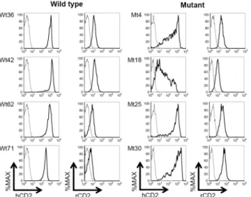
Flow cytometry analyses were carried out to initially assess the impact of target region-deletion on BAC reporter gene expression (Fig 3). Four wild type and four mutant reporter BAC trans-genic mouse lines were analyzed. T cells from wild type reporter BAC transtrans-genic mice expressed the upstream hCD2 reporter gene at high and relatively uniform levels on a per cell basis. In contrast, per cell hCD2 expression levels in the target region-deletion mutant BAC were more variegated in transgenic T cells. Expression of the downstream, rCD2 reporter gene was detected in all lines at low, but uniform levels in T cells. The deletion mutation did not appear to affect the uniformity of rCD2 expression levels.
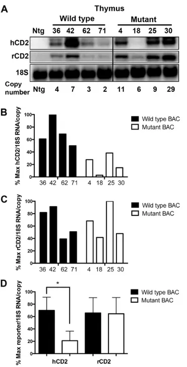
We next analyzed reporter gene mRNA levels in T cells isolated from wild type and mutant BAC transgenic mice. Northern blot analyses of transgenic thymus RNA samples (Fig 4)
Fig 2. Diagram (not to scale) of the TCRα/Dad1 dual-reporter BAC construct [14].Horizontal arrows indicate the orientation of the two reporter genes. A
Vα17 promoter drives expression of a genomic human CD2 reporter gene (ghCD2). The rat CD2 reporter is driven by the Dad 1 promoter. Vertical arrows indicate the location of the HS of the TCRαLCR (including the TCRαenhancer, Eα). The 3.9-kb region deletion in the mutant BAC is marked and runs from 38-bp 5’of the SacI site through 30-bp 5’of the end of Cαexon 1.
doi:10.1371/journal.pone.0132856.g002
Fig 3. Impaired hCD2 reporter expression in the absence of the deleted region.Flow cytometry analyses of human CD2 (hCD2) and rat CD2 (rCD2) reporter gene expression in spleen T cells (TCRβ+) from the indicated, independent wild type (Wt) and mutant (Mt) dual-reporter BAC transgenic mouse lines. Reporter gene expression in transgenic (solid line) and non-transgenic control (dashed line) cells is shown.
Fig 4. Absence of the deleted region impairs upstream, but not downstream reporter gene expression in thymocytes. (A)Northern blot analyses of human and rat CD2 reporter gene mRNA levels in thymocytes from the indicated lines of wild type and mutant reporter BAC transgenic mice. 18S rRNA signals are used as a loading control. Relative transgene copy number for each mouse line is indicated. The black line indicates excision of samples from the blot that are irrelevant to the present study. Panels B and C depict PhosphorImager analyses of the human CD2(B)and rat CD2(C)reporter mRNA signals detected by northern blots. The normalized mRNA levels (per transgene copy) from each wild type (black bars) and mutant (white bars) transgenic mouse line are graphed relative to each other (as % maximum).(D) Statistical analyses of the above data using the two-tailed students t test. Graph bars indicate the average (+/- S.E.) normalized mRNA levels among the lines. The asterisk indicates the statistical significance of the difference in hCD2 mRNA levels between wild type and mutant BAC (p= 0.012). In contrast, no significant difference in rCD2 mRNA levels was detected (p= 0.942).
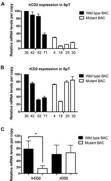
indicated that upstream hCD2 reporter mRNA levels (per transgene copy) driven by the mutant reporter BAC were, on average, about one-third of those observed from wild type reporter BAC. In contrast, downstream rCD2 reporter RNA levels were not affected by the mutation. Similar results were obtained from mRNA-level analyses of transgenic peripheral T cells (Fig 5). Per copy levels of hCD2 mRNA driven by the mutant BAC reporter were, on aver-age, nearly five-fold lower than those observed in wild type BAC transgenic T cells. Once again, the levels of rCD2 reporter mRNA per transgene copy were not altered by the deletion
Fig 5. The deleted region is functional in peripheral T cells.qRT-PCR analyses of human(A)and rat(B) CD2 reporter gene mRNA levels (+/- S.E.) in isolated spleen T cells (SpT) from the indicated lines of wild type (black bars) and mutant (white bars) transgenic mice. Observed reporter mRNA levels per copy from each transgenic line are graphed relative to each other (as % maximum). hCD2 reporter mRNA levels were normalized to endogenous TCRαmRNA levels, and rCD2 expression were normalized to endogenous Dad1 mRNA levels using primers that detect sequences not present in the reporter BAC. Three experiments (S1 Table) were performed in duplicates.(C)Statistical analyses of the above data using the two-tailed students t test. The asterisk indicates the statistical significance of the difference in hCD2 mRNA levels between wild type and mutant BAC (p = 0.016). In contrast, no significant difference in rCD2 mRNA levels was detected (p = 0.819). Graph bars indicate the average (+/- S.E.) normalized mRNA levels among the lines.
mutation. Overall the data demonstrate the function of the deleted region in regulating TCRα -based reporter transgene expression in both thymic and peripheral T cells.
Discussion
In this report we describe evidence for the presence of a novelcis-acting regulatory DNA region in the mouse TCRαgene locus. It is probable that this activity resides within the region of active chromatin we identified between the Jα2 and Cα1 exons. This DNase hypersensitive region of DNA would remain present in the locus after virtually any functional Vα-Jαgene rearrangement event. Thus, this regulatory complex could contribute to TCRαgene expression in the thymus and/or periphery. The residual TCRαgene activity observed in Eα/HS1/HS1’
region knockout mice [7] adds to the rational basis for this notion.
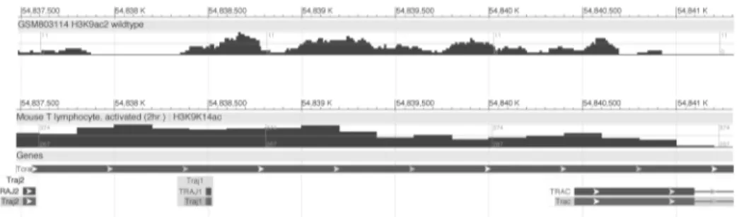
In addition, an early report presented evidence of transcriptional enhancer activity in the Jα-Cαintronic region of the human TCRαgene locus [20]. The enhancer activity described in that report was weaker (~14-fold) than that reported for the 3’Eαelement discovered subse-quently in the mouse TCRαlocus (>100-fold) [5]. This later report presented evidence sug-gesting that the corresponding Jα-Cαintronic region of the mouse TCRαlocus does not bear significant classical enhancer activity [5]. A contemporaneous study identifying the human counterpart to the 3’-Eαenhancer did not include examination of the human Jα-Cαintron for enhancer activity [21]. In any case, all these prior studies utilized transiently transfected reporter gene bearing plasmids in T cell tumor lines. Here we identifiedcis-acting regulatory DNA 5’of the Cαexons that is functional in the context of native T cell chromatin in whole mice. Publicly available data from chromatin immunoprecipitation-next generation sequenc-ing (ChIP-Seq) experiments corroborate these findsequenc-ings. The Jαregion displays epigenetic sig-natures of active chromatin, including acetylated histone H3, in T cells. In particular,
thymocyte chromatin displays discrete peaks of histone H3 acetylation in the DNA stretching from 5’of Jα1 into roughly the first third of the Cα1 exon. High histone acetylation levels across this DNA region are also observed in peripheral T cells (Fig 6).
A very recent report provided multiple lines of evidence indicating the inactivity of the Eα
element in peripheral T cells [11]. The conclusions of this report are consistent with a prior report indicating that the removal of Eαfrom a TCRαLCR-driven reporter transgene has vir-tually no impact on transgene expression in the spleen [6]. Taken together, these findings may help explain why TCRαLCR driven transgenes are expressed at lower levels in peripheral T
Fig 6. Signatures of active chromatin in the 3’Jαregion.Visualization of histone H3 acetylation marks in
CD4+,CD8+thymocytes (top track) and activated peripheral CD4+T lymphocytes (bottom track) assayed by ChIP-seq. Shown are screenshots of tracks obtained from publicly available data via the NCBI Epigenomics Browser. The region depicted spans the mouse TCRαgene locus DNA (chromosome 14) containing the Jα2 and Jα1 segments, and the Cα1 constant region exon. Top row: (Unpublished data). ChIP-seq in Mus musculus, strain 129SvJae x C57BL/6 (H3K9ac2). Accession number: ESX000004775. Bottom row: [22]. ChIP-seq in Mus musculus, strain C57BL/6 (H3K9K14ac). Accession number: ESX000001399.
cells than those observed in thymocytes [4], because the TCRαLCR includes Eαamong its functional components [6]. Despite apparent Eαinactivity, it is important to point out that the TCRαLCR, as a whole, remains active in the peripheral T cells, and this finding has been con-firmed in combination with four different reporter genes, each bearing its own distinct pro-moter [4,12,15,23]. Previous reports point to at least two distinct TCRαLCR sub-elements outside of Eα, named HS1’[6] and HS6 [17,24], that manifest activity in peripheral lymphoid organs. These elements may also contribute to TCRαmRNA levels in T cell subsets in which the Eαelement becomes inactive.
There is strong precedent for antigen receptor gene loci bearing multiple, importantcis -act-ing enhancer-like elements located both 5’and 3’of their constant region exons. The IgH [25], Igκ[26], TCRγ[27], TCRδ[1] loci all display a version of this arrangement. The findings pre-sented here would add the TCRαgene to this category. The literature has produced a consensus that the multiplecis-elements functioning within a particular gene locus can support both redundant and non-overlapping functions (e.g. [27–29]). A subset of these functions can have significant impact on immunity. A recent example of this comes from the IgH gene locus. The intronic Eμenhancer has long been known to play a key role in V-D-J recombination at the IgH locus [25]. But it was also found to have overlapping function with a 3’regulatory region complex [30]. Creation of a functional V-D-J knock-in/Eμknockout IgH allele bypassed Eμ’s role in recombination to enable investigation of its subsequent functions in mice. These studies revealed a surprising impairment of allelic exclusion at the targeted locus in the absence of Eμ [31]. The downstream consequences of this included faulty clonal selection and generation of autoimmune B cell clones [32]. These findings highlight the continuing importance of identify-ing and studyidentify-ingcis-acting elements that might, on the surface, appear either redundant or dis-pensable for antigen receptor gene function. The multiple genetic and epigenetic processes that occur at these complex gene loci regulate the assembly, timing, level and distribution of the proteins that form the basis of adaptive immunity. The present study adds a new region of the TCRαlocus to the collection of potential sources of regulation of these important processes.
Supporting Information
S1 Table. Data from triplicate experiments (Fig 5).
(PDF)
Acknowledgments
We thank L. Eckhardt for helpful comments on this manuscript, S. Cevallos, K. Wong and M. Kirvalidze for technical assistance, J. Kim for flow cytometry assistance, and D. Sant’Angelo, W. Mark and the Sloan-Kettering transgenic facility for the generation of transgenic mice.
Author Contributions
Conceived and designed the experiments: MKL SK BDO. Performed the experiments: MKL SK RIM. Analyzed the data: MKL RIM BDO. Wrote the paper: MKL BDO.
References
1. Krangel MS, Carabana J, Abbarategui I, Schlimgen R, Hawwari A. Enforcing order within a complex locus: current perspectives on the control of V(D)J recombination at the murine T-cell receptor alpha/ delta locus. Immunol. Rev. 2004; 200:224–32. PMID:15242408.
2. Shih HY, Hao B, Krangel MS. Orchestrating T-cell receptor alpha gene assembly through changes in chromatin structure and organization. Immunol. Res. 2011; 49(1–3):192–201. Epub 2010/12/04. doi:
3. Diaz P, Cado D, Winoto A. A locus control region in the T cell receptor alpha/delta locus. Immunity. 1994; 1(3):207–17. PMID:7889409
4. Harrow F, Ortiz BD. The TCRalpha locus control region specifies thymic, but not peripheral, patterns of TCRalpha gene expression. J. Immunol. 2005; 175(10):6659–67. PMID:16272321.
5. Winoto A, Baltimore D. A novel, inducible and T cell-specific enhancer located at the 3' end of the T cell receptor alpha locus. EMBO J. 1989; 8(3):729–33. PMID:2524381
6. Ortiz BD, Cado D, Winoto A. A new element within the T-cell receptor alpha locus required for tissue-specific locus control region activity. Mol. Cell. Biol. 1999; 19(3):1901–9. PMID:10022877
7. Sleckman BP, Bardon CG, Ferrini R, Davidson L, Alt FW. Function of the TCR alpha enhancer in alpha-beta and gammadelta T cells. Immunity. 1997; 7(4):505–15. PMID:9354471
8. Santoso B, Ortiz BD, Winoto A. Control of organ specific demethylation by an element of the T-cell receptor alpha locus control region. J. Biol. Chem. 2000; 275:1952–8. PMID:10636897
9. McMurry MT, Krangel MS. A role for histone acetylation in the developmental regulation of V(D)J recombination. Science. 2000; 287:495–8. PMID:10642553
10. Lahiji A, Kucerova-Levisohn M, Lovett J, Holmes R, Zuniga-Pflucker JC, Ortiz BD. Complete TCR-alpha gene locus control region activity in T cells derived in vitro from embryonic stem cells. J. Immunol. 2013; 191(1):472–9. Epub 2013/05/31. doi: jimmunol.1300521 [pii] doi:10.4049/jimmunol.1300521
PMID:23720809; PubMed Central PMCID: PMC3755507.
11. del Blanco B, Angulo U, Krangel MS, Hernandez-Munain C. T-cell receptor alpha enhancer is inacti-vated in alphabeta T lymphocytes. Proc. Natl. Acad.Sci. U.S.A. 2015; 112(14):E1744–E53. Epub 2015/ 04/02. doi:10.1073/pnas.1406551112PMID:25831496.
12. Kovalovsky D, Pezzano M, Ortiz BD, Sant'Angelo DB. A novel TCR transgenic model reveals that neg-ative selection involves an immediate, Bim-dependent pathway and a delayed, Bim-independent path-way. PloS One. 2010; 5(1):e8675. Epub 2010/01/15. doi:10.1371/journal.pone.0008675PMID:
20072628; PubMed Central PMCID: PMC2800196.
13. Kouskoff V, Signorelli K, Benoist C, Mathis D. Cassette vectors directing expression of T cell receptor genes in transgenic mice. J. Immunol. Methods. 1995; 180(2):273–80. PMID:7714342.
14. Lahiji A, Kucerova-Levisohn M, Holmes R, Zuniga-Pflucker JC, Ortiz BD. Adapting in vitro embryonic stem cell differentiation to the study of locus control regions. J. Immunol. Methods. 2014; 407:135–45. Epub 2014/04/01. doi:10.1016/j.jim.2014.03.012PMID:24681242; PubMed Central PMCID: PMC4037339.
15. Knirr S, Gomos-Klein J, Andino BE, Harrow F, Erhard KF, Kovalovsky D, et al. Ectopic T cell receptor-alpha locus control region activity in B cells is suppressed by direct linkage to two flanking genes at once. PloS One. 2010; 5(11):e15527. Epub 2010/12/03. doi:10.1371/journal.pone.0015527PMID:
21124935; PubMed Central PMCID: PMC2989920.
16. Gomos-Klein J, Harrow F, Alarcón J, Ortiz BD. CTCF-independent, but not CTCF-dependent, elements significantly contribute to TCRa locus control region activity. J. Immunol. 2007; 179(2):1088–95. PMID:
17617601
17. Harrow F, Amuta JU, Hutchinson SR, Akwaa F, Ortiz BD. Factors binding a non-classical Cis-element prevent heterochromatin effects on locus control region activity. J. Biol. Chem. 2004; 279(17):17842–9. PMID:14966120.
18. Deindl E. 18S ribosomal RNA detection on Northern blot employing a specific oligonucleotide. BioTech-niques. 2001; 31(6):1250, 2. Epub 2002/01/05. PMID:11768650.
19. Koop BF, Wilson RK, Wang K, Vernooij B, Zallwer D, Kuo CL, et al. Organization, structure, and func-tion of 95 kb of DNA spanning the murine T-cell receptor C alpha/C delta region. Genomics. 1992; 13 (4):1209–30. Epub 1992/08/01. PMID:1505954.
20. Luria S, Gross G, Horowitz M, Givol D. Promoter and enhancer elements in the rearranged alpha chain gene of the human T cell receptor. EMBO J. 1987; 6(11):3307–12. Epub 1987/11/01. PMID:3501368; PubMed Central PMCID: PMC553784.
21. Ho IC, Yang LH, Morle G, Leiden JM. A T-cell-specific transcriptional enhancer element 3' of C alpha in the human T-cell receptor alpha locus. Proc. Natl. Acad.Sci. U.S.A. 1989; 86(17):6714–8. PMID:
2788889
22. Ghoreschi K, Laurence A, Yang XP, Tato CM, McGeachy MJ, Konkel JE, et al. Generation of patho-genic T(H)17 cells in the absence of TGF-beta signalling. Nature. 2010; 467(7318):967–71. Epub 2010/10/22. doi:10.1038/nature09447PMID:20962846; PubMed Central PMCID: PMC3108066. 23. Ortiz BD, Cado D, Chen V, Diaz PW, Winoto A. Adjacent DNA elements dominantly restrict the
24. Ortiz BD, Harrow F, Cado D, Santoso B, Winoto A. Function and factor interactions of a locus control region element in the mouse T cell receptor-alpha/Dad1 gene locus. J. Immunol. 2001; 167(7):3836–
45. PMID:11564801
25. Jung D, Giallourakis C, Mostoslavsky R, Alt FW. Mechanism and control of V(D)J recombination at the immunoglobulin heavy chain locus. Annu. Rev. Immunol. 2006; 24:541–70. Epub 2006/03/23. doi:10. 1146/annurev.immunol.23.021704.115830PMID:16551259.
26. Schlissel MS. Regulation of activation and recombination of the murine Igkappa locus. Immunol. Rev. 2004; 200:215–23. Epub 2004/07/10. doi:10.1111/j.0105-2896.2004.00157.xPMID:15242407. 27. Xiong N, Kang C, Raulet DH. Redundant and unique roles of two enhancer elements in the TCRgamma
locus in gene regulation and gammadelta T cell development. Immunity. 2002; 16(3):453–63. Epub 2002/03/26. PMID:11911829.
28. Li F, Yan Y, Pieretti J, Feldman DA, Eckhardt LA. Comparison of identical and functional Igh alleles reveals a nonessential role for Emu in somatic hypermutation and class-switch recombination. J. Immu-nol. 2010; 185(10):6049–57. Epub 2010/10/13. doi:10.4049/jimmunol.0902992PMID:20937850; PubMed Central PMCID: PMC3280099.
29. Inlay MA, Gao HH, Odegard VH, Lin T, Schatz DG, Xu Y. Roles of the Ig kappa light chain intronic and 3' enhancers in Igk somatic hypermutation. J. Immunol. 2006; 177(2):1146–51. Epub 2006/07/05. PMID:16818772.
30. Birshtein BK. Epigenetic Regulation of Individual Modules of the immunoglobulin heavy chain locus 3' Regulatory Region. Front. Immunol. 2014; 5:163. Epub 2014/05/06. doi:10.3389/fimmu.2014.00163
PMID:24795714; PubMed Central PMCID: PMC4000994.
31. Li F, Eckhardt LA. A role for the IgH intronic enhancer E mu in enforcing allelic exclusion. J. Exp. Med. 2009; 206(1):153–67. Epub 2008/12/31. doi:10.1084/jem.20081202PMID:19114667; PubMed Cen-tral PMCID: PMC2626684.