Probing key DNA contacts in AraR-mediated
transcriptional repression of the
Bacillus subtilis
arabinose regulon
Irina Saraiva Franco
1, Luı´s Jaime Mota
1, Cla´udio Manuel Soares
2and
Isabel de Sa´-Nogueira
1,3,*
1
Laboratory of Microbial Genetics and 2Laboratory of Protein Modeling, Instituto de Tecnologia Quı´mica
e Biolo´gica, Universidade Nova de Lisboa. Av. da Repu´blica, Apt. 127, 2781-901 Oeiras and 3Faculdade de
Cieˆncias e Tecnologia, Universidade Nova de Lisboa, Quinta da Torre, 2829-516 Caparica, Portugal
Received May 11, 2007; Revised June 7, 2007; Accepted June 12, 2007
ABSTRACT
In the absence of arabinose, the AraR transcription factor represses the expression of genes involved in the utilization of arabinose, xylose and galactose inBacillus subtilis. AraR exhibits a chimeric organi-zation: the N-terminal DNA-binding region belongs to the GntR family and the C-terminal effector-binding domain is homologous to the GalR/LacI family. Here, the AraR–DNA-binding interactions
were characterized in vivo and in vitro. The effect
of residue substitutions in the AraR N-terminal domain and of base-pair exchanges into an AraR– DNA-binding operator site were examined by
assay-ing for AraR-mediated regulatory activityin vivoand
DNA-binding activity in vitro. The results showed
that residues K4, R45 and Q61, located in or near the winged-helix DNA-binding motif, were the most critical amino acids required for AraR function. In addition, the analysis of the various mutations in an AraR palindromic operator sequence indicated
that bases G9, A11 and T16 are crucial for AraR
binding. Moreover, an AraR mutant M34T was isolated that partially suppressed the effect of
mutations in the regulatorycis-elements. Together,
these findings extend the knowledge on the nature of AraR nucleoprotein complexes and provide insight into the mechanism that underlies the mode of action of AraR and its orthologues.
INTRODUCTION
The transcription factor AraR controls the utilization of carbohydrates in Bacillus subtilis. The control exerted by
AraR is modulated by the presence of the effector molecule arabinose leading to induction of expression of at least 13 genes, comprising the arabinose (ara) regulon, which includes thearaRgene (1–4). The products of these genes (araABDLMNPQ-abfA, araE, abnA and
xsa) include extracellular and intracellular catabolic enzymes involved in the degradation of arabinose, galactose and xylose containing polysaccharides, uptake of these sugars into the cell and further catabolism of L-arabinose and arabinose oligomers (1–3,5).
A key property of AraR is its ability to bind specific DNA sequences in the absence of the inducerL-arabinose, as determined by DNAse I footprinting analysis (4,6,7). AraR recognizes and binds at least eight palindromic operator sequences, located in the five known arabinose-inducible promoters. Three of these promoters contain twoaraboxes: the promoter of thearaABDLMNPQ-abfA operon (boxes ORA1 and ORA2), of araE (ORE1 and
ORE2) and of xsa (ORX1and ORX2). In the cases of the
genesaraR andabnA, a single ara box is present (ORR3
and ORB1). AraR binding to the promoters displaying two
araboxes is cooperative, requiring in phase and properly spaced operators, and involves the formation of a small loop in the DNA. These two mechanistically diverse modes of action of AraR result in distinct levels of transcriptional regulation, as cooperative binding to two
ara boxes results in a high level of repression while interaction with a single operator allows a more flexible control (4,6,7).
AraR is a 362 amino acid homodimeric protein that shows a chimeric organization, consisting of two func-tional domains with different phylogenetic origins (1,6,8): a small N-terminal DNA-binding domain (DBD) com-prising a winged helix–turn–helix (HTH) motif belonging to the GntR family of transcriptional regulators (9)
Present address:
Luı´s Jaime Mota. Imperial College London, Center for Molecular Microbiology and Infection, Armstrong Road, Flowers Building – 2nd floor, London SW7 2AZ, UK
*To whom correspondence should be addressed. Tel: +351 21 4469524; Fax: +351 21 4411277; Email: [email protected]
ß2007 The Author(s)
and a larger C-terminal domain homologous to that of the GalR/LacI family of bacterial regulators and sugar-binding proteins (10). AraR typifies one of the six GntR-subfamilies of proteins (11,12). Currently, there are 54 members of this rapidly growing class of proteins, which can be found in prokaryotes [CDART database; (13)].
Previously, a model for AraR was derived using comparative modelling based on crystal structures of
FadR (DBD) and PurR (COOH domain) from
Escherichia coli (8). We have used random and site-directed mutagenesis to map the functional domains of AraR required for DNA binding, dimerization and effector binding. The arabinose-binding pocket is com-posed of polar and charged residues, whereas the dimerization interface has a hydrophobic nature. In both cases, the residues are distributed along the primary sequence of the C-terminal domain (8). Based on crystal-lographic studies of structurally and functionally related proteins, binding of the effector to the COOH region in AraR is predicted to elicit a conformational change in the N-terminal region, leading to inhibition of binding to operator sequences, and allowing transcription from the arabinose-responsive promoters. This allosteric signal involves a switching mechanism for communicating structural changes triggered in the sensor domain to the regulatory domain, decreasing the affinity of the latter for DNA.
Winged helix motifs are functionally and mechanisti-cally versatile (14). They are primarily involved in DNA binding, but cases have been reported in which they participate in protein–protein interactions. Monomeric, homo- or heterodimeric protein–DNA complexes have been characterized and revealed quite distinct modes of binding to DNA, which can involve interactions between the recognition helix and the wing with the major and minor groove (15). Although the level of amino acid identity for the DBD of all members of the GntR superfamily is low (25%) they share this conserved structural topology (11). Global analysis of the conserva-tion of amino acid sequences in DNA-binding proteins concluded that residues interacting with the DNA back-bone establish a set of core contacts that provide stability for homologous protein–DNA complexes, and conse-quently are well conserved across all protein families. On the other hand, residues that interact with DNA bases have more variable levels of conservation (16). Previous mutagenic studies showed that AraR residues in the N-terminal region were required for DNA binding because mutations in these residues abolished its regula-tory functionin vivo(8). However, the precise contribution of the mutated amino acids to DNA-binding activity was unclear.
To understand the specific properties of the interaction AraR-operator sequences, we substituted amino acids, in or near the HTH motif, which according to the model were predicted to contact DNA. We determined the effects of these substitutions on the ability of AraR to function
in vivo and on the DNA-binding affinities in vitro. Conversely, mutational analysis of the AraR-binding sites was used to determine the base-specific requirements
for transcriptional regulation in vivo and DNA binding
in vitro. These experiments gave both expected and unexpected results, which together showed that specific AraR residues and operator bases are crucial to achieve a high level of regulatory activity, while others display variable contributions to DNA binding. In addition, an AraR mutant was isolated, which partially suppresses the loss of regulation observed in certain mutated DNA operators.
MATERIALS AND METHODS
Strains and growth conditions
Bacillus subtilis strains used in this work (Table 1) were grown in Luria–Bertani (LB) medium (17) or C minimal medium (18) and solid sugar-free agar (SFA) medium (LabM) or LB broth solidified with 1.6% agar. Chlor-amphenicol (5mg ml 1), kanamycin (10mg ml 1) and ery-thromycin (1mg ml 1) were added when appropriate. The Amy phenotype was tested by detection of starch hydro-lysis on tryptose blood agar base medium (Difco) plates, containing 1% of potato starch, with a I2–KI solution as
described previously (3). Escherichia coli DH5a (Gibco BRL) or XL1-blue were used for routine molecular cloning work andE. coliBL21 DE3 pLysS (19) for overexpression of mutant AraR proteins. Escherichia coli strains were grown on LB medium, with ampicillin (100mg ml 1), chloramphenicol (20mg ml 1), kanamycin (30mg ml 1) and IPTG (isopropyl-b-D-thiogalactopyranoside) (1 mM) added as appropriate. TheB. subtilisandE. colicells were transformed as described previously (7).
DNA manipulations and sequencing
DNA manipulations were carried out as described previously (20). Restriction enzymes were purchased from MBI Fermentas, New England Biolabs or Roche, and used according to manufacturer’s instructions. DNA was eluted from agarose gels using the GeneCleanII kit (Bio101) or the GFX DNA purification kit (GE Healthcare). PCRs were performed in a GeneAmp PCR system 2400 (Perkin-Elmer) and PCR products purified using QIAquick PCR purification kit (QIAgen). DNA was sequenced using an ABI PRIS BigDye terminator ready reaction cycle sequencing kit (Applied Biosystems).
Site-directed mutagenesis ofaraR
mutagenesis of pLS30, the amplified products were ligated, linearized and used to transform B. subtilis
(Table 1). Substitution R45L was obtained by chance when attempting to create mutation R45A (8) using an identical procedure. Presence of the mutations was verified by sequencing the araR allele in the resulting plasmids or strains.
Site-directed mutagenesis of operator regions
Plasmid pLM51 (6) a pBluescript II KS (+) derivative carrying the wild-type araABDLMNPQ-abfA promoter, was used as template for generating single-nucleotide substitutions in ORA1 or ORA2, using the QuikChange
(Stratagene) site-directed method and pairs of mutagenic oligonucleotides (listed in Supplementary Table 1).
Resulting plasmids contained the following mutations in ORA1: A1!C (pLM61), G5!T (pLM62), T6!G
(pLM58), C8!A (pLM63), G9!T [pLM54; (6)],
T10!G (pLM59), A11!C (pLM57), T16!G (pLM60);
or G9!T in ORA2 [pLM77; (7)]. The 204-bp BamHI–
EcoRI DNA fragment from these plasmids, containing the mutagenized operator region, was then subcloned in the same sites of pSN32 (6). This procedure generated respectively pLM68, pLM69, pLM65, pLM70, pLM56 (6), pLM66, pLM64, pLM67 and pLM78 (7), which bear transcriptional fusions of the araABDLMNPQ-abfA
promoter with single-point mutations to lacZ. After linearization, plasmids were used to transform B. subtilis
IQB215, giving rise to strains where these fusions were integrated at the amyE locus (Table 1). To analyse the Table 1. Bacillus subtilisstrains used in this work
Strain Genotype Relevant phenotype Source
IQB101 araAB0-lacZ erm Ara LacZ (1)
IQB350 araR::km araAB0-lacZ erm Ara LacZ+ pLM8
!IQB101a,b
IQB351 araR::km araAB0-lacZ ermamyE::araR cat LacZ pLS24!IQB350
IQB352 araR::km araAB0-lacZ ermamyE::araR cat LacZ pLS30!IQB350
IQB355 araR::km araAB0-lacZ ermamyE::araR F37S cat LacZ+ pIF1
!IQB350
IQB356 araR::km araAB0-lacZ ermamyE::araR Q61R cat LacZ+
pIF2!IQB350
IQB357 araR::km araAB0-lacZ ermamyE::araR L33S cat LacZ+ pIF3
!IQB350
IQB358 araR::km araAB0-lacZ ermamyE::araR13-65 cat LacZ+
pIF8!IQB350
IQB505 araR::km araAB0-lacZ ermamyE::araR H42A cat LacZ+ pIF41
!IQB350
IQB513 araR::km araAB0-lacZ ermamyE::araR R41A cat LacZ pSC16!IQB350
IQB563 araR::km araAB0-lacZ ermamyE::araR R45A cat LacZ+ PCRmut R45Ac
!IQB350
IQB564 araR::km araAB0-lacZ ermamyE::araR Q61A cat LacZ+ PCRmut Q61Ac
!IQB350
IQB568 araR::km araAB0-lacZ ermamyE::araR E30A cat LacZ+
PCRmut E30Ac!IQB350
IQB571 araR::km araAB0-lacZ ermamyE::araR Y5F cat LacZ+ PCRmut Y5Fc
!IQB350
IQB712 araR::km araAB0-lacZ ermamyE::araR K4A cat LacZ+
PCRmut K4Ac!IQB350
IQB530 araR::kmamyE::ORA0-lacZ cat LacZ+ pLM32!IQB215 (6)
IQB531 araR::kmDamyE::ORA1 (G9!T)0 -lacZ cat LacZ
+
pLM56!IQB215 (6)
IQB532 araR::kmamyE::ORA1 (A11!C)0-lacZ cat LacZ+ pLM64!IQB215
IQB533 araR::kmamyE::ORA1 (T6!G)0-lacZ cat LacZ
+
pLM65!IQB215
IQB534 araR::kmamyE::ORA1 (T10!G)0-lacZ cat LacZ+ pLM66!IQB215
IQB535 araR::kmamyE::ORA1 (T16!G)0-lacZ cat LacZ+ pLM67!IQB215
IQB536 araR::kmamyE::ORA1 (A1!C)0-lacZ cat LacZ+ pLM68!IQB215
IQB537 araR::kmamyE::ORA1 (G5!T)0-lacZ cat LacZ+ pLM69!IQB215
IQB538 araR::kmamyE::ORA2 (G9!T)0-lacZ cat LacZ
+
pLM78!IQB215
IQB257 araR::kmamyE::ORA1 (C8!A)0-lacZ cat LacZ pLM70!IQB215
IQB572 araR::kmamyE::ORA0-lacZ cat thrC::araR erm LacZ pIF76!IQB530
IQB573 araR::kmamyE::ORA1 (G9!T)0-lacZ cat thrC::araR erm LacZ+ pIF76!IQB531
IQB574 araR::kmamyE::ORA1 (A11!C)0-lacZ cat thrC::araR erm LacZ
+
pIF76!IQB532
IQB575 araR::kmamyE::ORA1 (T6!G)0-lacZ cat thrC::araR erm LacZ+ pIF76!IQB533
IQB576 araR::kmamyE::ORA1 (T10!G)0-lacZ cat thrC::araR erm LacZ pIF76!IQB534
IQB598 araR::kmamyE::ORA1 (C8!A)0-lacZ cat thrC::araR erm LacZ pIF76!IQB257
IQB599 araR::kmamyE::ORA1 (T16!G)0-lacZ cat thrC::araR erm LacZ+ pIF76!IQB535
IQB700 araR::kmamyE::ORA1 (A1!C)0-lacZ cat thrC::araR erm LacZ
+
pIF76!IQB536
IQB701 araR::kmamyE::ORA1 (G5!T)0-lacZ cat thrC::araR erm LacZ+ pIF76!IQB537
IQB702 araR::kmamyE::ORA2 (G9!T)0-lacZ cat thrC::araR erm LacZ
+
pIF76!IQB538
IQB583 araR::kmamyE::ORA0-lacZ cat thrC::araR M34T erm pIF85!IQB530
IQB708 araR::kmamyE::ORA1 (G9!T)0-lacZ cat thrC::araR M34T erm pIF85!IQB531
IQB709 araR::kmamyE::ORA1 (A11!C)0-lacZ cat thrC::araR M34T erm pIF85!IQB532
IQB582 araR::kmamyE::ORA1 (T6!G)0-lacZ cat thrC::araR M34T erm pIF85!IQB533
IQB710 araR::kmamyE::ORA1 (T10!G)0-lacZ cat thrC::araR M34T erm pIF85!IQB534
IQB704 araR::kmamyE::ORA1 (C8!A)0-lacZ cat thrC::araR M34T erm pIF85!IQB257
IQB703 araR::kmamyE::ORA1 (T16!G)0-lacZ cat thrC::araR M34T erm pIF85!IQB535
IQB705 araR::kmamyE::ORA1 (A1!C)0-lacZ cat thrC::araR M34T erm pIF85!IQB536
IQB706 araR::kmamyE::ORA1 (G5!T)0-lacZ cat thrC::araR M34T erm pIF85!IQB537
IQB707 araR::kmamyE::ORA2 (G9!T)0-lacZ cat thrC::araR M34T erm pIF85!IQB538
a
The arrows indicate transformation and point from donor DNA to recipient strain.
bTransformation was always carried out with linearized DNA. c
repression exerted by AraR on thearaboxes, integration of the araR allele at the thrC locus was accomplished by transformation with pIF76.
Isolation of AraR suppressor mutants
The insertion of a 1446-bpEcoRI–BamHI fragment from pLS30 (8), containing the araR allele, into pDG1664 (EcoRI–BamHI) (21) yielded plasmid pIF76. This plasmid was used as DNA template in random PCR mutagenesis, according to the method described by Leung et al. (22). Random PCR mutagenesis with oligos ARA6 and ARA73 amplified a 650-bp 50-end region of thearaR allele. After
digestion with BamHI–Eco47III, the fragment was sub-cloned in the same plasmid leading to the replacement of the equivalent region of the wild-type araR allele, from sites 227 to +251 relative to the transcription start site (containing the promoter and the first 75 codons of the
araRgene). The recombinant plasmids were transformed into E. coli DH5a yielding a library of araR mutations (contained in2200 transformants). A plasmid pool was used in separate experiments to transform B. subtilis
strains with aaraR amyE::ORA-lacZbackground, in
which the ORA operator sequence carried the mutations
described above (Table 1), leading to integration of AraR mutants at the thrC locus via double crossing-over. The constitutive expression of lacZ due to the presence of the mutatedaraboxes leads to a Lac+ phenotype in the receptor strains, reflected by a blue colour in SFA medium with X-gal. To isolate mutantaraRalleles suppressing the deleterious effect of the operator mutations, we screened for colonies displaying a weaker Lac phenotype (white/ light blue phenotype in the same medium). Chromosomal DNA from these colonies was used as template to amplify the mutagenized region of the araR allele, which was subsequently cloned back into pIF76 as described above and sequenced. The resulting plasmid, pIF85, bears a mutation leading to a single amino acid substitution, M34T.
b-Galactosidase assays
Bacillus subtilisstrains were grown in C minimal medium supplemented with 1% (w/v) casein hydrolysate in the presence and in the absence of L-arabinose 0.4% (w/v) as previously reported (1). Samples of cell culture were collected and analysed 2 h after the addition of L-arabinose. The ratio of b-galactosidase activity, determined as described (17) from cultures grown for 2 h in the presence and absence of inducer was taken as a measure of AraR repression in the analysed strains (Repression Index).
Imunoblotting of cell extracts
Bacillus subtilisstrains were grown as forb-galactosidase assays. Preparation of cell extracts and immunoblotting were performed as described (8). Blots were developed with anti-AraR-MBP2serum (6) using the ECL detection
system (Amersham Biosciences). Protein concentration was determined using a Bio-Rad kit.
Construction of plasmids for overexpression of AraR mutants and protein purification
Fusion of the C-terminus of AraR variants to six histidines in the plasmid pET30a(+) (Novagen) was engineered, placing the genes under the control of a T7 promoter. The construction of plasmid pLS16, carrying the wild-type allele, was described previously (7). Construction of plasmids carrying the AraR substitutions F37S, Q61R and L33S was accomplished in a similar manner. Briefly, the alleles containing these mutations were amplified with oligos ARA50 and ARA51 from the pLS30-derivatives pIF1, pIF2 and pIF3 obtained pre-viously (8). The 1112-bp PCR product was separately digested with AvaI–NdeI and AvaI–HindIII, the resulting 282-bp and a 805-bp fragments were inserted in pET30a(+) restricted with NdeI and HindIII, yielding plasmids pIF5, pIF6 and pIF7, respectively. To introduce mutations S53P, H42A or M34T, regions BglII–KpnI were obtained from plasmids pIF17, pIF41 and pIF85 described above, and used to substitute the same region in pIF7 generating pIF111, pIF123 and pIF121. For K4A, Y5F, E30A, R45A and Q61A B. subtilis chromosomal DNA from strains IQB712, IQB571, IQB568, IQB563 and IQB564 was used (Table 1). PCR products were digested with appropriate enzymes (NdeI–KpnI, BglII–KpnI or BglII–HindIII) and used to substitute the corresponding region in pIF7. These procedures yielded pIF124, pIF112, pIF78, pIF74 and pIF75, respectively. The presence of the mutations was verified by sequencing the araR alleles. For the purification of these AraR-his6 variants, E. coli
BL21 (DE3) pLysS (19) cells transformed with the corresponding pET30 derivatives were grown at 378C to an optical density at 600 nm of 0.6 in 1 l of LB medium, and then expression of the fusion proteins was induced by addition of IPTG to 1 mM. Incubation in the same conditions continued for additional 2 h. All subsequent steps were carried out similarly to the method described previously (7).
Electrophoretic mobility shift assays (EMSAs)
A DNA fragment carrying the ORA1–ORA2 region was
amplified from pLM51 using primers ARA262 and ARA263 (Supplementary Table 1). After purification,
the 126-bp PCR product was labelled with T4
Polynucleotide Kinase (MBI Fermentas) and [g-32P] dATP, followed by extraction with phenol/chloroform and precipitation with ethanol. Binding reactions con-tained 12 mM HEPES-KOH pH 7.6, 10 mM MgCl2, 0.5%
[w/v] BSA, 1 mM DTE, 10% Glycerol (v/v), 200 mM NaCl, 4 mM Na2HPO4, 4 mM NaH2PO4, 0.4 mM EDTA,
according to previous in vitro results (6) and Kd values
were obtained using the GraphPad Prism software. For competition DNA-binding experiments, various amounts of cold double-stranded oligonucleotides containing single mutations in the operator sequences (Supplementary Table 1) were added to the reaction in the presence of 40 nM AraR. As controls, we used oligonu-cleotides carrying the wild-type operator (ARA288 and ARA289) or a non-specific DNA sequence with the same length (ARA244 and ARA245). The following procedures were made as described above. The percentage inhibition in the presence of competitor DNA was determined similarly to the method described by Bera et al. (23). The radioactivity of bound DNA was quantified in the control without competitor, and in samples containing 500-fold molar excess of the distinct competitors. Inhibition (%) = 100[(bound)control (bound)sample/
(bound)control].
RESULTS AND DISCUSSION
In vivoeffect of amino acid substitutions in the DBD of AraR
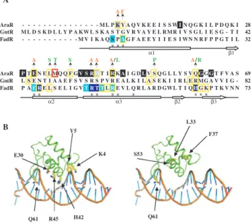
We have previously established that AraR interaction with DNA is achieved via a 70 amino acid N-terminal domain. These results were obtained by random and site-directed mutagenesis based on a 3D model of the DBD derived from the crystal structure of the E. coli regulator FadR (8). However, many of these mutations resulted in changes that could alter the protein structure or interfere with DNA binding. In order to conduct a more clarifying characterization of the role of specific AraR residues on its DNA-binding activity, we made several amino acid substitutions in or near the HTH motif. Residues were chosen based on the 3D model and/or primary sequence alignment of AraR-like proteins (8). The majority of the substituted amino acids was predicted to contact directly the bases of the DNA and consequently, would account for the specificity of the interaction with operator sequences. Positions K4, E30, R41, H42 and Q61 were exchanged to alanine and Y5 to phenylalanine (Figure 1A and B). The substitutions were designed to minimize local structure disruption and probe loss of contact with the DNA. In addition, in this work we analysed a mutation R45L generated by chance during the construction of mutant R45A, which was characterized in vivo in a previous work (8). Plasmid pLS30, carrying an araR
wild-type allele, and its derivatives harbouring the mutated araR alleles, obtained after site-directed muta-genesis, were integrated as single copy at theamyElocus ofB. subtilisreceptor strain IQB350. This strain bears an
araRnull mutation (araR) and a transcriptionalaraAB0– lacZ fusion (see Materials and Methods section) and therefore expresses constitutively b-galactosidase. In the resulting strains (Table 1), the levels of b-galactosidase expressed from the ara operon-lacZ fusion reflect the
in vivoregulatory activity of the AraR variant encoded by the allele integrated at theamyElocus.
The effect of each substitution was analysed by determining the levels of accumulated b-galactosidase in strains grown under inducing (presence of arabinose)
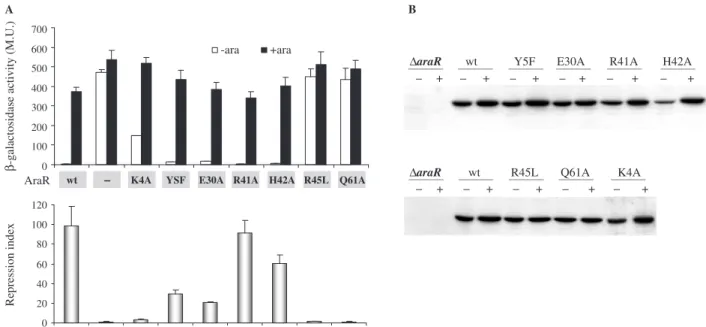
and non-inducing conditions (absence of arabinose). The results are summarized in Figure 2A. The regulatory
activity was quantified as the repression index
(Figure 2A). The receptor strain IQB350 (araR
araAB0-lacZ) and IQB352, a derivative carrying thearaR
wild-type allele at the amyElocus, were used as controls, yielding repression index values of 1 and 99, respectively, which correspond to the absence and maximal regulation exerted by the protein. Mutation R41A had no effect on the regulatory activity when compared to the wild type. Therefore, although amino acid R41 is conserved among all members of the GntR family (Figure 1A) and establishes interactions with bases in the major groove according to FadR-DNA data [R45 in FadR; (24)], it is dispensable for AraR bindingin vivo. Variant H42A displayed a minimal decrease on repression activity
A F
α1 β1
α2 α3 β2 β3
A
B Y5
E30
R45 H42 Q61
K4
L33
F37
S53
Q61
AraR - - - M L P K Y A Q V K E E I S S WIN Q G K I L P D Q K I 28 GntR M L D S K D L L Y P A KW L S K A S T G V R V A Y E L RMR I V S G L I E S G - T I 42 FadR - - - M V I K A Q S PAG F A E E Y I I E S I WN N R F P P G T I L 32
* * *
AraR PTEN E L MQ Q F GV S R HT IRKAI G DLV S Q G L L Y S V QGGGT F V A S 69 GntR L S E N T I A A E F S V S R S P V R E A L K I L A S E K I I R L E RMG A V V I G - 82 FadR P AE RE L S E L I G VT R T TLRE V L Q R L A R D GWL T I QHG K P T K V N N 73
* * * * * * * * * *
A S T S A A A/L P A/R
Figure 1. The DNA-binding domain of AraR and localization of mutations. (A) Sequence alignment of the N-terminal region of AraR, FadR and GntR. Residues that are conserved in the entire family GntR are shaded in black and residues characteristic of AraR homologous proteins in grey (8). Positions of mutations leading to a constitutive phenotype are boxed in yellow (8,26,29). A substitution yielding a suppressor phenotype is boxed in red. The introduced residues in AraR are shown above the sequence, coloured in orange when obtained through site-directed mutagenesis and green through random mutagen-esis (8). In FadR are coloured in light blue residues contacting the DNA backbone and in dark blue the ones contacting the DNA bases, according to crystallographic data (28); an asterisk below the sequence indicates amino acids within contact distance of DNA (24). The secondary structure (arrows representing beta-strands and bars alpha-helices) of FadR (amino acid residues 1–73) is shown below the alignment according to van Aaltenet al. (24). The microorganisms of source and accession numbers are: AraR from B. subtilis (P96711);
GntR from B. subtilis (P10585); FadR from E. coli (P09371).
(1.6-fold). Moderate effects were observed with Y5F and E30A, 3.3- and 4.8-fold, respectively. The more drastic effects were seen with K4A, a 30-fold decrease in repression, and with R45L and Q61A the regulatory activity was completely abolished. The lack of regulation of R45L is identical to that observed with R45A (8). Together, the results suggest that these three residues play the most important roles in DNA binding of the ones analysed here.
Because the observed decrease in repression could be the result of deficientin vivo accumulation of the mutant proteins, as a consequence of lower stability and proteolysis, we measured the abundance of each AraR variant. The strains were grown as for theb-galactosidase assays and the level of AraR estimated by western immunobloting using equivalent amounts of their soluble cell extracts (Figure 2B). The cellular level of all mutant proteins was comparable to that seen with wild-type AraR, ruling out the possibility of deregulation originated by degradation of the repressor.
Analysis of the wild-type AraR and mutant proteins DNA-binding affinityin vitro
The apparent affinity constants (Kd) of AraR mutants for
operator sequences were determined by EMSAs using a
32
P-labelled 126-bp DNA fragment, which carried both operators of the metabolic operon, ORA1 and ORA2
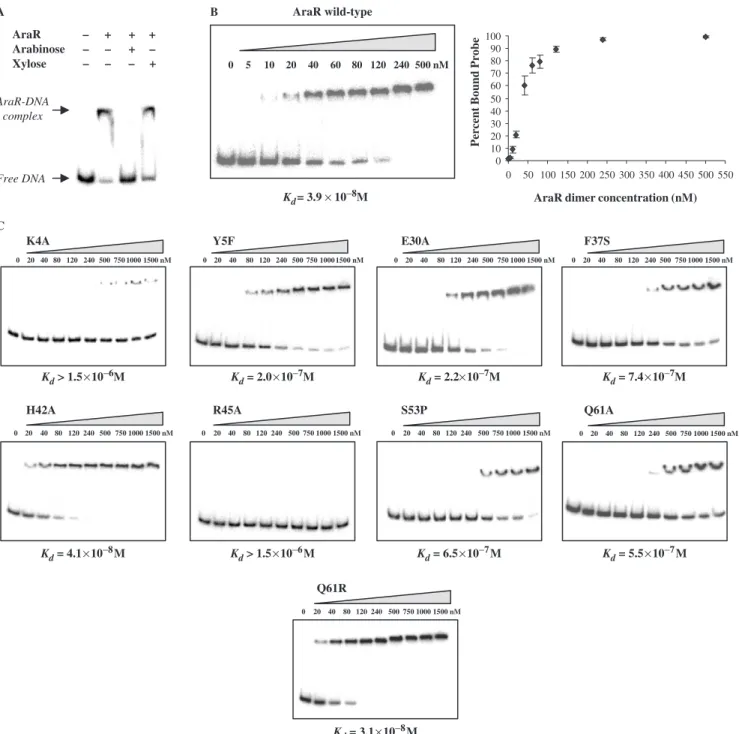
(depicted in Figure 5). Binding of AraR to this DNA fragment was specific, as the presence of the inducer arabinose but not xylose prevented the formation of the
protein–DNA complex (Figure 3A). Titration of 1 nM of DNA with increasing concentrations of wild-type repres-sor (Figure 3B) allowed the determination of an apparent
Kd3.910 8M, which is defined as the amount of protein
necessary to shift 50% of the labelled probe (25). This value is comparable to that previously calculated for each individual box using DNase I quantitative footprinting experiments, 3.410 8M and 4.710 8M for ORA1and
ORA2, respectively (6).
All the mutant proteins that displayed an effectin vivo
were overexpressed inE. coliand purified to homogeneity (see Materials and Methods section). Binding to DNA was assayed by EMSA and their respective apparent affinity constants determined (Figure 3C). Variant H42A, which showed the minimal loss of repression in vivo (2-fold) bound DNA with an apparentKdof 4.110 8M, similar
to the wild-type protein. The most severe effects were displayed by Q61A (Kd5.510 7M), K4A and R45A,
both showing an apparentKd41.510 6M. These three
mutants were also unable to perform a regulatory activity
in vivo. Residues K4 and H42 are completely conserved among AraRs (8) and in a contact distance of the DNA according to the model (Figure 1A and B), however, mutation of these residues had different outcomes. Since these two residues are not conserved among the members of the entire GntR family they may contribute in very different extents to the DNA-binding specificity of AraR-like proteins.
R45 is a conserved amino acid in the GntR family members. In the regulator FadR from E. coli, the
0 20 40 60 80 100 120 0 100 200 300 400 500 600 700
A B
WT Dar a R K4A Y5 E30A R41A H42A R45L Q61A
-ara +ara
Repression index
β
-galactosidase activity (M.U.)
AraR
∆araR wt Y5F E30A R41A H42A
+ + + + + +
∆araR wt R45L Q61A K4A
+ + + + +
wt – K4A YSF E30A R41A H42A R45L Q61A
substitution of the corresponding residue (R49) has also a drastic effectin vivo (26). Moreover, the crystal structure of the FadR–DNA complex (27,28) shows that R49 locates in the recognition helix of the winged HTH and interacts with a phosphate group, not specifically a base. According to the predictions of the tertiary structure of AraR, R45 is also located in the recognition helix
(Figure 1B), which is generally more responsible for the interaction with DNA, in particular the positively charged residues. Q61 belongs to the predicted wing of the DNA-binding motif (Figure 1A and B). The corresponding residue in both FadR and in GntR from B. subtilis is also positively charged (Figure 1A) and substitutions led to loss of DNA-binding ability (26,29). In FadR, H65
AraR wild-type
0 5 10 20 40 60 80 120 240 500 nM
0 10 20 30 40 50 60 70 80 90 100
0 50 100 150 200 250 300 350 400 450 500 550
AraR dimer concentration (nM)
Percent Bound Probe
AraR A
C
B
– + Arabinose –
– – –
+
+ – +
– + Xylose
Free DNA AraR-DNA
complex
Kd= 3.9 × 10⁻⁸M
K4A
H42A R45A S53P
Q61R
Q61A
Y5F E30A F37S
Kd> 1.5×10⁻⁶M
Kd= 4.1×10⁻⁸M Kd> 1.5×10⁻⁶M
Kd= 3.1×10⁻⁸M
Kd= 6.5×10⁻⁷M Kd= 5.5×10⁻⁷M
Kd= 2.0×10⁻⁷M Kd= 2.2×10⁻⁷M Kd= 7.4×10⁻⁷M
0 20 40 80 120 240 500 750 1000 1500 nM
0 20 40 80 120 240 500 750 1000 1500 nM 0 20 40 80 120 240 500 750 1000 1500 nM 0 20 40 80 120 240 500 750 1000 1500 nM
0 20 40 80 120 240 500 750 1000 1500 nM
0 20 40 80 120 240 500 750 1000 1500 nM 0 20 40 80 120 240 500 750 1000 1500 nM 0 20 40 80 120 240 500 750 1000 1500 nM
0 20 40 80 120 240 500 750 1000 1500 nM
Figure 3. Binding of AraR toaraABDLMNPQ-abfA promoter (operators ORA1-ORA2) in EMSA. AraR was incubated with the 50-end-labelled
is part of the wing and makes specific contacts with an adenine (24,28).
Intermediate decreases of DNA binding were observed
with Y5F and E30A, with Kd 2.010
7
M and
2.310 7M, respectively, similarly to that seen in vivo. Therefore, these exchanges led to a comparable effect both
in vivoandin vitro(Figures 2 and 3). Moreover, the nature of the mutation Y5F revealed the importance of the OH group in the interaction with DNA. Both residues are conserved in the GntR-family proteins, and the corresponding residues in FadR, A9 and E34, were shown to contact the DNA backbone (24,28). The latter also contacts nearby amino acids, contributing presum-ably to the stabilization of residues that interact specifi-cally with the DNA bases.
Additionally, four other AraR mutants, L33S, F37S, S53P and Q61R, obtained by random mutagenesis and characterizedin vivo in a previous work [(8); Figure 1B), were studied by EMSA. Both substitutions F37S and S53P had led to derepressionin vivo, of24- and 2-fold, and in mutant L33S the regulatory activity was almost comple-tely abolished (8). These values could be explained by the observed instability of the proteins (8). However, purified mutants F37S and S53P showed decreased DNA-binding affinities,Kd7.410 7M and 6.510 7M, respectively,
that may also contribute to the deregulationin vivo. These results could be explained by the nature and localization of these substitutions, which suggest implications in the folding of the DBD. Overexpression and purification of L33S yielded only small amounts of protein. Nevertheless, at the maximal concentration that we could use in EMSA assays, 50 nM of the mutant, no DNA binding was observed (data not shown).
Interestingly, while the Q61A substitution completely abolished regulation in vivo and DNA binding in vitro, the change to arginine in the same position showed only a 1.6-fold decrease of regulatory activity in vivo (8), and the affinity to the DNA probein vitro, Kd 3.110 8M,
was even slightly higher than that displayed by wild-type AraR. Noteworthy, in GntR the inverse of AraR mutation Q61R (i.e. GntR R75Q) leads to a significant loss of regulationin vivo(29). Based on these observations, we may speculate that the rise of positive charge as a result of AraR substitution Q61R increased the overall (non-specific) affinity for DNA, leading in vivo to a titration of the protein.
In summary, K4, R45 and Q61, were the most critical AraR residues in achieving specific DNA binding, and Y5 and E30 also play an important role, and overall there was a good correlation between the effects of the mutations in the binding affinities to thearaoperon promoterin vitro
and in the regulatory activityin vivo.
Effect of base-pairs mutations in the operator sequences on transcriptional regulation by AraR
AraR recognizes and binds at least eight palindromic operator sequences, located in the five known arabinose-inducible promoters. Three of these promoters contain twoaraboxes: the promoter of thearametabolic operon (boxes ORA1 and ORA2), ofaraE(ORE1 and ORE2) and
of xsa(ORX1and ORX2). In the cases of the genesaraR
andabnA, a single arabox is present (ORR3and ORB1).
AraR binding to the promoters displaying two
araboxes is cooperative and involves the formation of a small loop in the DNA. In fact, for fullin vivorepression, communication between repressor molecules bound to two properly spaced operators is required, as shown by the analysis of mutations designed to prevent cooperative binding of AraR (6,7). An alignment of the eight ara
boxes, identified by DNase I footprinting and/or mutagenesis, showed the 16-bp consensus sequence
50-ATTTGTACGTACAAAT-30 and highlighted the
conserved nucleotides at each position (Figure 4A). This operator consensus presents the typical signature for cis-acting elements recognized by GntR family members 50-(N)x-GT-N(0-15)-AC-(N)x-30(11).
In a previous work, we showed that G9is important for
AraR binding because the substitution G9!T in
both boxes ORA1 and ORA2 caused defect in the
regulatory activity of AraR in vivo and prevented cooperative binding (6). To further investigate which nucleotides within the consensus sequence were necessary for protein binding, single-nucleotide exchanges were made in ORA1 at the promoter of the ara operon. The
most conserved bases were substituted and mutations were designed to introduce transversions from AT to CG and CG to AT: A1!C, G5!T, T6!G, C8!A, T10!G,
A11!C and T16!G. The mutated promoters
transcrip-tional fused to the lacZ gene were independently integrated at the amyE locus of the B. subtilis receptor strain (see Materials and Methods section and Table 1). Strain IQB572, bearing a transcriptional fusion to the wild-type operator (araR amyE::ORA(wt)-lacZ thrC::araR) was used as a control to assay the repres-sion exerted by AraR. The levels of accumulated
b-galactosidase activity measured in all strains are shown in Figure 4B. Mutations having the most drastic effect on AraR binding were G9!T, both in ORA1 and
ORA2 [as previously determined; (7)], A11!C and
T16!G, leading to a decrease in the regulatory activity 49-fold compared to the control. A moderate effect of deregulation, varying from 2.4- to 4.4-fold, was observed for A1!C, G5!T and T6!G, and substitution T10!G
had no effectin vivo.
Surprisingly, C8!A abolishedlacZ expression both in
inducing and non-inducing conditions. One possibility to explain this result would be an increase in the affinity of AraR for the mutated operator leading to a tight binding of the repressor (even in the presence of arabinose), thus preventing transcription by RNA polymerase. To test this hypothesis, we investigated the Lac phenotype in araR -null mutants (araR) bearing the transcriptional fusion
ORA1(C8!A)–lacZandORA(wt)–lacZ, strains IQB257 and
IQB530 (Table 1), respectively. The results obtained in solid medium with X-gal and arabinose indicated that the lack of lacZ expression in mutant C8!A (Position +4,
Analysis of operator mutations on AraR–DNA affinityin vitro
The effect of the operator mutations was also analysed
in vitro by EMSA competition assays. The experiments were performed in the presence of 1 nM of the ORA1–
ORA2DNA probe described above, 40 nM of AraR, and
increasing concentrations of a double-stranded 38-bp competitor oligonucleotide (50–500 nM) containing the wild-type or the described mutations in ORA1or ORA2. In
addition, oligonucleotides carrying all possible substitu-tions at the highly conserved base pairs, G9, A10and T16
were also used. We compared the ability of these cold DNAs to titrate binding of AraR to the labelled probe, reflected in the decrease of the intensity of the protein– DNA complex band. The wild-type ORA1box was able to
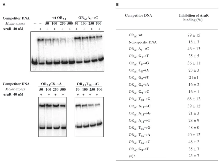
compete for AraR binding in a concentration-dependent manner, with a 79% loss of band shift at 500-fold excess competitor DNA (Figure 5A). In contrast, a non-specific oligonucleotide (equivalent in length; Supplementary Table 1) used as control disrupted only 18% of the binding (data not shown). Inhibition of binding in the presence of 500 nM of the different competitors was quantified and the results are summarized in Figure 5B.
The DNA containing mutation T10!G in ORA1
competed in levels similar to that obtained for the wild-type box (68 and 79% inhibition, respectively). In contrast, AraR was unable to bind the boxes with single base-pair substitutions in G9, either to T (the mutation
tested in vivo, previously), to A and C (inhibition values between 21 and 16%). A notorious decrease in binding to A11!C was also observed, which was more
pro-nounced when A was exchanged for G or T. However,
oligonucleotides containing a mutation at T16(either to G,
A or C) were still able to partially compete for the repressor. The three mutations, A1!C, G5!T and
T6!G, leading to a partial de-repression in vivo also
showed an intermediate effect. Taken together, the results indicate a good correlation between the in vivo and
in vitro, but the exchanges at T16 and the mutation
A11!C, comparatively to the regulatory activity in vivo
were expected to bind to AraR less tightly. This could be due to the more reduced sensitivity of the competition assays. Interestingly, the mutation C8!A, that affected
transcription even in the presence of inducer, did not compete (23%) for AraR, indicating that the mutation has an effect on AraR binding that could not be testedin vivo. In previous work, a search for AraR operator sequences in the B. subtilis genome (6,30) identified a putative binding sequence in the open reading frame ydjK
[identified as a myo-inositol transporter, iolT; (31)]. The sequence 50-TTTTTACGTACAATT-30[+27 relative
to the translation start site; (31)] displayed only two deviations (underlined) from the consensus sequence: A1and G5. Construction and analysis of transcriptional
fusions of the promoter region and 50-end ofydjKtolacZ
showed that expression is not AraR dependent, thus the potential operator is not functional in vivo (Ina´cio,J.M. and I.S.-N., unpublished data). To determine the ability of AraR to bind this sequence in vitro, competition assays were performed as described above but no competition was detected (Figure 5B). Since T at Position 1 is present in functionally active AraR boxes (Figure 4A) these observations suggest, in accordance to the mutagenic
0 10 20 30 40 50 60 0 500 1000 1500 2000 2500
A B
+ara −ara
β
-galactosidase activities (M.U.)
Repression index
C T G A T G C G
ORA1 T A A A A T T G T T C G T A C A A A T A T T T A
ORA2 C A T T A G T A C G T A T C T T T T G T A
ORE1 A T A T T T G T A C G T A C T A A T T A
ORE2 T A T A A G T A C G T A C A A T T
ORR3 A A A T T T G T C C G T A T A C A T T T T
ORX1 A A T A C A T A C G T A C A A A T A T A A A
ORX2 G A C A T G T A C G A A C A T A T A T A A T OR
B1 T T T T T G T C T G T A C A A A T T A C
A1
C
T6
G
G9
T T10
G A11
C T16®
G C8
A
wt G9
(ORA2)
1 2 3 4 5 6 7 8 9 10 11 12 13 14 15 16
G5
T T
Consensus
↑ ↑ ↑ ↑ ↑ ↑ ↑ ↑
↓ ↓ ↓ ↓ ↓ ↓ ↓ ↓ ↓
Figure 4. Ara operator sequences. (A) Alignment of the eight AraR boxes and a picture representing the conservation of bases at each position in the inferred consensus 16-bp palindromic operator sequence [generated by WebLogo 2.8.2 software (33)].Araboxes are located in the promoter of the
araABDLMNPQ-abfAmetabolic operon (ORA1 and ORA2),araE(ORE1and ORE2),araR(ORR3),xsa(ORX1and ORX2) andabnA(ORB1). The
conserved nucleotides in at least seven boxes are shaded. Bases substituted in ORA1or ORA2by site-directed mutagenesis used forin vivoanalysis
are indicated, and the new base is shown above. (B) Effect ofarabox mutations onin vivo regulation by AraR.b-Galactosidase activities were determined inB. subtilisstrains carryingORA–lacZfusions integrated at theamyElocus and wild-typearaRat thethrClocus grown in the absence or presence of arabinose (upper panel, in white and black bars, respectively). The repression index (lower panel) reflects the regulation exerted on each ara box (Figure 2). Nucleotide substitutions (obtained by site-directed mutagenesis) are indicated for the mutated position in the
analysis described above, that G at Position 5 plays an important role in AraR binding.
In conclusion, we found that bases in both arms of the palindrome of the AraR boxes are involved in AraR– DNA contacts. The in vivo and in vitro studies together with sequence analysis of the eight functionally active AraR B. subtilis boxes indicated that bases G9, A11 and
T16 are crucial for AraR binding, and that A1 and T6
play also an important role. Furthermore, Position 5 required a purine (Pu) for functionality in vivo, and sequence analysis suggested that the corresponding mirror base (Position 12) in the other arm of the palindrome is always a pyrimidine (Py). An alignment of all putative AraR-binding sequences based on a search of the consensus 50-ATTTGTACGTACAAAT-30
in genomes of bacteria from the Bacillus/Clostridium
group that contain AraR orthologues also highlighted the majority of the invariable positions: Pu5, T6 and the
correspondent mirror A11, Py12 and G9 at the centre of
the palindrome (Supplementary Figure 1).
Mutation M34T partially restores AraR binding to mutated DNA operators bothin vivoandin vitro
In order to isolate AraR mutants that could suppress the loss of regulation caused by the single nucleotide substitutions in thearaboxes, anin vivoscreening method was developed (see Materials and Methods section). Briefly, random mutagenesis of the 50-end of the araR
allele was performed by PCR and the resulting library of plasmids carrying the mutated alleles was used to transform B. subtilis strains, allowing its integration at the non-essentialthrClocus via a double-crossover event. The receptor strains possessed a araR amyE::ORA -lacZ background, and carried different mutations in the ORA operator sequence (Table 1). The constitutive
expression of lacZ due to the inability of the wild-type AraR to bind the mutated operator leads to a Lac+ phenotype in the absence of inducer. However, if the integrated mutant araR allele encodes a protein that suppressed the deleterious effect of the operator mutation
Competitor DNA
A B
wt ORA1 ORA1A1→C
ORA1T10→G
Molar excess − − 50 100 250 500 50 100 250 500 AraR 40 nM
Competitor DNA
Molar excess
AraR 40 nM
− + + + + + + + + +
ORA1C8→A 50 100 250 500
+ + + +
50 100 250 500 + + + +
Competitor DNA Inhibition of AraR binding (%)
ORA1wt 79 ± 15
Non-specific DNA 18 ± 3
ORA1 A1→C 46 ± 13
ORA1 G5→T 35 ± 5
ORA1 T6→G 36 ± 11
ORA1 C8→A 23 ± 3
ORA1 G9→T 21 ± 1
ORA1 G9→A 16 ± 2
ORA1 G9→C 16 ± 1
ORA1 T10→G 68 ± 12
ORA1 A11→C 39 ± 12
ORA1 A11→G 21 ± 3
ORA1 A11→T 28 ± 9
ORA1 T16→G 48 ± 0
ORA1 T16→A 40 ± 12
ORA1 T16→C 48 ± 2
ORA2G9→T 35 ± 7
ydjK 25 ± 7
Figure 5. In vitro analysis of AraR binding to mutated ara boxes. (A) Competition EMSA experiments using double-stranded oligonucleotides containing mutatedaraboxes. AraR (40 nM) was incubated with the 32P-labelled OR
A1–ORA2 region (1 nM) in the presence or absence of the
a Lac phenotype is displayed indicating recovery of regulation. Thus, we screened for transformants with decreased b-galactosidase production in the absence of arabinose.
One transformant of strain IQB533, containing the fusion ORA1(T6!G)–lacZ, displayed a gain-of-function
phenotype, and the sequencing of thearaRallele revealed the substitution M34T, located in the HTH motif of the protein. To determine if this effect was specific or also affected AraR binding to the other mutated promoters, the allele was integrated at the thrC locus of the corresponding B. subtilis strains and b-galactosidase activities were measured (Figure 6A). Interestingly, the repression exerted by mutant M34T was higher than that exerted by wild-type AraR in almost all operators, in particular with mutant boxes G9!T (both in ORA1 and
ORA2), T6!G, A11!C and T16!G. Since a higher level
of intracellular accumulation of this AraR variant could explain this phenotype, we determined the accumulation of the wild-type AraR and mutant M34T in strains IQB572 and IQB583, respectively (Table 1). The observed cellular levels of protein were similar in both strains (Figure 6B), indicating that the phenotype displayed by M34T was not due to increased concentration of protein. In fact, EMSA assays performed as described above showed that the mutant displays an increased affinity to theara operon promoter probe, with an apparentKd of
1.010 8M (Figure 6C), which is almost 4-fold lower than that of the wild-type protein (Kd=3.910 8 M).
The substitution M34T is located in the first helix of the winged HTH motif (Figure 1A). According to a study on protein–DNA interactions based on structures of 129 complexes (32), threonine is responsible for a far larger number of protein–DNA bonds than methionine, although almost all are made with the DNA backbone and not the bases. This is in agreement with the results we obtained, that show an increase in the repression exerted over all mutated boxes, suggesting an increased DNA affinity of M34T through non-specific contacts.
CONCLUDING REMARKS
Previous studies have mapped the functional domains of AraR and characterized the C-terminal region involved in effector binding and dimerization (8). In this work, we focused on two additional and crucial components of the transcription process, the DBD and cis-acting elements. Guided by molecular modelling combined with multiple primary sequence alignment of AraR orthologues and GntR family members, we identified amino acids poten-tially involved in DNA binding. The effect of their substitution was analysedin vivoandin vitroand revealed key residues necessary for the regulatory activity. In addition, important bases for AraR–DNA interactions in both arms of the palindromic operator sequences were also identified. We obtained both expected and unex-pected results highlighting the uniqueness of protein– DNA interactions in each particular system. A future determination of the structure of AraR, in its unbound form or in complex with the inducer or DNA, would allow a more detailed analysis of the mechanism by which AraR binds its cognate operator sequences and how the conformational change triggered by the binding of arabinose prevents this interaction.
SUPPLEMENTARY DATA
Supplementary Data are available at NAR Online.
ACKNOWLEDGEMENTS
This work was partially supported by grants POCTI/
BME/36164/00 and POCI/BIA-MIC/61140/04 from
Fundac¸a˜o para a Cieˆncia e Tecnologia (FCT) and FEDER to I.S.-N. I.S.F. is the holder of Ph.D. fellowship SFRH/BD/5233/01 from FCT. Funding to pay the Open Access publication charges for this article was provided by Fundac¸a˜o para a Cieˆncia e Tecnologia.
Conflict of interest statement. None declared.
0 10 20 30 40 50 60
A B C
AraR wt AraR M34T
Repression index
wt M34T
+ −
− +
0 5 10 20 40 60 80 120 240 500 nM
M34T
Kd = 1.0×10⁻⁸M
A1 T6 C8 G9 T10 A11 T16
wt G9
T G C G T A G T C
(ORA2) G5
↓ ↓ ↓ ↓ ↓ ↓ ↓ ↓ ↓
Figure 6.Characterization of suppressor mutant M34T. (A) Comparison of the repression exertedin vivoby AraR wild type or mutant M34T over
araboxes.b-Galactosidase activities were determined inB. subtilisstrains carrying the differentaraA–lacZfusions integrated at theamyElocus and the wild-type or M34TaraRallele integrated at thethrClocus, grown in inducing and non-inducing conditions. The bars represent the repression index obtained for wild-type AraR (white bars) or variant M34T (grey bars) (Figure 2). Values are the average of three independent experiments, each assayed in duplicate. Error bars represent the SD. (B) Intracellular accumulation of AraR wild type and mutant M34T by western immunoblot analysis from cultures grown in the absence ( ) or presence (+) of inducer (Figure 2). (C) Binding of AraR M34T to araABDLMNPQ-abfA
REFERENCES
1. Sa´-Nogueira,I., Nogueira,T.V., Soares,S. and de Lencastre,H. (1997) TheBacillus subtilisL-arabinose (ara) operon: nucleotide sequence, genetic organization and expression.Microbiology,143 (Pt 3), 957–969.
2. Sa´-Nogueira,I. and Ramos,S.S. (1997) Cloning, functional analysis, and transcriptional regulation of theBacillus subtilis araEgene involved in L-arabinose utilization.J. Bacteriol.,179, 7705–7711.
3. Sa´-Nogueira,I. and Mota,L.J. (1997) Negative regulation of L-arabinose metabolism inBacillus subtilis: characterization of the
araR(araC) gene.J. Bacteriol., 179, 1598–1608.
4. Raposo,M.P., Ina´cio,J.M., Mota,L.J. and de Sa´-Nogueira,I. (2004) Transcriptional regulation of genes encoding arabinan-degrading enzymes inBacillus subtilis.J. Bacteriol.,186, 1287–1296. 5. Krispin,O. and Allmansberger,R. (1998) TheBacillus subtilisAraE
protein displays a broad substrate specificity for several different sugars.J. Bacteriol.,180, 3250–3252.
6. Mota,L.J., Tavares,P. and Sa´-Nogueira,I. (1999) Mode of action of AraR, the key regulator of L-arabinose metabolism inBacillus subtilis.Mol. Microbiol.,33, 476–489.
7. Mota,L.J., Sarmento,L.M. and de Sa´-Nogueira,I. (2001) Control of the arabinose regulon inBacillus subtilisby AraR in vivo: crucial roles of operators, cooperativity, and DNA looping.J. Bacteriol., 183, 4190–4201.
8. Franco,I.S., Mota,L.J., Soares,C.M. and de Sa´-Nogueira,I. (2006) Functional domains of theBacillus subtilistranscription factor AraR and identification of amino acids important for nucleoprotein complex assembly and effector binding.J. Bacteriol.,188, 3024–3036.
9. Haydon,D.J. and Guest,J.R. (1991) A new family of bacterial regulatory proteins.FEMS Microbiol. Lett.,63, 291–295.
10. Weickert,M.J. and Adhya,S. (1992) A family of bacterial regulators homologous to Gal and Lac repressors.J. Biol. Chem.,267, 15869–15874.
11. Rigali,S., Derouaux,A., Giannotta,F. and Dusart,J. (2002) Subdivision of the helix-turn-helix GntR family of bacterial regulators in the FadR, HutC, MocR and YtrA subfamilies.
J. Biol. Chem.,277, 12507–12515.
12. Lee,M.H., Scherer,M., Rigali,S. and Golden,J.W. (2003) PlmA, a new member of the GntR family, has plasmid maintenance functions inAnabaenasp. strain PCC 7120.J. Bacteriol.,185, 4315–4325.
13. Geer,L.Y., Domrachev,M., Lipman,D.J. and Bryant,S.H. (2002) CDART: protein homology by domain architecture.Genome Res., 12, 1619–1623.
14. Clark,K.L., Halay,E.D., Lai,E. and Burley,S.K. (1993) Co-crystal structure of the HNF-3/fork head DNA-recognition motif resembles histone H5.Nature,364, 412–420.
15. Gajiwala,K.S. and Burley,S.K. (2000) Winged helix proteins.
Curr. Opin. Struct. Biol.,10, 110–116.
16. Luscombe,N.M. and Thornton,J.M. (2002) Protein-DNA interactions: amino acid conservation and the effects of mutations on binding specificity.J. Mol. Biol.,320, 991–1009.
17. Miller,J. (1972)Experiments in Molecular Genetics.Cold Spring Harbor Laboratory Press, Cold Spring Harbor, NYC, USA. 18. Pascal,M., Kunst,F., Lepesant,J.A. and Dedonder,R. (1971)
Characterization of two sucrase activities inBacillus subtilis
Marburg. Biochimie,53, 1059–1066.
19. Studier,F.W., Rosenberg,A.H., Dunn,J.J. and Dubendorff,J.W. (1990) Use of T7 RNA polymerase to direct expression of cloned genes.Methods Enzymol.,185, 60–89.
20. Sambrook,J., Fritsch,E. and Maniatis,T. (1989)Molecular cloning: a laboratory manual. Cold Spring Harbor Laboratory Press, New York, USA.
21. Guerout-Fleury,A.M., Frandsen,N. and Stragier,P. (1996) Plasmids for ectopic integration inBacillus subtilis.Gene,180, 57–61. 22. Leung,D., Chen,E. and Goeddel,D.V. (1989) A method for random
mutagenesis of a defined DNA segment using a modified polymerase chain reaction.Technique,1, 11–15.
23. Bera,A.K., Zhu,J., Zalkin,H. and Smith,J.L. (2003) Functional dissection of theBacillus subtilis puroperator site.J. Bacteriol.,185, 4099–4109.
24. van Aalten,D.M., DiRusso,C.C. and Knudsen,J. (2001) The structural basis of acyl coenzyme A-dependent regulation of the transcription factor FadR.EMBO J.,20, 2041–2050.
25. Riggs,A.D., Suzuki,H. and Bourgeois,S. (1970) Lac repressor-operator interaction. I. Equilibrium studies.J. Mol. Biol.,48, 67–83. 26. Raman,N., Black,P.N. and DiRusso,C.C. (1997) Characterization
of the fatty acid-responsive transcription factor FadR: biochemical and genetic analyses of the native conformation and functional domains.J. Biol. Chem.,272, 30645–30650.
27. van Aalten,D.M., DiRusso,C.C., Knudsen,J. and Wierenga,R.K. (2000) Crystal structure of FadR, a fatty acid-responsive transcription factor with a novel acyl coenzyme A-binding fold.
EMBO J.,19, 5167–5177.
28. Xu,Y., Heath,R.J., Li,Z., Rock,C.O. and White,S.W. (2001) The FadR.DNA complex. Transcriptional control of fatty acid metabolism inEscherichia coli.J. Biol. Chem.,276, 17373–17379. 29. Yoshida,K., Fujita,Y. and Sarai,A. (1993) Missense mutations in theBacillus subtilis gntrepressor that diminish operator binding ability. J. Mol. Biol.,231, 167–174.
30. Rodionov,D.A., Mironov,A.A. and Gelfand,M.S. (2001) Transcriptional regulation of pentose utilisation systems in the
Bacillus/Clostridiumgroup of bacteria.FEMS Microbiol. Lett.,205, 305–314.
31. Yoshida,K., Yamamoto,Y., Omae,K., Yamamoto,M. and Fujita,Y. (2002) Identification of two myo-inositol transporter genes of
Bacillus subtilis.J. Bacteriol.,184, 983–991.
32. Luscombe,N.M., Laskowski,R.A. and Thornton,J.M. (2001) Amino acid-base interactions: a three-dimensional analysis of protein-DNA interactions at an atomic level.Nucleic Acids Res.,29, 2860–2874. 33. Crooks,G.E., Hon,G., Chandonia,J.M. and Brenner,S.E. (2004)

![Figure 4. Ara operator sequences. (A) Alignment of the eight AraR boxes and a picture representing the conservation of bases at each position in the inferred consensus 16-bp palindromic operator sequence [generated by WebLogo 2.8.2 software (33)]](https://thumb-eu.123doks.com/thumbv2/123dok_br/16667710.742555/9.918.89.845.109.416/operator-sequences-alignment-representing-conservation-consensus-palindromic-generated.webp)