Formation and Dorsal-Ventral Patterning in Limb
Development
Endika Haro1, Irene Delgado1¤, Marisa Junco1, Yoshihiko Yamada2, Ahmed Mansouri3,4,5, Kerby C. Oberg6, Marian A. Ros1,7*
1Instituto de Biomedicina y Biotecnologı´a de Cantabria (CSIC-UC-SODERCAN), Santander, Spain,2Laboratory of Cell and Developmental Biology, NIDCR, National Institutes of Health, Bethesda, Maryland, United States of America,3Max Planck Institute for Biophysical Chemistry, Department of Molecular Cell Biology, Am Fassberg, Go¨ttingen, Germany,4Department of Clinical Neurophysiology, University of Go¨ttingen, Go¨ttingen, Germany,5Genome and Stem Cell Center, GENKOK, Erciyes University, Kayseri, Turkey,6Department of Pathology and Human Anatomy, Loma Linda University, Loma Linda, California, United States of America,7Departamento de Anatomı´a y Biologı´a Celular, Facultad de Medicina, Universidad de Cantabria, Santander, Spain
Abstract
The formation and maintenance of the apical ectodermal ridge (AER) is critical for the outgrowth and patterning of the vertebrate limb. The induction of the AER is a complex process that relies on integrated interactions among the Fgf, Wnt, and Bmp signaling pathways that operate within the ectoderm and between the ectoderm and the mesoderm of the early limb bud. The transcription factors Sp6 and Sp8 are expressed in the limb ectoderm and AER during limb development.Sp6
mutant mice display a mild syndactyly phenotype whileSp8mutants exhibit severe limb truncations. Both mutants show defects in AER maturation and in dorsal-ventral patterning. To gain further insights into the role Sp6 and Sp8 play in limb development, we have produced mice lacking both Sp6 and Sp8 activity in the limb ectoderm. Remarkably, the elimination or significant reduction inSp6;Sp8gene dosage leads to tetra-amelia; initial budding occurs, but neitherFgf8norEn1are activated. Mutants bearing a single functional allele of Sp8 (Sp62/2;Sp8+/2) exhibit a split-hand/foot malformation
phenotype with double dorsal digit tips probably due to an irregular and immature AER that is not maintained in the center of the bud and on the abnormal expansion ofWnt7aexpression to the ventral ectoderm. Our data are compatible with Sp6 and Sp8 working together and in a dose-dependent manner as indispensable mediators of Wnt/ catenin and Bmp signaling in the limb ectoderm. We suggest that the function of these factors links proximal-distal and dorsal-ventral patterning.
Citation:Haro E, Delgado I, Junco M, Yamada Y, Mansouri A, et al. (2014) Sp6 and Sp8 Transcription Factors Control AER Formation and Dorsal-Ventral Patterning in Limb Development. PLoS Genet 10(8): e1004468. doi:10.1371/journal.pgen.1004468
Editor:Mark Lewandoski, National Cancer Institute, United States of America
ReceivedDecember 6, 2013;AcceptedMay 14, 2014;PublishedAugust 28, 2014
This is an open-access article, free of all copyright, and may be freely reproduced, distributed, transmitted, modified, built upon, or otherwise used by anyone for any lawful purpose. The work is made available under the Creative Commons CC0 public domain dedication.
Funding:Supported by grant BFU2011-24972 from the Spanish Government (MAR) and by National Organization of Rare Diseases (KCO) and by the Intramural Research Program at the National Institute of Dental and Craniofacial Research, NIH, USA (YY). The funders had no role in study design, data collection and analysis, decision to publish, or preparation of the manuscript.
Competing Interests:The authors have declared that no competing interests exist. * Email: [email protected]
¤ Current address: Departamento de Desarrollo y Reparacio´n Cardiovascular, Centro Nacional de Investigaciones Cardiovasculares, CNIC, Madrid, Spain
Introduction
The apical ectodermal ridge (AER), a specialized thickened epithelium at the distal edge of the developing limb bud, is a major signaling center for limb development (for a review, see [1]). The AER, through the production of several members of the Fibroblast growth factor (Fgf) family, controls survival, proliferation and appropriate gene expression in the subjacent mesoderm [2–5].
The AER is formed through a complex and not completely understood process that starts with the induction of the AER precursor cells that are marked by their expression ofFgf8. In the mouse, these precursors are specified in the ventral ectoderm of the early limb bud to progressively compact at the tip of the bud to form the mature AER [6,7]. The mature AER is a linear and regular band of polystratified (in mouse) and pseudostratified (in chick) epithelium rimming the distal dorsal-ventral boundary of the limb bud. Once the digit primordia have formed, the AER flattens and expression of Fgfsceases, first over the interdigital
spaces and later over the digit tips [8]. Cell lineage analysis has demonstrated that the AER is a transitory structure formed by a self-sustaining cell population that is exhausted before birth [9].
prior to AER induction,Fgf8is never activated and the AER does not form resulting in amelic phenotypes [16,17]. However, when Bmp signaling is removed from the limb ectoderm afterFgf8and AER induction, the expression ofFgf8 is prolonged in the AER leading to syndactyly [16]. Bmp signaling has been proposed to act both upstream and downstream of Wnt/ catenin signaling and, despite intensive study, the interactions between these pathways in the induction and maintenance of the AER remains only partially understood [14,15].
Very interesting is the connection between the AER and the establishment of dorsal-ventral (DV) patterning [18]. During normal development the position of the mature AER always coincides with the DV boundary. However, it is well known that a normal functional AER can form in the absence of normal DV patterning. For instance, in theeudiplopodiachick mutant an extra AER appears within the dorsal ectoderm leading to extra double dorsal limb outgrowth [19]. Also, in the double Wnt7a;En1
(Engrailed1) mutant a virtually normal AER forms despite disturbed DV patterning [20,21]. It has been suggested that the coordination between the position of the AER and the DV boundary depends on BMP signaling because, besides its above mentioned role on AER induction and maintenance, it also regulates DV patterning through the induction ofEn1, which in turn restrictsWnt7ato the dorsal ectoderm [17,21–23].
Sp6 and Sp8, also known as epiprofin and buttonhead, respectively, are two members of the Sp transcription factor family that have been implicated in AER induction and maintenance [24–27]. Both share similar patterns of expression in the limb bud ectoderm and AER and function downstream of Wnt/ catenin signaling and upstream of Fgf8. Based on their overlapping patterns of expression and on their individual loss-of function phenotypes, we suspected that these two factors act in a complementary manner in the induction and maintenance of the AER downstream of Wnt/ catenin [26]. Therefore, in order to further elucidate the functions and potential redundancy of these two genes, we generated double Sp6;Sp8 null mutants. We also generatedSp6-null;Sp8-conditional mutants using an Sp8floxed allele with both the AP2aCre and the Msx2Cre deleter lines. Interestingly, mutant embryos that lacked the fourSp6;Sp8alleles or that retained a single Sp6 allele were tetra-amelic. Initial budding occurred, but Fgf8 was not activated in the limb ectoderm preventing further development. Mutants bearing a single functional copy of Sp8 displayed a split-hand/foot malformation phenotype (SHFM) with dorsalization of the digital tips. The phenotypic data together with the molecular defects
identified in mutant limb buds indicate that Sp6 and Sp8 are together absolutely necessary for AER development and DV patterning.
Results
BothSp6andSp8are expressed in the entire prospective limb ectoderm and progressively become confined to the AER as the limb bud emerges. Loss of function ofSp6[26,28] results in soft tissue syndactyly in the forelimb and osseous syndactyly to complete phalangeal synostosis in the hindlimb, whereas the inactivation of Sp8 [24,25] results in variable limb truncations most frequently at the level of the elbow/knee. Both mutations show a deficit in the maturation of the AER. Also, dorsalization of the ventral digit tips is a characteristic feature ofSp6mutants and the molecular analysis ofSp8 mutants indicates that early limb buds become progressively dorsalized [24–26]. Although the individual inactivation of eitherSp6orSp8does not interfere with the initial activation of Fgf8 in the AER, several studies have demonstrated that both factors function downstream of Wnt/ catenin signaling and that Sp8 is able to bind and activate the
Fgf8 promoter [26,27,29,30]. This, together with their similar expression patterns in the limb ectoderm, led us to propose that Sp6 and Sp8 transcription factors have a redundant function in the Wnt/ catenin dependent induction ofFgf8in the AER [26]. We have previously shown thatSp8expression is maintained in the absence ofSp6[26] and here we found thatSp6is expressed in the absence ofSp8although at a lower level than normal, and is progressively downregulated in concert with the downregulation of
Fgf8expression (Figure S1 and [24,25]). Thus,Sp6may directly or indirectly require Sp8 to maintain a normal level of expression. Nevertheless, the expression ofSp6even at a reduced level in the
Sp8 mutant could account for the induction and partial maintenance ofFgf8supporting our hypothesis that both factors function in a redundant manner during limb development.
Limb phenotype of doubleSp6;Sp8mutants
To test our hypothesis we analyzed limb development in doubleSp6;Sp8mutants (Figure 1). For this genetic approach, we used the Sp6 (Sp62; [28]) and theSp8 (Sp8CreERT2, hereafter
referred to as Sp82; [24]) null alleles and we analyzed the
progeny from crosses between Sp6+/2;Sp8+/2 double
heterozy-gous mice (Figure 1). Sp6+/2;Sp8+/2 double heterozygous mice
showed no obvious defect in either limb patterning or skeletogenesis, yet displayed subnormal fertility. Skeletal prepa-rations of the neonates recovered from these crosses were used to characterize the limb phenotype; other phenotypic traits will be considered elsewhere.
Animals singly mutant for Sp6 or for Sp8 exhibited their previously described phenotypes including exencephaly and spina bifida inSp8mutants [24–26]. In our crosses, the majority ofSp8
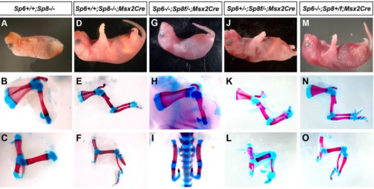
mutant limbs were truncated at the level of the elbow/knee with the olecranon also present in half of the specimens. Remarkably, in 100% of newborn double mutants both forelimbs and hindlimbs were absent (Figure 1A–C; 3 out of 102). In these mutants, no skeletal elements formed distal to the scapula (Figure 1B). Caudal lumbar vertebrae were highly disorganized and the body appeared truncated caudal to the sacrum with only rudimentary cartilage contributing to the pelvis (Figure 1C and Figure S2). Also, animals in which both copies of theSp8gene and one copy of theSp6gene had been removed (Sp6+/2;Sp82/2) were always tetra-amelic
(Figure 1D–F; 10 out of 102). However, in contrast to double mutants, the pelvic girdles showed undeveloped iliac and ischial anlagen (Figure 1F and Figure S2). The effect of a single Author Summary
In this report we examined the functional roles ofSp6and
Sp8 during limb development using compound loss-of-function mutants. Sp6and Sp8, two members of theSp
gene family, are expressed in the limb bud ectoderm and function downstream of WNT/ catenin signaling forFgf8
functional copy ofSp6in the morphogenesis of the pelvic girdle is shown in detail in Figure 2.
Mutant mice in which both copies of theSp6gene and one copy of the Sp8 gene had been inactivated (Sp62/2;Sp8+/2) had
proximal-distal (PD) complete, but extremely malformed, limbs (Figure 1G–I, 11 out of 102). Consistently, the forelimb paw had the ‘‘claw-like’’ appearance typical of split-hand/foot malforma-tion (SHFM) in which anterior digits were hypoplastic or missing and posterior digits were frequently fused (Figure 1G–H–H9). The radius was occasionally absent (Figure 1H9). Hindlimbs showed a more severe phenotype with the zeugopod constantly abnormal (Figure 1I). Although there was some variability, the majority of specimens displayed a misshaped and frequently truncated tibia and a thin fibula surmounted by one or two rows of small skeletal rods that we interpreted as digits (Figure 1I). The phenotype was variable among different animals and within individuals each paw
showing specific deficiencies. No left or right severity preference was identified. This phenotype is comparable to the human SHFM, a highly variable malformation that has also been termed ectrodactyly, split hand, cleft hand and lobster claw hand [31–34]. Of most interest, the digits in both fore and hindlimbs of
Sp62/2;Sp8+/2 mutants were bidorsal exhibiting circumferential
nails and lacking ventral pads (Figure 1J–K).
In summary, our genetic analysis shows that Sp6/Sp8 transcription factors are together absolutely required for limb development. Furthermore, the data support our hypothesis that Sp6 and Sp8 perform complementary functions in the limb ectoderm. Interestingly, one single functional allele of Sp6 is insufficient, in the absence of an Sp8 allele, to support limb development. In contrast, one single functional allele ofSp8, in the absence of anSp6 allele, permits development of all three PD segments, although displaying a SHFM phenotype.
Figure 1. Effects of inactivatingSp6andSp8in limb development.The external aspect (top row) and skeletal preparations of the forelimb (middle row) and hindlimb (bottom row) of newborns are shown for each genotype (genotypes indicated at the top). In the absence of the four functional alleles ofSp6andSp8(A–C), or when only one functional allele ofSp6remains (D–F) no limbs form. Underdeveloped hip bones with rudimentary ilium and ischium form when one functional allele ofSp6is present (F). Animals with only one functional allele ofSp8(G–I) display a split hand/foot malformation phenotype with occasional absence of the radius (H9) and more severe phenotype in the hindlimb (I). The digit tips in these limbs show conical nails (J), compare with normal digits (K). Abbreviations: s, scapula; h, humerus; r, radius; u, ulna, f, femur, t, tibia, fi, fibula, is: ischium il, ilium.
Sp8is expressed at higher level thanSp6 in the limb ectoderm
SinceSp6andSp8display similar temporal and spatial patterns of expression in the limb ectoderm [24–26], one possible explanation for the difference in their functional capacity as described above is thatSp8has specific functions thatSp6cannot accomplish. However, it is also possible that these functional differences are due to differences in their levels of expression. Thus, to quantify theSp8andSp6levels of expression in the limb ectoderm we performed a quantitative RT-PCR assay in E10.5 control embryos. Our results showed thatSp8was expressed more robustly thanSp6during limb development (Figure 2). Expression ofSp8 was 3 fold higher than expression ofSp6in the forelimb and 5 fold higher in the hindlimb. Our quantitative analysis also showed that the expression of bothSp6andSp8was higher in the forelimb than in the hindlimb, although it should be noted that the development of the hindlimb is delayed compared to that of the forelimb at this stage, which could account for the forelimb/ hindlimb disparity.
To investigate the basis for the differential level of expression of
Sp6 and Sp8 in the limb, we performed anin silicoanalysis of their putative promoter regions (Figure S3). To enhance the identification of functionally relevant regulatory sequences, we limited our evaluation to regions 59of the coding sequences that were conserved across divergent species as determined by the mVista browser [35]. We further screened the conserved regions for potential transcription factor binding sites using Alibaba 2.1 and Sequencher 4.8 and then confirmed conservation between mouse and human. Our analysis identified 12 potential catenin/ Lef1 binding sites 59to theSp8coding sequence, whereasSp6had only five. This finding provides a potential mechanism for the increased level ofSp8transcription during limb development. In addition, the presence of 29 potential Sp binding sites in the region containing the putativeSp6promoter and the 12 present inSp8
supports a possible cross-regulation between Sp transcription factors as suggested by the lowerSp6expression in absence of Sp8 (Figure S1). Based on our quantitative and in silico analysis we speculate that Sp8 makes a more substantial contribution to limb
development than Sp6 because of a higher level of transcription, a speculation that requires further investigation.
Ap2aCreinactivation ofSp8on anSp6deficient background
When performing the crosses between double heterozygous we found a reduced frequency of pregnancies in double heterozygous females and also that the fraction of double mutant offspring was significantly below the expected 1/16 Mendelian frequency. To circumvent these issues and avoid the neural phenotype fromSp8
null mutants, we used anSp8floxed conditional allele (Sp8f; [36])
to remove it specifically from the limb ectoderm. Among the available lines with Cre activity in the limb ectoderm (Msx2Cre
[37];Brn4Cre[17];RARCre[38];AP2aCre[39];Mox2Cre[40]), we selected theAP2aCreline because it has been reported to drive very early Cre function in both fore and hindlimbs, at least before activation ofFgf8[41]. BecauseSp8is already expressed at E7.5 in the embryonic ectoderm ([24,25] and authors’ personal observations), we decided to determine in more detail the activity of theAP2a;Cretransgenic line using the ROSA26 reporter strain (R26R; [42]). Our analysis showedAP2a;Creactivity in the early embryonic ectoderm at E8.5 indicating that the removal of the
Sp8floxed allele would occur before limb initiation (Figure S4). Thus, we used theAP2a;Creline, the conditional allele ofSp8 and theSp6null allele to generate the combined loss of function of
Sp6andSp8in the limb ectoderm (Figure S5). As to be expected, the double mutants (Sp62/2;Sp8f/2;AP2aCre) and the mutants that retained a single allele ofSp6(Sp6+/2;Sp8f/2;AP2aCre) were 100% tetra-amelic and showed similar phenotypes to those described above for the double ubiquitous deletions (Figure S5A–F). Also as expected, theSp62/2;Sp8f/+;AP2
aCregenotype exhibited the SHFM phenotype with its typical variability (compare Figure 1H–I with Figure S5H–I). In sum, the limb phenotypes obtained using theSp8floxed allele and theAP2aCre line replicated exactly the phenotypes obtained with the consti-tutive deletions. Finally, it should be noted that the neural phenotype was not rescued in conditional mutants (Figure S5A, 5D) indicating an unanticipated wide overlap between the expression ofAP2aandSp8in the neural tube (Figure S4). Msx2Creinactivation ofSp8on anSp6deficient background
We also inactivated Sp8 from the limb ectoderm using the
Msx2;Creline simultaneously with the inactivation of Sp6. This
Msx2;Cretransgenic line has been extensively monitored using the ROSA26 reporter strain [15,37] and it is known that it drives Cre activity beforeFgf8activation of expression in the hindlimb but afterFgf8 expression and initiation of limb development in the forelimb. We reasoned that the use of this conditional mutant would provide information on the requirement ofSp8once the early stages of limb initiation have occurred.
First of all we compared the phenotype of the limb conditional
Sp8mutant (Sp8f/2;Msx2Cre) with that of the Sp8 null mutant
(Sp82/2) in both forelimbs and hindlimbs (Figure 3A–F).
Not-withstanding the variability, the phenotypes using the conditional allele were on average milder than those using the constitutive null allele [24,25,30] (Figure 3A–F). In the conditionalSp8f/2;Msx2Cre
mutant, an initial burst of Sp8 expression permitted normal forelimb development up to the wrist and furthermore one or two incomplete posterior digits were formed (Figure 3E). In the hindlimbs, one posterior digit was always present although the tibia frequently appeared truncated (Figure 3F). This improvement in the phenotype (compare Figure 3A–C with Figure 3D–F) indicates
Figure 2. RT-qPCR quantification ofSp6andSp8transcripts in the limb ectoderm of E10.5 control embryos. Histogram bars represent the average expression values after normalization to the ubiquitously expressed 18s-RNA (standard deviation shown as error bars).Sp8(red) exhibits a higher level of expression thanSp6(blue) both in forelimbs (FL) and in hindlimbs (HL) and both factors are expressed at higher level in the forelimb than in the hindlimb.
that a transient early expression ofSp8has a considerable impact on both fore and hind limb development.
The conditional removal of Sp8 in the absence of Sp6
(Sp62/2;Sp8f/2;Msx2Cre) resulted in a forelimb truncated at the elbow while the hindlimbs didn’t develop (Figure 3G–I). When one copy of Sp6 remained (Sp6+/2;Sp8f/2;Msx2Cre) the
pheno-type notably improved with truncations at the level of the wrist/ ankle associated with the formation of an incomplete digit (Figure 3K–L). Finally, when a functional copy ofSp8 remained besides the Sp8 floxed allele (Sp62/2;Sp8+/f;Msx2Cre
) the phenotype obtained was SHFM (Figure 3M–O).
In summary, when the phenotypes of our allelic series are classified according to severity, a clear correlation with the total dosage of Sp6and Sp8is observed (Figure 1, 3 and Figure S5). The more parsimonious explanation is that both transcription factors are functionally equivalent during limb development, although Sp8 makes a greater contribution than Sp6 presumably due to a higher level of expression (Figure 2). Our study also suggests that there is a threshold of expression below which no limb forms and that the level ofSp6expression attained by a single allele ofSp6is below this threshold.
A functional AER does not develop when the gene dosage ofSp6 andSp8 is significantly reduced or completely eliminated
Since both Sp6 and Sp8 are involved in the Wnt/ catenin dependent induction ofFgf8, it seems reasonable to presume that the amelic phenotype of double mutants may rely on a failure to induce a functional AER. Therefore we examined embryonic limbs at the stages when the limb bud is emerging and the AER is being induced. For this analysis we used the Sp6 and Sp8
constitutive null alleles. By E9.5, in the normal limb bud, several genes includingFgf8,Bmp4andMsx2are expressed in the ventral limb ectoderm forming the preAER [17,22,43]. These AER precursors will become progressively confined to the distal tip as the AER matures [7,20] (Figure 4A, C, G, I, K, O).
However, in the absence of the fourSp6;Sp8alleles (Sp62/2;
Sp82/2) or when only one functional allele of Sp6 remained
(Sp6+/2; Sp82/2),Fgf8was never detected in the limb ectoderm at
any of the stages analyzed (Figure 4B for E9.5 (25–30 somites); Figure 4J for E10.5 (36–40 somites) and Figure 4P for E11.5). Because these two genotypes always showed identical expression patterns for all the genes analyzed, only the results ofSp62/2;
Sp82/2 mutants are shown in the Figures. In contrast toFgf8,
Bmp4andBmp2expression was found to occur normally at E9.5 both in the limb ectoderm and limb mesoderm of double mutants and mutants with a single functional allele ofSp6(Figure 4C–D). This was confirmed by the expression ofMsx2, abona fidetarget of Bmp signaling [44,45] (Figure 4G–H). However, neitherBmp4
(Figure 4K–L) norMsx2were maintained in the limb ectoderm by E10.5. Disregarding the absence ofFgf8expression, initiation of limb development was normal in Sp62/2;Sp82/2 and Sp6+/2;
Sp82/2compound mutants with the formation of a small bulge;
thus by E9.5 the phenotype was not yet evident (Figure 4A–H). The current view considers that Fgf10 signaling from the limb mesoderm induces Wnt/ catenin signaling in the ectoderm and this leads toFgf8 activation and therefore AER induction in the ectoderm. Subsequently, Fgf8 from the ectoderm signals back to the mesoderm to maintain Fgf10, establishing an Fgf10-Fgf8 positive feedback loop necessary for further outgrowth [11]. Consistent with Sp6 and Sp8 acting downstream ofFgf10 and Wnt/ catenin signaling, double mutant limb buds normally
Figure 3.Msx2Creremoval ofSp8on aSp6deficient background.The external aspect (top row) and skeletal preparations of the forelimb (middle row) and hindlimb (bottom row) of newborns are shown for each genotype (genotypes indicated at the top).Msx2Creconditional removal allows transient expression ofSp8in both forelimbs and hindlimbs which results inSp8conditional mutant (D–F) displaying a milder limb phenotype than ubiquitous mutants (A–C). One single conditional allele ofSp8in the forelimb (G–H) seems to be equivalent to both functional alleles ofSp6(A– B) while in the hindlimb is not sufficient for limb development (C, I). This conditional allele ofSp8in addition to one single allele ofSp6permits the formation of the three PD segments of the limb although with a single digit (J–L). Finally, one conditional allele ofSp8plus a normal alleleSp8results in SHFM (M–O).
activatedFgf10expression in the limb mesenchyme (Figure 4E–F at E9.5). However, due to the failure to activate Fgf8, the emergent limb buds cannot maintainFgf10in the limb mesoderm (Figure 4M–N) and regress so that by E11.5 no trace of the limb bud remained (Figure 4O–P).
These results demonstrate the absolute requirement of Sp6/Sp8 forFgf8activation in the limb ectoderm and are consistent with Sp6/Sp8 being necessary mediators of Wnt/ catenin induction of
Fgf8. Finally, our results also show thatBmp4expression in the limb ectoderm, which requires catenin [14,15], can occur in the
Figure 4.Fgf8is not detected in doubleSp6;Sp8mutants.ISH to transverse sections through the level of the forelimbs at the stage indicated at the top and with the probe indicated on the left. Genotypes are also marked at the top of the figure. In the absence ofSp6andSp8,Fgf8expression in the limb ectoderm is never detected as shown at E9.5 (A–B), E10.5 (I–J) and E11.5 (O–P). However,Bmp4(C–D),Fgf10(E–F) andMsx2(G–H) are normally activated at E9.5 but not maintained at later stages (K–N). Note that the initial budding of the double mutant is similar to normal (A–H) but further growth is impaired (I–N) and complete regression has occurred by E11.5 (O–P). In all panels dorsal is up and distal to the right.
total absence of Sp6 and Sp8 (this work, see Figure 4C–D) as well as in the absence of significant AER-relatedFgfexpression [46].
In the absence or significant reduction ofSp6/Sp8, limb development initiates, but later regresses by apoptosis
Next we investigated the reason of the regression of the emerging limb bud in double mutants. The phenotype of the double mutants is reminiscent of the chick mutant limbless. In
limbless the limb bud arises normally, but due to the inability to form an AER, the entire bud undergoes cell death and disappears [47,48]. Also, cell death is a constant feature after the surgical removal of the AER [3,4] or genetic attenuation of Fgf signaling from the AER [38,46,49,50]. Therefore, we analyzed cell death by TUNEL in our double mutant limb buds.
Abnormal cell death, compared with control littermates, was not detected at E9.5 in Sp62/2;Sp82/2 and Sp6+/2;Sp82/2
compound mutants. However, extensive apoptosis was apparent both in the mesoderm and ectoderm of these mutant limb buds by E10.5 (Figure 5A–B). Cell death started and was most prominent in the central region of the bud but apoptotic cells were also observed in the ectoderm particularly at dorsal proximal and ventral level (Figure 5B). This extensive apoptosis can account for the regression of the limb bud and the amelic phenotype as in
limbless[47,48].
AER morphogenesis initiates even in the complete absence or significant reduction ofSp6 andSp8
In the histological sections of double mutant limb buds we noticed a thickening of the ventral ectoderm that was particularly evident in the TUNEL assays because of the abundant cell death in this region (Figure 5A–B). To analyze this thickening with maximum detail, we performed semithin sections (1 micron thick) of araldite embedded embryos. Transverse sections through double mutant (Sp62/2;Sp82/2) andSp6+/2;Sp82/2embryos at
the level of the forelimbs showed an irregular thickening of the ventral ectoderm by E10.5 (Figure 5C–D). The thickening didn’t span the whole ventral ectoderm but was patchy and sometimes protruded into the mesoderm; it had the appearance of a ventrally positioned and immature AER, in which the apoptotic images were very abundant. To confirm that this thickening was of ectoderm origin, we used immunohistochemistry and confocal microscopy to localize E-Cadherin (Cdh1), which is an epithelial marker, and laminin, a major component of the basement membrane. The double immunohistochemistry demonstrated that the thickening was ectodermal as it expressed Cdh1 and was underlined by a laminin marked basement membrane (Figure 5E– F). To assess the functionality of this thickened ectoderm, we analyzed the expression of Connexin 43 (Cx43), a gap junction protein encoded by theGja1gene and considered a marker of the specialized AER ectoderm [51]. In contrast to the high expression present in the wild type AER, Cx43 was not detected above background in the thickened ectoderm of double mutants (Figure 5G–H).
Taken together our results indicate that Sp6/Sp8 factors are absolutely required for a functional AER, but dispensable for initial AER morphology confirming an independence between AER morphology and function.
Absence or significant reduction of Sp6/Sp8 activity in the limb ectoderm disrupts DV patterning
The known relationship between the specification of the AER and DV patterning together with the DV phenotypic alterations present in Sp62/2 and Sp62/2;Sp8+/2 mutants prompted us to
analyze the state of DV patterning in our mutants. Furthermore, the ventral position of the mutant AER indicates a failure in the normal morphogenetic movements of the ectoderm that compact the AER, a process in which En1 has been implicated [6,20]. Thus, we analyzed the expression of two genes relevant to DV patterning, Wnt7a and En1, in consecutive serial limb bud sections.
In the emerging limb bud (E9.25; 22–23 So), before En1
expression is detectable,Wnt7ais normally expressed in the dorsal ectoderm exceeding the mid-distal point of the bud and extending
into the ventral ectoderm [7,23] (author’s personal observations). Shortly afterwards, the expression of the pre-AER markersFgf8
and Bmp4 in the ventral ectoderm and of En1 in the more proximal ventral ectoderm progressively restricts Wnt7a to the dorsal ectoderm (Figure 6A, C).
In double mutant Sp62/2; Sp82/2 and Sp6+/2;Sp82/2
embryos, the initial extended expression of Wnt7a was never restricted to the dorsal ectoderm and its expression persisted covering almost the entire limb ectoderm while En1expression was not detected in the ventral ectoderm (Figure 6B–D, F–H). These results reveal that the absence or significant reduction of
Sp6/Sp8 dosage interferes with the normal specification of DV patterning resulting in double dorsal distal limb buds. Our results also show that a virtually normal Bmp signaling in the early limb bud (Figure 4C–D and Figure 4G–H) is not sufficient for En1
expression in the absence ofSp6andSp8.
Mutants retaining a single functional allele ofSp8exhibit a split-hand/foot malformation
The presence of a single allele ofSp8(Sp62/2;Sp8+/2orSp62/2;
Sp8f/+
;AP2aCreorSp62/2;Sp8f/+;Msx2Cre), was sufficient to allow the elaboration of all three segments along the PD axis, although the autopod was characterized by the loss or malformation of central elements creating a SHFM.
To understand the molecular basis of this phenotype, we analyzed the expression of Fgf8 during limb development in
Sp62/2;Sp8+/2 mutants. This analysis showed that the AER
precursors were irregularly specified in the ventral ectoderm. The whole mount in situ hybridization at E10 showed obvious gaps
and irregularities in the area in whichFgf8should be uniformly expressed (Figure 7A). During further development, the expression ofFgf8became robust in the posterior AER, but was absent in the central-anterior areas except for a typical spot of residual anterior expression (Figure 7B–C). The expression of Bmp4 in the ectoderm always replicated the same abnormal pattern as Fgf8
(Figure 7A–C). Furthermore, the compaction and maturation of the AER was defective as it remained flat and broad with occasional extensions into the ventral ectoderm (arrow in Figure 7C and 7E9). Thus, in harmony with previous reports [32,52,53], the SHFM phenotype in ourSp62/2;Sp8+/2mutants
derives from a failure to properly establish and maintain the AER, preferentially in the central to anterior limb region.
InSp62/2;Sp8+/2mutants the expression ofWnt7a and En1
was consistently abnormal but highly variable even within a single limb bud.Wnt7awas always found to abnormally extend into the ventral ectoderm to a variable degree that always correlated with a complementary ventral expression of En1(Figure 7D, D9, D0). This was easily appreciated when consecutive sections of the same limb bud were hybridized for Wnt7a and En1 as shown in Figure 7D9 and 7D0. Accordingly, the expression of Lmx1b, the downstream target of Wnt7a responsible for the dorsalization of the dorsal mesoderm [54], was found to variably extend under the flattened and broad AER into the ventral mesoderm distally (Fig. 7E–E9). These molecular alterations explain the bidorsal tips of Sp62/2;Sp8+/2 mutants. To ascertain possible DV defects at
more proximal levels we performed a histological analysis on transversal sections of E15.5 mutant and control limbs. Our results showed that DV patterning of muscles and tendons were preserved
Figure 6. Effects of inactivatingSp6andSp8genes on dorsal-ventral limb patterning.ISH to transverse sections through the level of the forelimbs at the stage indicated at the top and with the probe indicated on the left. Genotypes are also marked at the top. Note that, contrary to controls (A, E),Wnt7ais not restricted to the dorsal ectoderm in double mutant embryos (B, F). Accordingly,En1expression is undetectable in the ventral limb ectoderm of mutant embryos (D, H) compared to controls (C, G). The arrowheads and arrows mark the distal limit ofWnt7aandEn1 expression, respectively.
Figure 7. Molecular and morphological analysis ofSp62/2;Sp8+/2mutant limb buds.(A–C) WMISH forFgf8andBmp4showing irregular activation in the limb bud ectoderm ofSp62/2;Sp8+/2E10 (A), E10.5 (B) and E11.5 (C) forelimb buds compared to control littermates. Note the
irregular early activation and predominant posterior maintenance ofFgf8andBmp4expression, except for a residual focus of anterior expression (red arrowheads). (D, D9, D0) ISH forWnt7aandEn1to consecutive (7 microns apart) sections of control and mutant E10.5 forelimb buds (D, D9and D0). Note the variable expansion ofWnt7ainto the ventral ectoderm always associated with a corresponding proximal restriction ofEn1(D9, D0) indicated by red arrowheads (D9) and red arrows (D0). (E–E9) ISH forLmx1bandFgf8in consecutive sections of control andSp62/2;Sp8+/2E11.5 forelimb buds. TheLmx1b expression invades the ventral mesoderm distally under the broad and flat AER. (F–G) Hematoxylin-Eosin stained transverse histological sections at the autopod and zeugopod level of E15.5 control (F) andSp62/2;Sp8+/2(G) limbs. Some of the individual muscles and tendons are labeled. Abbreviations: EC, extensor digitorium communis; FDS, flexor digitorium sublimis; FDP, Flexor digitorium Profundus; ECR, extensor carpi radiallis; m, metacarpal; R, radius; U, ulna.
for the most part in the stylopod and zeugopod, but were less well defined in the autopod (Figure 7F–G).
In humans, isolated or non-syndromic SHFM is a genetically heterogeneous developmental disorder of which six loci have been identified [31–34]. SHFM Type I, the most frequent variety, is due to a mutation on chromosome 7, in a region that contains the two homeobox genesDLX5andDLX6[55–57]. SHFM Type IV maps to chromosome 3 and it has been shown thatTP63is the gene involved [31,58]. Furthermore, it has been shown thatDlx5
and Dlx6 are transcriptional targets of Tp63 [59,60]. Tp63 is a member of the p53 family of transcription factors crucial for stratified epithelial differentiation [61,62] and Dlx5 and Dlx6 are members of the family of distalless-related homeodomain transcription factors (Dlx1–Dlx6) that play key roles in limb development. Therefore, we analyzed the expression ofTp63and
Dlx5andDlx6in our SHFM mutants, to determine whether the Tp63 pathway was involved. Our analysis showed thatTp63and
Dlx5 and Dlx6 were normally expressed in the Sp62/2;Sp8+/2
mutant except for the flattened AER morphology (Figure 8A–D). Finally, the analysis of double mutants showed that Tp63,Dlx5
and Dlx6 were initially expressed normally in the complete absence ofSp6andSp8(Figure 8E–F) suggesting that if Sp6/Sp8 are components of the Tp63 network, they act downstream of Dlx5 and Dlx6. The expression of Tp63 in Sp62/2;Sp82/2
mutants was further confirmed by immunohistochemistry (Fig-ure 8G–H).
Discussion
Sp6andSp8 play complimentary functions in limb development
There are numerous examples in limb development of related genes with similar patterns of expression playing redundant functions and therefore providing robustness to the system. Among these are members of the Fgf, Bmp, and Hox gene families in which the overall final gene dosage is the key parameter for normal morphology [49,63,64]. Here, by using a variety of
loss-of-function alleles we have identified thatSp6andSp8control AER development and DV patterning in a redundant and dose-dependent manner. However, both genes do not contribute equally which may in part be due to their differential levels of transcription.
Notwithstanding the phenotypic variation associated with each particular genotype, when the predominant phenotypes obtained from the allelic series of compound Sp6 and Sp8 mutants are categorized in order of increasing severity, a strong correlation with gene dosage is observed (schematically shown for the forelimb phenotypes in Figure 9). A progressive reduction in the dose of
Sp6andSp8gene products leads to predictable morphology, from syndactyly, to SHFM, oligodactyly, truncation and finally amelia. This comparative analysis shows that the amount of gene product provided by a single functional allele ofSp8permits the complete development of the PD axis while one functional allele ofSp6does not, most likely because the gene product provided is below the critical threshold required for AER induction. Both alleles ofSp6
provide less gene product than a single allele ofSp8and equivalent to a transient expression of one copy of Sp8, as occurs in the forelimb when the Msx2;Cre deleter line is used. Collectively, the data from our allelic series indicate thatSp6andSp8are, for the most part, functionally equivalent and work in concert during limb development.
We found that the putative Sp8 promoter has an increased number of potential catenin/Lef1 binding sites compared toSp6, which might account for the higher levels of Sp8 expression. Interestingly, another member of the Sp family also expressed in the limb ectoderm, Sp9 [27], is unable to promote limb development in the absence of Sp6/Sp8 possibly because of its low level of expression [27]. Supporting this notion, there is a decreased number of catenin/Lef1 binding sites within theSp9
putative promoter region when compared toSp6.
We also considered whether the differences in Sp functional capacity could be due to structural differences. Comparative analysis of known protein domains and multiple alignment of Sp6, Sp8 and Sp9 revealed variability in the amino ends with the only
Figure 8.Tp63andDlx5expression in mutant limb buds.(A–F)Tp63andDlx5expression is normally detected in the limb ectoderm of control (A, B),Sp62/2;Sp8+/2(C, D) andSp62/2;Sp82/2(E, F) mutants althoughDlx5is downregulated. (G–H) Immunostaining for Tp63 (green) showing expression in theSp62/2;Sp82/2double mutant limb bud similar to wild type littermate. All the panels show longitudinal sections of E10.5 forelimb
buds.
common domains shared by these transcription factors being the zinc finger domains located in the carboxy ends. No structure-function correlation in the variable amino terminal domains was evident. For example, Sp6 and Sp8 are structurally disparate, but function in a complementary fashion. In contrast, Sp9 is structurally more similar to Sp8, than Sp6 is to Sp8 and yet does not show a complementary function in the limb. Therefore, even though these factors differ in their amino terminal domains, which may be functional in a different context [65], it is reasonable to speculate that in the limb, their functional capacity relies on their level of expression; this remains to be demonstrated.
Sp6andSp8 are absolutely necessary forFgf8induction and maintenance
Two of the main phenotypic features in our allelic series are truncations and SHFM. Studies in different mouse models and experimental manipulations in chick have established that these phenotypes can result from perturbations in AER functioning [1] and, accordingly, our analysis showed thatSp6/Sp8are required for the formation and maintenance of a functional AER.
The first phase in the formation of the AER is the induction of AER precursor cells in the limb ectoderm characterized by the expression of Fgf8. This depends on at least three important signaling inputs: i) Fgf10 produced in the limb mesoderm and signaling through the Fgf receptor 2b (Fgfr2b) expressed within the ectoderm [12,13,66–70], ii) Wnt/ catenin signaling produced in the limb ectoderm and signaling preferentially to the ventral limb ectoderm [14,15] and iii) Bmp signaling, mainly from the limb ectoderm, but also possibly from the limb mesoderm that signals through the Bmpr1a receptor in the limb ectoderm [16,17,22,71,72]. Although the crosstalk between these three inputs is complex and not completely understood, both the Fgf10
and the Bmp signaling pathways have been shown to act upstream of Wnt/ catenin signaling in the induction of the AER [14,16].
The analysis we have performed shows that when the dose of
Sp6/Sp8is significantly reduced,Fgf8is not activated, disregard-ing initial normalFgf10expression and Bmp signaling. Because both Sp6 and Sp8 have been shown to function downstream of Wnt/ catenin signaling [26,27,30], and Sp8 has been shown to bind and activate theFgf8 promoter [29], our results fit with a model in which Sp6 and Sp8 function as transcriptional activators of Fgf8 downstream of Wnt/ catenin signaling in the limb ectoderm (Figure 10). Sp6 and Sp8 function together and in a dose-dependent manner as necessary mediators of the Wnt/ catenin-Fgf8 regulatory loop. Our phenotypic and molecular studies indicate that the level of gene product produced by a single
Sp8allele is around the minimum dose required for the activation and maintenance ofFgf8 expression while that produced by a singleSp6allele does not reach this minimum.
It is known that the Wnt/ catenin signaling pathway is not only required for AER induction, but also for its maintenance. The limb truncations observed when, in the absence of Sp6, Sp8 is removed from the forelimb ectoderm after the AER has been induced (Sp62/2;Sp8f/2;Msx2Cre), indicate an ongoing role for
Sp8 in AER maintenance, further supporting our model. Most interestingly, our analysis shows that the complete absence of Sp6 and Sp8 transcription factors does not prevent the initiation of AER morphology confirming the independence between AER function and morphology. This is in high contrast to catenin loss-of-function mutants in the limb ectoderm that completely lack any evidence of a morphological AER or ectoderm thickening [14,15]. This difference may reflect the requirement of catenin for a proper AER morphology as has already been suggested [15,30,49] and corroborates that the Wnt3/ catenin-Sp6/Sp8-Fgf8 regula-tory loop is not a simple lineal one. Tp63, a crucial factor for AER morphology andFgf8maintenance of expression [61,62], and a
Figure 9. Illustration showing the correlation between theSp6/Sp8gene dose and the severity of the limb phenotype.Blue boxes represent theSp6alelles and red boxes theSp8alelles. Grey boxes represent null alleles and boxes with a red to grey graduation represent conditionally removal with theMsx2Creallele.
well-established target of Wnt/ catenin in the ectoderm [73] may be at the root of this difference. Characterization of separate Sp6/ Sp8 and Tp63 mediated pathways may help to uncouple
catenin’s multiple roles in AER formation and function. catenin is also necessary for the expression of other AER markers (i.e.Bmp4,Msx2[14,15]) in addition toFgf8.Sp6and
Sp8are necessary for the expression ofFgf8, butBmpligands and
Msx2 are normally activated in the total absence of Sp6/Sp8. Collectively, these data demonstrate that Sp6 and Sp8 mediate only part of the catenin functions in the limb ectoderm, principally the induction ofFgf8.
Recently, it has been shown that a conserved Wnt-Sp8-Fgf8 genetic cassette is also used to regulate the outgrowth of other body appendages such as the genital tubercle [30]. This work identified Sp8 as partially mediating the regulation ofFgf8by the canonical Wnt/ catenin pathway, a function that we demonstrate here that is fully accomplished by Sp6 and Sp8 together. Their result showing the failure of forced expression ofSp8in the AER (R26Sp8;Msx2) to rescue the phenotype of catenin loss-of-function in the limb ectoderm is very likely due to Sp8 not
reaching, in these experiments, the minimum level of expression required forFgf8induction.
Role ofSp6andSp8in dorsal-ventral patterning
During normal development the AER forms at the DV boundary of the limb bud reflecting a tight link between AER formation and DV patterning. Based on the analysis of the
limbless,En1mutants and on misexpression experiments in chick, it was hypothesized that the expression of En1 in the ventral ectoderm might function to establish a DV interface as a prerequisite for AER induction [74,75]. However, there are several examples of normal AERs forming in the absence of a DV boundary, such aseudiplopodia, the double Wnt7a;En1mutant and experiments in chick creating bidorsal limbs [19–21,76].
Here we report that DV patterning is also disrupted when the
Sp6/Sp8gene dose is perturbed. In the amelic phenotypes, even if the limb does not form, the molecular analysis of the emerging limb buds indicates that they are bi-dorsal asWnt7aexpression is extended along most of the limb ectoderm while En1 is not detected. Interestingly, the failure to activateEn1occurs despite
Figure 10. Regulatory pathways mediated by Sp6 and Sp8.Sp6andSp8are necessary mediators of the Wnt/bcatenin-dependent induction of Fgf8in the limb ectoderm. In addition, these two factors also collaborate with BMP signaling in the induction ofEn1in the ventral limb ectoderm. Finally, Sp6 and Sp8 may also act downstream ofTp63andDlxgenes.
normal expression of Bmp ligands in the limb ectoderm and mesoderm. In the SHFM phenotypes the digital tips display conical nails. In these limb buds the AER is irregularly induced and where maintained it remains flat, broad and immature. This correlates with an extension ofWnt7aexpression into the ventral ectoderm and a proximally restricted expression of En1
[20,21,23,77].Lmx1bexpands into the ventral mesoderm distally explaining the bi-dorsal phenotypic traits in the digits ofSp62/2; Sp8+/2mutants, while DV patterning is largely preserved at more
proximal levels.
We found that in the absence of a sufficient amount of Sp6/Sp8 gene products Bmp signaling is not sufficient to induceEn1. Sp family members are known to bind and interact with other transcription factors, including Smads. Thus, we hypothesized that Sp6/Sp8 transcription factors interact/cooperate with Smad proteins downstream of Bmp signaling to mediateEn1activation [78,79] (Figure 10). This interaction could occur at the protein level or by summative or synergistic effects on theEn1promoter. Interestingly, the putativeEn1promoter exhibits 25 potential Sp binding sites and 12 Smad binding sites that are conserved between human and mouse. Further investigation will be required to clarify this relationship (Figure S6).
TheSp6;Sp8double mutant limbs are reminiscent of those of thelimbless mutation in chicken.Limblessis a simple Mendelian autosomal recessive mutation characterized by tetra-amelia in the homozygous condition [80]. The mutation causes defects in no other organs, although it is effectively lethal because the chicks are unable to hatch without legs [47,48]. Limb development initiates inlimbless embryos and the early limb buds are morphologically indistinguishable from normal embryos until stage 19. However, the early limb buds are bidorsal and don’t form an apical ridge [74,75,81]. The limb bud mesoderm undergoes cell death beginning in the mid-distal mesoderm at stage 19–20 so that by stage 24, no signs of limb buds remain [47,75]. The similarities between our mutant and the limbless mutation may indicate a common target gene. After chromosomal mapping of thelimbless
mutation, Robb and coworkers [82] suggestedSp8 as a priority candidate. This is reinforced by the fact that Sp6 seems to be absent in chickens (Figure S3). However, further studies to validate this suspicion have not been done. Interestingly,limblessdoes not display the neural phenotype characteristic ofSp8 mutants, i.e., except for the limb phenotype the embryo is normal. This could be explained by a defect in a limb specificSp8regulatory element in
limbless. However, the lack of any AER morphology inlimbless, in contrast to the doubleSp6;Sp8mutants, decreases the likelihood thatSp8is the gene targeted.
Sp6andSp8 and split hand/foot malformation
In humans, the SHFM is a genetically heterogeneous congenital malformation characterized by a deficit in the formation of the central elements of the hands and feet that results in a central cleft associated with fusion and malformations of the remaining digits. The phenotype is highly variable, even between the limbs of a single affected individual, and ranges from a mild central syndactyly to severe loss of elements with oligodactyly and sometimes even affecting the zeugopod. It is currently accepted that this phenotype is the result of a premature regression of the central part of the AER [52,53,59]. Remarkably, the limb phenotype of the embryos that develop with a single copy of
Sp8 reproduces the human SHFM condition. The molecular analysis of these mutant limb buds indicates that the product obtained from one allele of Sp8, in the absence of Sp6, barely reaches the threshold required forFgf8induction. This is based on the low levels of Fgf8 transcription achieved and also on the
irregular expression domain that likely results from a cell autonomous effect of the mutation. Due to normal biologic variation, the level of Sp8 attained may reach the threshold required for Fgf8 induction in some cells, but not in others. Interestingly, at later stagesFgf8expression is not maintained in central regions suggesting that this later deficit inFgf8expression is the cause of the SHFM phenotype in Sp6;Sp8 compound mutants. Since the irregular early activation ofFgf8has not been observed in other models of SHFM, its possible contribution to the phenotype remains to be investigated [52,53,59].
Removal of all known AER-related Bmp ligands (Bmp2,Bmp4
andBmp7) from the AER usingMsx2Cre also results in SHFM [71]. However, inSp62/2;Sp8+/2mutants,Bmp4is still expressed
in the remaining AER suggesting that this SHFM phenotype is not caused by the loss of Bmp expression in the AER. In fact, since Bmp signalling is required for the induction ofFgf8, the SHFM phenotype following AER-related Bmp removal can also be explained by an irregular induction ofFgf8.
Of great interest is the recent genetic analysis ofFgf8regulation that has identified nearly 50Fgf8-regulatory modules in a 220 Kb region centromeric to the gene [83]. All the AER-specific enhancers, many of them embedded in the FBXW4 gene, drive expression all along the AP extension of the AER. Interestingly, SHFM type III [84,85] is caused by duplications of this genomic region that disrupt the normal architecture of the multiple enhancers likely affectingFgf8expression [83]. Therefore, SHFM type III is likely the result ofFgf8misregulation [83].
As previously mentioned, despite the identification of 6 loci involved in SHFM, onlyTP63(SHFM type IV) andDLX5and
DLX6(SHFM type I) have been unequivocally associated with this malformation [34]. Mutations inWNT10B(SHFM type VI) were also identified to be causative for SHFM, although there is some doubt on whether these mutations are sufficient for the phenotype [86–88]. Since similar phenotypes are frequently caused by disruption of different components of a regulatory network, we have considered the possibility thatSp6andSp8genes might be part of the Tp63 network. Indeed, the phenotypes of our mutants are identical, including the DV component, to those recently reported in a new identified human mutation in DLX5 [57]. However, the fact that Tp63, Dlx5 and Dlx6 have essentially normal expression patterns in the earlySp6/Sp8mutant limb bud indicates that, if Sp6/Sp8 transcription factors act within the Tp63 network, they function downstream of Tp63 and Dlx factors. Tp63 is necessary for the formation and maintenance of a normal epidermal layer [61,62]. In mouse, removal ofTp63 results in several abnormalities including limb truncations that are most similar to the Sp8-null phenotype [24,25,61,62] suggesting that Tp63 may preferentially controlSp8, but notSp6in mice. In any case, the relationship between the Tp63-Dlx and the Sp-Fgf8 regulatory modules, both downstream of Wnt/ catenin, add an extra level of complexity to limb development that requires further investigation.
Conclusions
This study provides compelling evidence for the absolute requirement of Sp6 and Sp8 for limb development as in their complete absence, or substantial reduction, no limbs form. By using a variety of loss-of-function alleles to remove the activity of
Sp6andSp8genes, we reveal that these two factors work together and in a dose-dependent manner as necessary mediators for AER development and DV patterning.
En1establishing a link between proximal-distal and dorsal-ventral patterning.
Materials and Methods
Ethics statement and mouse strains
All animal procedures were conducted accordingly to the EU regulations and 3R principles and reviewed and approved by the Bioethics Committee of the University of Cantabria. Mutant mouse lines were described previously: Sp6null allele [28];Sp8
null allele [24];Sp8floxed allele [36];AP2aCre[39] andMsx2Cre lines [37]; R26R [42]. Mice and embryos were genotyped by PCR, using genomic DNA extracted from tail biopsies and yolk sacs, respectively.
Skeletal preparation
After removing skin and viscera, mouse embryos were fixed in 95% ethanol. Alizarin Red and Alcian blue skeletal staining was performed according to standard protocols, cleared by KOH treatment and stored in glycerol.
In situ hybridization
In situ hybridization (ISH) was performed in whole-mount and in sections following standard procedures using the previously describedBmp4 [64],Dlx5and Dlx6 [53],En1[23]Fgf8 [43],
Fgf10 [89], Lmx1b[90], Msx2 [91], Tp63[62], Sp6 [28] and
Wnt7a[92] antisense riboprobes.
RNA quantification by real-time PCR
Embryonic fore and hind- limb buds were dissected in cold RNAse-free PBS from E10.5 wild type embryos. Total RNA was isolated separately from 3 pools of 8 forelimbs or 8 hindlimbs each. cDNA synthesis was done using standard conditions.
Real-time RT-PCR was carried out on an Mx3005P cycler, using the SYBRGreen PCR Master Mix (Invitrogen) and the data were analyzed using the MxPro software (Stratagene). Results were tested statistically performing ANOVA and Student-T test, being statistically significant when p,0.05.
Expression of Sp6 and Sp8 was normalized to that of housekeeping gene 18sRNA. The primers used (59 to 39
orientation) were: Sp6-F: tgctaaccgctgtctgtgg; Sp6-R:ctggtatgtctg-gagaggttgc; Sp8-F: ttatctccaaggtgcacacg; Sp8-R:gcttgaaccaggact-catacg; 18sRNA-R: ttggcaatgtttcgctc;18sRNA-F: cgccgctagaggt-gaaattt.
Cell death assay
Detection of cell death was performed in sections of paraffin embedded tissue using terminal deoxynucleotidyl transferase mediated dUTP nick-end labelling (TUNEL) with the Apoptag Fluorescein Direct In Situ Apoptosis Detection Kit (Intergen) following the manufacturer’s instructions.
-gal reporter analysis
For detection of -galactosidase activity,R26R;Ap2aCredouble transgenic embryos were fixed for 30 min, rinsed in PBS and incubated in the presence of X-gal as described [93].
Immunohistochemistry
Immunohistochemistry was performed in paraffin sections using the anti E-cadherin (Byoscience, # 610182), anti Laminin (Abcam, #ab11575), anti Tp63 (Abcam, #Ab53039) and anti Connexin43 (Abcam, # ab11370) primary antibodies. Antigen retrieval was performed by incubation with proteinase K (10mg/
ml) for E-cadherin and laminin or with citrate buffer in pressure cooker for Tp63 and Connexin43. AlexaH488 and TexasRED fluorescently tagged secondary antibodies were used. Vecthasield containing DAPI for nuclear counter staining was used as mounting medium. Confocal images were acquired in a SP-5 laser-scan confocal microscope (Leica Microsystems).
In silicoanalysis
Conservation ofEn1,Sp6andSp8loci between mouse, human, opossum, chicken and zebrafish was determined using pairwise alignment software (mVista browser, http://genome.lbl.gov/vista/). Conserved noncoding regions were further analyzed for potential transcription factor binding sites using AliBaba 2.1 (http://www. generegulation.com/pub/programs/alibaba2/index.html) and Se-quencher 4.8 (Gene Codes Inc.) informatic software.
Supporting Information
Figure S1 Expression ofSp6in the limb ectoderm ofSp8mutants. Whole mount in situ hybridization for Sp6 in limb buds ofSp8
mutant and control littermates. Stage and genotypes as indicated. (TIF)
Figure S2 Pelvic girdle morphology in Sp6;Sp8 mutants. Caudal body skeletal preparations of newborns. Genotypes indicated on the left. In the complete absence ofSp6 and Sp8, the pelvis is reduced to a small rudimentary cartilage element. One single functional allele of Sp6 (Sp6+/2;Sp82/2) leads to the formation of a misshaped ileum and ischium. A schematic drawing showing the three hip bones in different colors (pubis: yellow; ischium: orange and ileum: brown) accompanies each figure. (TIF)
Figure S3 Analysis 59 upstream of Sp6 and Sp8 (putative promoter regions). Multiple pairwise alignments of theSp6(A) and
Sp8 (B) loci comparing human and the species indicated. Light blue corresponds to the untranslated regions of the gene, dark blue to the coding sequence and pink to noncoding regions with at least 70% conservation. Note that only a portion of the chickenSp6
coding sequence is present in Genebank. Conserved regions within the first intron and the region 59 to the transcription start site containing binding sites are enclosed in red boxes (numbered 1–5 or 1–3, respectively). These conserved regions are illustrated (59R39) below the mVista analysis as lines (the actual size is noted above each illustration) and depict the relative positions of potential transcription factor binding sites (see legend within the figure). The motifs used to identify potential binding sites are shown in the boxed insert [94].
(TIF)
Figure S4 Cre reporter activity under the Ap2alocus in the pre-limb ectoderm. (A) Lateral and (B) dorsal views of E8.5 embryo showing ROSA26 reporter activity. (C) transversal section of the same embryo at the level indicated in B. ROSA26 activity was detected in the entire ectoderm at E8.5 (A,B), including the pre-limb ectoderm (black arrowhead in C) and also in the dorsal neural tube. (TIF)
Figure S5 AP2aCre removal of Sp8 on an Sp6 deficient background. The external aspect (A, D, G) and skeletal preparations of the forelimb (B, E, H) and hindlimb (C, F, I) of newborns are shown for each genotype (genotypes indicated at the top). Note that the phenotypes are similar to those of the ubiquitous deletions shown in Figure 1. Abbreviations as in Figure 1.
Figure S6 Analysis 59 upstream of En1 (putative promoter region). Multiple pairwise alignments of theEn1locus comparing human and the species indicated. Light blue corresponds to the untranslated regions of the gene, dark blue to the coding sequence and pink to noncoding regions with at least 70% conservation. Conserved regions within the first intron and the region 59to the transcription start site containing binding sites are enclosed in red boxes (numbered 1–3). These conserved regions are illustrated (59R39) below the mVista analysis as lines (the actual size is noted above each illustration) and depict the relative positions of potential transcription factor binding sites (see legend within the figure). The motifs used to identify potential binding sites are shown in the boxed insert [94].
(TIF)
Acknowledgments
We are very grateful to MaFe´lix Bastida and Marian Ferna´ndez-Tera´n for critical comments on the manuscript and to Ann Moon and Gail Martin for theAP2a;Cre andMsx2;Cre lines, respectively. In addition, we thank Mar Rodriguez and Vı´ctor Campa for excellent technical assistance and members of the Ros’s lab for continuous support.
Author Contributions
Conceived and designed the experiments: MAR. Performed the exper-iments: EH ID MJ KCO MAR. Analyzed the data: EH ID MJ YY AM KCO MAR. Contributed reagents/materials/analysis tools: YY AM. Wrote the paper: EH KCO MAR.
References
1. Fernandez-Teran M, Ros MA (2008) The Apical Ectodermal Ridge: morphological aspects and signaling pathways. Int J Dev Biol 52: 857–871. 2. Niswander L, Jeffrey S, Martin GR, Tickle C (1994) A positive feedback loop
coordinates growth and patterning in the vertebrate limb. Nature 371: 609–612. 3. Dudley AT, Ros MA, Tabin CJ (2002) A re-examination of proximodistal
patterning during vertebrate limb development. Nature 418: 539–544. 4. Rowe DA, Cairns JM, Fallon JF (1982) Spatial and temporal patterns of cell
death in limb bud mesoderm after apical ectodermal ridge removal. Dev Biol 93: 83–91.
5. Fallon JF, Lopez A, Ros MA, Savage MP, Olwin BB, et al. (1994) FGF-2: apical ectodermal ridge growth signal for chick limb development. Science 264: 104– 107.
6. Kimmel RA, Turnbull DH, Blanquet V, Wurst W, Loomis CA, et al. (2000) Two lineage boundaries coordinate vertebrate apical ectodermal ridge formation. Genes Dev 14: 1377–1389.
7. Bell SM, Schreiner CM, Scott WJ (1998) The loss of ventral ectoderm identity correlates with the inability to form an AER in the legless hindlimb bud. Mech Dev 74: 41–50.
8. Salas-Vidal E, Valencia C, Covarrubias L (2001) Differential tissue growth and patterns of cell death in mouse limb autopod morphogenesis. Dev Dyn 220: 295–306.
9. Guo Q, Loomis C, Joyner AL (2003) Fate map of mouse ventral limb ectoderm and the apical ectodermal ridge. Dev Biol 264: 166–178.
10. Kengaku M, Capdevila J, Rodriguez-Esteban C, De La Pena J, Johnson RL, et al. (1998) Distinct WNT pathways regulating AER formation and dorsoventral polarity in the chick limb bud. Science 280: 1274–1277. 11. Ohuchi H, Nakagawa T, Yamamoto A, Araga A, Ohata T, et al. (1997) The
mesenchymal factor, FGF10, initiates and maintains the outgrowth of the chick limb bud through interaction with FGF8, an apical ectodermal factor. Development 124: 2235–2244.
12. Min H, Danilenko DM, Scully SA, Bolon B, Ring BD, et al. (1998) Fgf-10 is required for both limb and lung development and exhibits striking functional similarity to Drosophila branchless. Genes Dev 12: 3156–3161.
13. Sekine K, Ohuchi H, Fujiwara M, Yamasaki M, Yoshizawa T, et al. (1999) Fgf10 is essential for limb and lung formation. Nat Genet 21: 138–141. 14. Soshnikova N, Zechner D, Huelsken J, Mishina Y, Behringer RR, et al. (2003)
Genetic interaction between Wnt/beta-catenin and BMP receptor signaling during formation of the AER and the dorsal-ventral axis in the limb. Genes Dev 17: 1963–1968.
15. Barrow JR, Thomas KR, Boussadia-Zahui O, Moore R, Kemler R, et al. (2003) Ectodermal Wnt3/beta-catenin signaling is required for the establishment and maintenance of the apical ectodermal ridge. Genes Dev 17: 394–409. 16. Pajni-Underwood S, Wilson CP, Elder C, Mishina Y, Lewandoski M (2007)
BMP signals control limb bud interdigital programmed cell death by regulating FGF signaling. Development 134: 2359–2368.
17. Ahn K, Mishina Y, Hanks MC, Behringer RR, Crenshaw EB, 3rd (2001) BMPR-IA signaling is required for the formation of the apical ectodermal ridge and dorsal-ventral patterning of the limb. Development 128: 4449–4461. 18. Zeller R, Duboule D (1997) Dorso-ventral limb polarity and origin of the ridge:
on the fringe of independence? Bioessays 19: 541–546.
19. Goetinck PF (1964) Studies on Limb Morphogenesis. Ii. Experiments with the Polydactylous Mutant Eudiplopodia. Dev Biol 10: 71–91.
20. Loomis CA, Kimmel RA, Tong CX, Michaud J, Joyner AL (1998) Analysis of the genetic pathway leading to formation of ectopic apical ectodermal ridges in mouse Engrailed-1 mutant limbs. Development 125: 1137–1148.
21. Cygan JA, Johnson RL, McMahon AP (1997) Novel regulatory interactions revealed by studies of murine limb pattern in Wnt-7a and En-1 mutants. Development 124: 5021–5032.
22. Pizette S, Abate-Shen C, Niswander L (2001) BMP controls proximodistal outgrowth, via induction of the apical ectodermal ridge, and dorsoventral patterning in the vertebrate limb. Development 128: 4463–4474.
23. Loomis CA, Harris E, Michaud J, Wurst W, Hanks M, et al. (1996) The mouse Engrailed-1 gene and ventral limb patterning. Nature 382: 360–363. 24. Treichel D, Schock F, Jackle H, Gruss P, Mansouri A (2003) mBtd is required to
maintain signaling during murine limb development. Genes Dev 17: 2630–2635. 25. Bell SM, Schreiner CM, Waclaw RR, Campbell K, Potter SS, et al. (2003) Sp8 is crucial for limb outgrowth and neuropore closure. Proc Natl Acad Sci U S A 100: 12195–12200.
26. Talamillo A, Delgado I, Nakamura T, de-Vega S, Yoshitomi Y, et al. (2010) Role of Epiprofin, a zinc-finger transcription factor, in limb development. Dev Biol 337: 363–374.
27. Kawakami Y, Esteban CR, Matsui T, Rodriguez-Leon J, Kato S, et al. (2004) Sp8 and Sp9, two closely related buttonhead-like transcription factors, regulate Fgf8 expression and limb outgrowth in vertebrate embryos. Development 131: 4763–4774.
28. Nakamura T, Unda F, de-Vega S, Vilaxa A, Fukumoto S, et al. (2004) The Kruppel-like factor epiprofin is expressed by epithelium of developing teeth, hair follicles, and limb buds and promotes cell proliferation. J Biol Chem 279: 626– 634.
29. Sahara S, Kawakami Y, Izpisua Belmonte JC, O’Leary DD (2007) Sp8 exhibits reciprocal induction with Fgf8 but has an opposing effect on anterior-posterior cortical area patterning. Neural Dev 2: 10.
30. Lin C, Yin Y, Bell SM, Veith GM, Chen H, et al. (2013) Delineating a conserved genetic cassette promoting outgrowth of body appendages. PLoS Genet 9: e1003231.
31. Guerrini L, Costanzo A, Merlo GR (2011) A symphony of regulations centered on p63 to control development of ectoderm-derived structures. J Biomed Biotechnol 2011: 864904.
32. Duijf PH, van Bokhoven H, Brunner HG (2003) Pathogenesis of split-hand/ split-foot malformation. Hum Mol Genet 12 Spec No 1: R51–60.
33. Elliott AM, Evans JA (2006) Genotype-phenotype correlations in mapped split hand foot malformation (SHFM) patients. Am J Med Genet A 140: 1419–1427. 34. Gurrieri F, Everman DB (2013) Clinical, genetic, and molecular aspects of
split-hand/foot malformation: An update. Am J Med Genet A 161: 2860–2872. 35. Frazer KA, Pachter L, Poliakov A, Rubin EM, Dubchak I (2004) VISTA:
computational tools for comparative genomics. Nucleic Acids Res 32: W273– 279.
36. Zembrzycki A, Griesel G, Stoykova A, Mansouri A (2007) Genetic interplay between the transcription factors Sp8 and Emx2 in the patterning of the forebrain. Neural Dev 2: 8.
37. Sun X, Lewandoski M, Meyers EN, Liu YH, Maxson RE, Jr., et al. (2000) Conditional inactivation of Fgf4 reveals complexity of signalling during limb bud development. Nat Genet 25: 83–86.
38. Moon AM, Capecchi MR (2000) Fgf8 is required for outgrowth and patterning of the limbs. Nat Genet 26: 455–459.
39. Macatee TL, Hammond BP, Arenkiel BR, Francis L, Frank DU, et al. (2003) Ablation of specific expression domains reveals discrete functions of ectoderm-and endoderm-derived FGF8 during cardiovascular ectoderm-and pharyngeal develop-ment. Development 130: 6361–6374.
40. Delgado I, Dominguez-Frutos E, Schimmang T, Ros MA (2008) The incomplete inactivation of Fgf8 in the limb ectoderm affects the morphogenesis of the anterior autopod through BMP-mediated cell death. Dev Dyn 237: 649– 658.
41. Boulet AM, Moon AM, Arenkiel BR, Capecchi MR (2004) The roles of Fgf4 and Fgf8 in limb bud initiation and outgrowth. Dev Biol 273: 361–372. 42. Soriano P (1999) Generalized lacZ expression with the ROSA26 Cre reporter
strain. Nat Genet 21: 70–71.
43. Crossley PH, Martin GR (1995) The mouse Fgf8 gene encodes a family of polypeptides and is expressed in regions that direct outgrowth and patterning in the developing embryo. Development 121: 439–451.