www.elsevier.com/locate/carbpol
0144-8617/$ - see front matter 2006 Elsevier Ltd. All rights reserved. doi:10.1016/j.carbpol.2006.08.018
In vivo
growth-inhibition of Sarcoma 180 tumor by alginates
from brown seaweed
Sargassum vulgare
Alessandra Paula Alves de Sousa
a, Márcia Rocha Torres
a, Cláudia Pessoa
a,
Manoel Odorico de Moraes
a, Francisco Dário Rocha Filho
b,
Ana Paula Negreiros Nunes Alves
a, Letícia Veras Costa-Lotufo
a,¤a Departamento de Fisiologia e Farmacologia, Faculdade de Medicina, Universidade Federal do Ceará, P.O. Box-3157, 60430-270 Fortaleza, Ceará, Brazil b Laboratório de Patologia Prof. Livínio Pinheiro, Instituto do Câncer do Ceará, Fortaleza, Ceará, Brazil
Received 4 November 2005; accepted 29 August 2006 Available online 6 October 2006
Abstract
Previous studies had demonstrated that alginates from Sargassum sp. (Phaeophyta) showed a considerable activity against various murine tumors. The aim present study is to investigate the in vivo antitumor activity of two alginates (SVHV and SVLV) with diVerent viscosity extracted from brown seaweed Sargassum vulgare C. Agardh against Sarcoma 180 cells transplanted in mice. Both alginates inhibited growth of sarcoma 180. The oral route of administration was more eVective for both alginates, leading to an inhibition of 51.8 and 74.8% for SVLV at the doses of 50 and 100 mg/m2/day, respectively, and of 66.2 and 88.8% for SVHV at the same doses. SVLV was 2.04 times more active after oral administration, while SVHV was 1.89, both at the dose of 100 mg/m2/day. Alginates-antitumor activity
was related to the tumor proliferation rate inhibition, as observed by reduction of Ki67 staining in tumor of the treated-animals. The his-topathological analysis of liver and kidney showed that both organs were aVected by SVHV and SVLV treatment. However, only SVLV led to acute tubular necrosis. Alginates cause the enlargement of the white pulp of the spleen of treated animals, suggesting that the observed antitumor activity could be related to alginates immunomodulatory properties.
2006 Elsevier Ltd. All rights reserved.
Keywords: Alginate; Antitumor; Seaweed
1. Introduction
Seaweeds have caused an emerging interest in the bio-medical area due to their content on pharmacologically bioactive substances with great chances to be employed against bacteria, viruses, other pathogens and tumors (Blunden, 1993; Ireland et al., 1993; Smit, 2004). Despite the
ascending number of new Wndings about seaweed
metabo-lites possessing biological activity on the last three decades
few products having actual potential have been identiWed
or developed (Smit, 2004). Among those substances that
received most attention from pharmaceutical companies for development of new drugs are the sulfated
polysaccha-rides (antitumors and antivirals), the halogenated fura-nones (antifouling compounds) and the kahalalide F
(anti-cancer and anti-aids compounds) (reviewed by Smit, 2004).
Polysaccharides represent a very interesting class of macromolecules, widely spread in nature, and they have attracted more attention recently in the biochemical and medical areas due to their immunomodulatory and
anti-cancer eVects (Ooi & Liu, 2000). In fact, seaweeds could be
considered a great source of polysaccharides, which have
attained commercial signiWcance due to their physical
prop-erties such as gelling, water-retention and their ability to
emulsify (Moe, Draget, Skjåk-Bræk, & Smidsrød, 1995;
Renn, 1997; Smit, 2004). In this context, alginates, struc-tural components in marine brown algae (Phaeophyta), possessed a huge importance with an annual production of
about 30,000 metric tons (Moe et al., 1995).
* Corresponding author. Tel.: +55 85 4009 8255; fax +55 85 4009 8333.
Alginates are unbranched polymers of polysaccharide
with gel-forming properties composed of -(1-4)-D
-mannu-ronic acid (M), -(1-4)-L-guluronic acid (G) and alternating
(MG) blocks (Moe et al., 1995). Alginates are used as
ingre-dients in food, pharmaceuticals and diverse consumer
prod-ucts and industrial processes (Moe et al., 1995). They present
many biomedical and biotechnological applications. They have been used for immobilization of Langerhans islets in
the treatment of experimental diabetes mellitus in rats (Fan
et al., 1990; Tze & Tai, 1982) and for microencapsulation of hormone-producing cell for the treatment of diabetes
melli-tus and parathyroid disease (Darquy & Sun, 1987;
Soon-Shiong et al., 1994). It has been shown that alginates led to monocytes and macrophages stimulation, resulting in an
increase of cytokine production (Otterlei et al., 1991; Son,
Moon, Rhee, & Pyo, 2001; Thomas, Harding, & Moore,
2000). The macrophage stimulation seems to be related to
alginates ability to enhance the healing process (Thomas
et al., 2000). Experimental data suggested that low-molecu-lar weight alginates could be useful in the prevention of
obesity, hypercholesterolemia, and diabetes (Kimura,
Watanabe, & Okuda, 1996). Alginates also presented
antico-agulant activity (Ronghua, Yumin, & Jianhong, 2003).
Concerning antitumor activity, alginates from
Sargas-sum sp. (Phaeophyta) showed a considerable activity against various murine tumors, such as Sarcoma 180 (solid and ascitic types), Ehrlich ascites carcinoma and IMC
car-cinoma (Fujiihara, Izimma, Yamamoto, & Nagumo, 1984;
Fujiihara & Nagumo, 1992, 1993). Fujiihara and Nagumo (1992, 1993) suggested that the antitumor activity is highly
inXuenced by the composition of the homopolymer, being
alginates with a higher MM-block content stronger active.
As established for other polysaccharides (Ooi & Liu, 2000),
the authors investigated whether macrophage stimulation could be responsible for alginates antitumor activity,
how-ever, experimental data failed to conWrm this hypothesis
(Fujiihara & Nagumo, 1993).
The aim present study is to investigate the in vivo
antitu-mor activity of two alginates with diVerent viscosity
extracted from brown seaweed Sargassum vulgare C.
Agardh against Sarcoma 180 cells transplanted in mice. The histopathological and morphological analyses of the tumor and the animal organs, including liver, spleen and kidney were performed in order to evaluate toxicological aspects of the alginates treatment.
2. Experimental
2.1. Extraction and puriWcation of alginates
Specimens of the brown seaweed S. vulgare were
col-lected in August/2004 at the Atlantic coast of Brazil (Pach-eco Beach, Caucaia, Ceará), cleaned of epiphytes, washed in
distilled water and stored at ¡20 °C. Alginates were
extracted according to the method of Calumpong, Maypa,
and Magbanua (1999) with some modiWcations. The dried
algae (300 g) were extracted at 60 °C, Wxed in 2%
formalde-hyde for 24 h, and then washed in water. The thalli were put into 0.2 N HCl for 24 h and washed again in water before extraction with 2% sodium carbonate. This mixture was homogenized, incubated in a water bath under continuous stirring at temperature of 60 °C and then two samples were collected after 1 and 5 h incubation. Crude extracts were collected after the centrifugation of these solutions whose precipitates were discarded, and then, they were precipi-tated by the addition of ethanol in order to obtain the algi-nate fractions. These fractions were washed twice in acetone and dried out. The intrinsic viscosities of the two samples obtained were comparatively analyzed, and the alginates obtained after 1 and 5 h incubation were
denomi-nate SVLV (S. vulgare low viscosity) and SVHV (S. vulgare
high viscosity), respectively. The alginate fractions (SVLV
and SVHV) were puriWed after dissolving them in distilled
water, followed by an ethanol precipitation. The precipi-tates were then washed twice in acetone and dried out.
The SVLV and SVHV alginates were physico-chemical
characterized by Torres (2003). The intrinsic viscosity of
SVLV alginate was 5.26 dL/g while that of SVHV was
9.10 dL/g. The alginates presented diVerent molecular
masses, being 19.3£104g/mol that of SVLV and
24.8£104g/mol that of SVHV. 1H NMR spectra have
shown the SVLV alginate posses features of a high mannu-ronic acid (M blocks) content while the SVHV one have shown to be of an intermediary kind (MG blocks). The SVLV alginate had shown a higher proportion of M/G (1.38) than the SVHV alginate (1.04).
2.2. Assay of antitumor activity
A total number of 185 Swiss mice (female, 20–30 g) obtained from the central animal house of the Federal Uni-versity of Ceará, Brazil were used. Animals were housed in cages with free access to food and water. All animals were kept under a 12:12 h light–dark cycle (lights on at 6:00 a.m.). Sarcoma 180 tumor cells were maintained in the peri-toneal cavities of the swiss mice obtained from the central animal house of the Federal University of Ceará.
Six-day-old Sarcoma 180 ascites tumor cells (5£105 cell/
500l) were implanted subcutaneously into the right hind groin of the mice. One day after inoculation, SVLV and
SVHV (50 and 100 mg/m2) or 5-FU (25 mg/m2) were
dis-solved in NaCl 0.9% and administered intraperitoneally or orally for 7 days. The negative control was injected with
physiological saline. On day 10, the mice were sacriWced.
Tumors, livers, spleens and kidney were extirpated, weighed
and Wxed in formaldehyde 10%. Inhibition ratio (%) was
cal-culated by the following formula: inhibition ratio
(%)D[(A¡B)/A]£100, where A is the tumor weight average
of the negative control, and B is that of the treated group.
2.3. Histopathology and morphological observations
After being Wxed with formaldehyde 10%, tumors, livers,
changes and hemorrhage. Then, a portion of the tumor, liver, spleen and kidney were cut into small pieces and later, the histological sections were stained with hematoxylin and eosin. Histological analyses were performed by light microscopy.
2.4. Ki67 immunohistochemical detection
Tumor sections were deparaYnized with xylene and
dehydrated with ethanol. The slides were then immersed in water for 10 min. For antigen retrieval, the slides were
boiled in citrate buVer (pH 6.0) for 15 min in a microwave
and subsequently cooled for 20 min. Afterward, the slides were washed in TBS, and then the endogenous peroxidase was blocked by 0.3% hydrogen peroxide for 15 min. After washing with TBS, the sections were incubated overnight at 4 °C with mouse antibodies to Ki67 at a concentration of 1:50. After 24 h, the slides were washed and incubated with a multilink antibody for 20 min, washed in TBS and incu-bated for 20 min with avidin–biotin–peroxidase complex. After washing with TBS, the slides were incubated for 3 min
with diaminobenzidine, and Wnally, counter-stained with
hematoxylin prior to mounting. The percentage of prolifer-ating neoplastic cells was evaluated directly at the optical microscope. To quantify the amount of proliferation, all
Ki67-positive cells were counted in 4–6 random Welds per
slide.
2.5. Statistical analysis
Data are presented as means§SEM. The diVerences
between experimental groups were compared by ANOVA
followed by Student–Newman–Keuls (p< 0.05).
3. Results and discussion
3.1. Antitumor activity
The search for potential polysaccharides as antitumor agents have gained more attention in the last few decades, since these macromolecules have shown promising
immu-nomodulatory properties associated with antitumor eVects
(Ooi & Liu, 2000). The enhancement of host defenses is now considered an alternative to the traditional cancer cytotoxic chemotherapy, because it involved lesser side
eVects.
The aim of the present study was to evaluate antitumor
eVects of alginates (SVHV and SVLV) isolated from
S. vulgare and some of the toxicological eVects related to their administration. Both alginates inhibited growth of
sarcoma 180. The inhibition rates are showed at Table 1.
The oral route of administration was more eVective for
both alginates, leading to an inhibition of 51.8% and 74.8%
for SVLV at the doses of 50 and 100 mg/m2/day,
respec-tively, and of 66.2% and 88.8% for SVHV at the same doses. SVLV was 2.04 times more active after oral administration,
while SVHV was 1.89, both at the dose of 100 mg/m2/day.
Oral ingestion is the most common method of drug admin-istration, because it is the safest, most convenient, and most
economical (Benet, Kroetz, & Sheiner, 1996). In the cancer
area, the availability of chemotherapeutic drugs adminis-trable by oral route represents a step forward in the
man-agement of patients (Gebbia & Puozzo, 2005).
Previous work had discussed the inXuence of the content
of D-mannuronic and L-guluronic acid blocks in alginates
antitumor activity (Fujiihara & Nagumo, 1992, 1993).
According to these authors, the higher content of MM-blocks in alginate correlate with the higher antitumor activ-ity. The SVLV alginate had shown a higher proportion of M/G (1.38) than the SVHV (1.04). The SVHV alginate
showed fractions of FGGGD0.214; FMM+MGD0.667;
FGGM+MGMD0.118, while the SVLV alginate have shown
fractions with FMMD0.6 and FGGGD0.4, and GGM and
MGM fractions were not observed (Torres, 2003).
Panik-kar and Brasch (1996) proposed a classiWcation system with three kinds of alginates based on the monomers
composi-tion: alginates with “high M blocks content” (where, FM is
greater or equal to 0.7); alginates with “high G blocks
con-tent” (FM is less or equals to 0.6) and intermediate alginates
possessing MG blocks (FM between 0.6 and 0.7). Based only
in this classiWcation system, the SVHV is as an alginate of
intermediate type (0.67), while the SVLV (0.6) is as a high G
Table 1
The inhibition rate of alginates (SVLV and SVHV) isolated from Sargassum vulgare on mice transplanted Sarcoma 180 tumor
Data are presented as means§SEM from N experiments. SigniWcant diVerences from control group were evaluated by ANOVA followed Student–New-man–Keuls: ap< 0.05.
Drug Dose (mg/m2/day) Liver (g) Spleen (g) Kidney (g) Tumor (g) Inhibition (%) N
Control – 1.67§0.04 0.19§0.01 0.36§0.01 2.12§0.17 – 26
5-FU 25 1.49§0.05 0.12§0.01a 0.30
§0.01a 0.51
§0.17a 75.5
§8.1a 17 SVLV 50 (v.o.) 1.73§0.07 0.21§0.02 0.36§0.01 1.02§0.19a 51.8
§9.2a 18 100 (v.o.) 1.55§0.05 0.14§0.01a 0.37
§0.01 0.53§0.13a 74.8
§6.1a 20 50 (i.p.) 2.04§0.07a 0.31
§0.03a 0.43
§0.01a 1.34
§0.22a 36.8
§10.5a 15 100 (i.p.) 1.78§0.06 0.26§0.02a 0.36
§0.01 1.34§0.17a 36.6
§8.3a 18
block content one. However, the block structure particular to SVLV represented a high content of manuronic acid in
opposition to the classiWcation of Panikkar and Brasch
(1996). However, contrary to the expectation, SVHV
seemed to be slightly more eVective than SVLV.
Histopathological analyses of the tumors extirpated from control mice showed groups of large, round and polygonal cells, with pleomorphic shapes, hyperchromatic nuclei and binucleation. Several degrees of cellular and nuclear pleomorphism were seen. Mitosis, muscle invasion and coagulation necrosis were also noticed. In the tumors extirpated from animals treated with 5-FU, SVLV and SVHV, extents areas of coagulative necrosis were observed. Ki67 staining for cell proliferation was performed in the tumors removed on day 10 from the animals untreated and
treated with 5-FU (25 mg/m2/day), SVLV (100 mg/m2/day,
after oral and intraperitoneal route of administration) or
SVHV (100 mg/m2/day, after oral and intraperitoneal route
of administration). Nuclear staining and good preservation of morphological details were observed in all tumors sec-tions immunostained with Ki67 antibody. In some of the examined samples, cells in mitosis showed, in addition to the expected Ki67 chromosomal labeling, also a strong cytoplasmic positivity. Background staining had seen in all
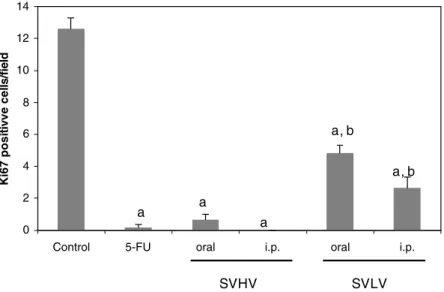
the cases. Fig. 1 shows the amount of Ki67 positive cells in
the analyzed slides. The results from this analysis show that the relative number of Ki67-positive tumor cells was sub-stantially less in tumors from mice treated with 5-FU, SVHV and SVLV when compared with control tumors
(p< 0.05).
Immunohistochemical staining of cells for
proliferation-associated proteins oVers information about the tumor
pro-liferation rate. The monoclonal antibody Ki67, described by Gerdes, Schwab, Lemke, and Stein (1983), is a mouse
monoclonal antibody that identiWes a nuclear antigen
asso-ciated with G1, S, G2 and M phases. This molecule is
expressed along all cell cycle, except in G0 and early G1 (Gerdes et al., 1984). Thus, results obtained with Ki67 stain-ing showed that the antitumor activities of both SVHV and SVLV are associated with a reduction in the tumor prolifer-ation rate. Moreover, SVHV staining inhibition was higher than SVLV, corroborating the observed in tumor weight
(p< 0.05).
3.2. Toxicological eVects
After the treatment with alginates SVHV and SVLV (25 mg/kg/day), the kidney, livers and spleens weights were
signiWcantly altered (p< 0.05, Table 1). As expected for a
chemotherapeutic drug, 5-FU treatment resulted in liver,
spleen and kidney weight decreasing (p< 0.05, Table 1).
The spleens removed from control mice showed normal and preserved white pulp, and megakaryocytes with
hyper-lobulated nuclei without morphologic alterations (Fig. 2A).
In contrast, alginates, especially SVHV when orally injected, led to a hyperplasia of white pulp in the spleen of
treated animals (Fig. 2C), which suggests an
immunostimu-lant activity of these compounds. The hematopoiesis occurs in spleen mainly during the neonatal life, however its
hema-topoietic ability is preserved during the whole life (Banks,
1992; Coelho, 2002). The alginates observed antitumor activity seems to be related to the enhancement of host defenses, since the studied alginates failed to exhibit direct cytotoxicity against tumor cell lines in culture (data not shown). In fact, it is postulated that the antitumor activity of polysaccharides is associated with their
immunomodula-tory properties (Ooi & Liu, 2000). As previously mentioned,
in the case of alginates, the literatures remains controversy. It is also observed a discrete dysmegakaryopoiesis in
algi-nates treated animals (Fig. 2D), however there is no
alter-ation in the number of circulating platelets (data not shown). Most chemotherapy drugs, including 5-FU, are
Fig. 1. EVect of 5-FU, SVHV and SVLV (100 mg/m2/day) on Sarcoma 180 cell proliferation using Ki67 antibody. Ki67-positive cells from 4 Welds/tumor were counted, and the mean§SEM of positive cells was calculated. a, p< 0.05, compared to control, and b, p< 0.05, compared to 5-FU, ANOVA followed by Student–Newman–Keuls.
0 2 4 6 8 10 12 14
Control 5-FU oral i.p. oral i.p.
K
i6
7
p
o
s
itivv
e
c
e
lls/field
SVHV SVLV
a a
a
a, b
immunosuppressive and have negative side eVects ( Takigu-chi et al., 2001). Indeed, in 5-FU treated animals, it was observed hypoplasia of white pulp and small lymphoid
aggregates as well as megakaryocytes (Fig. 2B).
Histopathological analyses of the kidneys from both SVLV and SVHV-treated animals showed several degrees of hydropic changes and vacuolization of the cytoplasm of proximal tubular epithelium and glomerular and tubular hemorrhage, but the glomeruli structure was essentially
preserved. These Wnding were observed in treated animals
despite the administration route used. Moreover, the cells lining the tubules of kidney removed from SVLV-treated
animals (50 and 100 mg/m2/day), only after intraperitoneal
injection, are necrotic (Fig. 3). This necrosis is characterized
by no nuclear staining and a deeply eosinophilic cytoplasm
(Curran, 1990). Thus, SVLV could be considered more toxic to the kidney.
Necrosis of the epithelium of the renal tubule may occur
on a large scale as a consequence of diVerent classes of
chemicals administration (Olsen & Solez, 1994). The
conse-quence is sudden onset of anuria or severe oliguria and acute renal failure. However, the epithelium has a consider-able power of regeneration and renal function can be stored. Despite the intensity of the cell damage, the regener-ation depends also on the integrity of the interstitial tissues (Curran, 1990; Olsen & Solez, 1994). It is worthwhile to mention that the histopathological analyses of SVLV treated animals demonstrated that the interstitial tissues
are preserved, without edema or lymphocyte inWltration,
which suggest the possibility of regeneration.
Fig. 2. Histopathology of the spleen from mice transplanted Sarcoma 180 tumor. (A) Control group; (B) 5-FU (25 mg/m2/day); (C) SVHV (100 mg/m2/day, v.o.) treated group. The detail of (A) shows the megakaryocytes on the spleen of control mice. White arrows indicate the megakaryocytes in the spleen of SVHV-treated animals. The white circles delineate the white pulps.
Despite the observed eVects in kidneys, the histopatholo-gical analyses indicated that the liver was also a target
organ for alginates toxicity. The observed eVects included
KupVer cells hyperplasia, portal tracts and centriolobular
vennus congestion, focal inWltrate of chronic inXammatory
cells, intense ballooning degeneration of hepatocytes, sinu-soidal hemorrhage and focal areas of steatosis
microvesicu-lar. These eVects were observed in all treated-animals.
The drugs should be considered as a possible cause of any liver lesion found on a biopsy. A large number of drugs
of diVerent chemical structure and with widely diVering
pharmacological actions occasionally give rise to important
liver lesions (Scheuer & Lefkowitch, 2000). Incriminated
substances include antituberculous drugs, non-steroidal
anti-inXammatory drugs, anaesthetics, herbal remedies,
methotrexate, chlorinated hydrocarbons and many others (Kummar, Abbas, & Fausto, 2004; Rang & Dale, 1991; Scheuer & Lefkowitch, 2000). Regardless, the liver shows great adaptive and regeneration abilities. For example, the increase in endoplasmic reticulum produced by long-term treatment with anticonvulsant drugs is commonly regarded as an adaptive phenomenon. On the other hand, regenera-tion of hepatic tissues occurs in many diseases, except in the most deleterious ones. Even when the hepatocellular necro-sis is present, but the conjunctive tissue is preserved, the
regeneration is almost complete (Kummar et al., 2004;
Scheuer & Lefkowitch, 2000). The hepatic alterations observed after alginates treatment could be considered
reversible (Kummar et al., 2004; McGee, Isaacson, &
Wright, 1992; Scheuer & Lefkowitch, 2000).
Acknowledgements
The authors are grateful to the Brazilian Agencies FINEP, CAPES, CNPq, BNB/FUNDECI, PRONEX, and
FUNCAP for fellowship and Wnancial support. Silvana
França dos Santos and Francisco José Queiroz de Oliveira provided excellent technical assistance.
References
Banks, W. J. (1992). Histologia Veterinária Aplicada. São Paulo: Editora Manole Ltda.
Benet, L. Z., Kroetz, D. L., & Sheiner, L. B. (1996). Pharmacokinetics. The dynamics of drug absorption, distribution and elimination. In J. G. Hardman, L. E. Limbird, P. B. MoliniV, R. W. Ruddon, & A. G. Gil-man (Eds.), Goodman & Gilman’s The pharmacological basis of thera-peutics (pp. 3–28). New York: The McGraw-Hill Companies, Inc. Blunden, G. (1993). Marine algae as sources of biologically active
com-pounds. Interdisciplinary Science Reviews, 18, 73–80.
Calumpong, P. H., Maypa, P. A., & Magbanua, M. (1999). Population and alginate yield and quality assessment of four Sargassum species in Negros Island, central Phillipines. Hydrobiologia, 398/399, 211–215. Coelho, H. E. (2002). Patologia Veterinária. São Paulo: Editora Manole
Ltda.
Curran, R. C. (1990). Color atlas of histopathology. New York: Oxford University Press.
Darquy, S., & Sun, A. M. (1987). Microencapsulation of parathyroid cells as a bioartiWcial parathyroid in vitro studies. ASAIO Journal, 33, 356– 358.
Fan, M. Y., Lum, Z. P., Fu, X. W., Levesque, L., Tai, I. T., & Sun, A. M. (1990). Reversal of diabetes in BB rats in transplantation of encapsula-tion pancreatic islets. Diabetes, 39, 519–522.
Fujiihara, M., Izimma, N., Yamamoto, I., & Nagumo, T. (1984). PuriW ca-tion and chemical and physical characterizaca-tion of an antitumor poly-saccharide from the brown seaweed Sargassum fulvellum.
Carbohydrate Research, 125, 97–106.
Fujiihara, M., & Nagumo, T. (1992). The eVect of the content of D-mannu-ronic acid and L-guluD-mannu-ronic acid blocks in alginates on antitumor activ-ity. Carbohydrate Research, 224, 343–347.
Fujiihara, M., & Nagumo, T. (1993). An inXuence of the structure of algi-nate on the chemotactic activity of macrophage and the antitumor activity. Carbohydrate Research, 243, 211–216.
Gebbia, V., & Puozzo, C. (2005). Oral versus intravenous vinorelbine: clin-ical safety proWle. Expert Opinion on Drug Safety, 4, 915–928. Gerdes, J., Lemke, H., Baisch, H., Wacker, H. H., Schwab, U., & Stein, H.
(1984). Cell cycle analysis of a cell proliferation associated human nuclear antigen deWned by the monoclonal antibody Ki-67. Journal of Immunology, 133, 1710–1715.
Gerdes, J., Schwab, U., Lemke, H., & Stein, H. (1983). Production of a mouse monoclonal antibody reactive with a human nuclear antigen associated with cell proliferation. International Journal of Cancer, 31, 13–20.
Ireland, C. M., Copp, B. R., Foster, M. P., McDonald, L. A., Radisky, D. C., & Swersey, C. (1993). Biomedical potential of marine natural prod-ucts. In D. H. Attaway & O. R. Zaborsky (Eds.), Marine biotechnology: Pharmaceutical and bioactive natural products (pp. 1–37). New York: Plenum Publishing Corporation.
Kimura, Y., Watanabe, K., & Okuda, H. (1996). EVects of soluble sodium alginate on cholesterol excretion and glucose tolerance in rats. Journal of Ethnopharmacology, 54, 47–54.
Kummar, V., Abbas, A., & Fausto, N. (2004). Robbins & Cotran pathology basis of disease. China: WB Saunders.
McGee, J. O. D., Isaacson, P. A., & Wright, N. A. (1992). Oxford textbook of pathology: Pathology of systems. New York: Oxford University Press.
Moe, S. T., Draget, K. I., Skjåk-Bræk, G., & Smidsrød, O. (1995). Alginates. In A. M. Stephen (Ed.), Food polysaccharides (pp. 245–286). New York: Marcel Dekker, Inc.
Olsen, S., & Solez, K. (1994). Acute tubular necrosis and toxic renal injury. In C. C. Tisher & B. M. Brenner (Eds.), Renal pathology: With clinical and functional correlations (pp. 769–809). Philadelphia: J.B. Lippincott Company.
Ooi, V. E. C., & Liu, F. (2000). Immunomodulation and anti-cancer activ-ity of polysaccharide-protein complexes. Current Medicinal Chemistry, 7, 715–729.
Otterlei, M., Ostgaard, K., Skjak-Braek, G., Smidsrod, O., Soon-Shiong, P., & Espevik, T. (1991). Induction of cytokine production from human monocytes stimulated by arginate. Journal of Immunotherapy, 10, 286– 291.
Panikkar, R., & Brasch, D. J. (1996). Composition and block structure of alginates from New Zealand brown seaweeds. Carbohydrate Research, 293, 119–132.
Rang, H. P., & Dale, M. M. (1991). Pharmacology. Hong Kong: Churchill Livingstone.
Renn, D. W. (1997). Biotechnology and the red seaweed polysaccharide industry: status, needs and prospects. Trends in Biotechnology, 15, 9– 14.
Ronghua, H., Yumin, D., & Jianhong, Y. (2003). Preparation and in vitro anticoagulant activities of alginate sulfate and its quaterized deriva-tives. Carbohydrate Research, 52, 19–24.
Scheuer, P. J., & Lefkowitch, J. H. (2000). Drugs and Toxins. In P. J. Sche-uer & J. H. Lefkowitch (Eds.), Liver biopsy interpretation (pp. 134–150). London: WB Saunders.
Smit, A. J. (2004). Medicinal and pharmaceutical uses of seaweed natural products: a review. Journal of Applied Phycology, 16, 245–262. Son, E. H., Moon, E. Y., Rhee, D. K., & Pyo, S. (2001). Stimulation of
acid-containing alginate (HMA) exposure in vivo. International Immu-nopharmacology, 1, 147–154.
Soon-Shiong, P., Henitz, R. E., Merideth, N., Yao, Q. X., Zheng, T., Mur-phy, M., et al. (1994). Insulin independence in a type I diabetic patient after encapsulated islet transplantation. Lancet, 343, 950–951. Takiguchi, N., Saito, N., Nunomura, M., Kouda, K., Oda, K., Furuyama,
N., et al. (2001). Use of 5-FU plus hyperbaric oxygen for treating malignant tumors: evaluation of antitumor eVect and measurement of 5-FU in individual organs. Cancer Chemotherapy Pharmacology, 47, 11–14.
Thomas, A., Harding, K. G., & Moore, K. (2000). Alginates from wound dressings activate human macrophages to secrete tumour necrosis fac-tor-. Biomaterials, 21, 1797–1802.
Torres, M.R. (2003). PuriWcação, caracterização físico-química, atividades inXamatória e imunoadjuvante de alginatos da alga marinha Sargas-sum vulgare C. Agardh. Tese apresentada a Coordenação do Curso de Pós-Graduação em Bioquímica da Universidade Federal do Ceará. Tze, W. J., & Tai, J. (1982). Biocompatibility and immunological studies of