Short Report
S
Printed in Brazil - ©2015 Sociedade Brasileira de Química 0103 - 5053 $6.00+0.00*e-mail: [email protected]
Collision-Induced Dissociation Analysis of Brevianamide A and C in Electrospray
Ionization Mass Spectrometry
Ana Lígia L. de Oliveira,a Ricardo Vessecchi,b Norberto P. Lopesa and Hosana M. Debonsi*,a
aNúcleo de Pesquisa em Produtos Naturais e Sintéticos (NPPNS), Departamento de Física e
Química, Faculdade de Ciências Farmacêuticas de Ribeirão Preto, Universidade de São Paulo, Avenida do Café s/n, 14040-903 Ribeirão Preto-SP, Brazil
bDepartamento de Química, Faculdade de Filosofia, Ciências e Letras de Ribeirão Preto,
Universidade de São Paulo, 14040-901 Ribeirão Preto-SP, Brazil
Brevianamides A and C are isomeric cyclic peptides with several reported biological activities, isolated from diverse microorganisms. Currently, there has been no previous investigation of brevianamide fragmentation utilizing electrospray ionization mass spectrometry (ESI-MS). In this work experiments were carried out in the positive mode using two different spectrometers (low and high resolution) with an ESI source. Computational chemistry studies helped identify the protonation sites based upon atomic charges, proton affinities and molecular orbitals, computed using the B3LYP/6-31++G (d,p) model. The data suggests that the presence of the allylic position of the lactamic N in brevianamides C governs its fragmentation pathways. Distinguishing between brevianamides A and C using positive ion electrospray tandem mass spectrometry (ESI(+)MS/MS) is made possible by the spectral difference of each isomer and offers an alternative to other spectroscopic techniques.
Keywords: diketopiperazine, fragmentation mechanisms, brevianamide, natural products
Introduction
Diketopiperazines are cyclic natural dipeptides that have been isolated from microorganisms, sponges, sea stars, tunicates (ascidians) and red algae.1 These
compounds show significant pharmacological effects and have been classified as promising leads in drug discovery. Recently, Wang et al.2 reviewed and summarized the
bioactivity of diketopiperazines and have presented the analysis of their structural moieties. The authors concluded that the chemical simplicity of these compounds, their rigid structure, chiral nature and the possibility to contain a large variety of side chains enable diketopiperazines to act on several different receptors, giving them a range of medicinal applications.2 Brevianamides are a class
of diketopiperazines that normally have an additional bicyclo[2.2.2]diazoctane ring system.3 Their biological
activity has inspired several researchers to investigate their biosynthesis and pharmacological benefits, as well as new synthetic routes.4-6
Over the last few years molecular networking based on tandem mass spectrometry (MS/MS) has emerged as a way to visualize complex data for further analysis and interpretation.7 Thorough the application of MS/MS
analysis, Winnikoff et al.7 were able to analyze 20 crude
extracts. They identified high chemical diversity and proved accurateness of the technique in both confirming the presence of specific bioactive metabolites and dereplicating other culture-based sources.7 Electrospray ionization (ESI)
is one of the most utilized MS techniques for dereplication
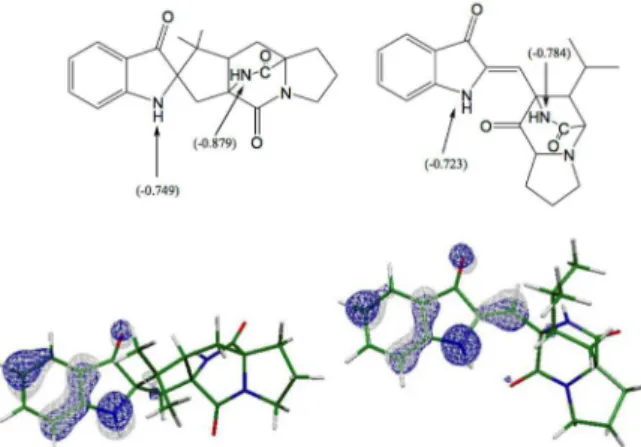
Figure 1. Structures of brevianamides A (C21H22N3O3, [M+H]+ 366,1810)
and metabolomic studies.8 ESI is able to induce soft
ionization, typically via acid-base or metal complex formation through which little fragmentation takes place.9
To obtain structural information it is necessary to have a second stimulus, normally ion activation by collision-induced dissociation (CID).10 The activation procedure
increases the internal ion energy and stimulates multiple dissociation processes.11 The defining of the fragmentation
pathways provides powerful information for dereplication studies.8
Furtado et al.12 furnished a complete MS/MS
analysis of a group of diketopiperazines with the same structural core.The authors observed a common loss of CO directly from the protonated molecule. The definitions of the protonation sites were derived from computational chemistry and confirmed by the fragmentation mechanism. These results also defined a series of ions that were diagnostic for the substituents at C(4) and C(9), which contributed to their use as an analytical tool during structural elucidation and characterization.12 Despite this important advance
in understanding the mechanism of fragmentation of diketopierazines, the presence of the bicyclo [2.2.2] moiety in brevianamides may induce additional reactions in which the previously published pathways would not be applicable. In order to rationalize the differences in the fragmentation pathways of these two compounds, electrospray ionization mass spectrometry analysis (ESI-MS) was conducted and their dissociation patterns were studied using tandem (MS/MS) and sequential (MSn) mass spectrometers. In
order to interpret the results, the protonation sites were estimated using the B3LYP/6–31++G(d,p) model for two isomeric brevianamides.
Methodology
Samples preparation
Brevianamides A and C (Figure 1) were isolated as previously described14 and the spectroscopic data was fully
in agreement with the published structures.13,14 Solutions
were prepared daily to a final concentration of 10 µg mL-1
of each compound with MeOH/H2O (1:1).
Mass spectrometry (MS) and tandem mass spectrometry analysis (MS/MS)
The analyses were conducted using ESI in low and high-resolution instruments. Low resolution analyses were performed using AmaZon SL (Bruker®) and applying N
2 as
a nebulizing (70 psi) and drying gas (10 L min-1, 200 ºC)
while the capillary high voltage was set to 3.5 kV and the
end plate to 500 V. Analysis conducted in high-resolution was carried out in a Bruker Daltonics micrOTOF™-Q II (hybrid QTOF). Instrument settings included a capillary voltage applied at 3.5 kV, a source block and desolvation temperature set to 180 ºC, a mass resolution set to 10,000 full width at half maximum (FWHM) and N2 was used as the
collision gas. The high-resolution analyses were achieved using the mass standard [TFA+Na]+.
Computational studies
In order to understand the protonation and fragmentation mechanism for alkaloids, the molecules were studied using computational chemistry. The use of proton affinity, gas-phase basicity, molecular orbital analysis and Fukui functions were decisive in early studies with diketopiperazines,12 where the protonation sites were used
to describe the reactivity of these compounds under CID conditions.15
In the present study the initial step for structure confirmation was obtained by a MM2 force field, after which, the compounds were optimized via the Gaussian 03 program16 using the B3LYP/6-31++G(d,p) model.17,18 The
vibrational frequencies for optimized structures were calculated using the same model to obtain only positive values, which indicates the minimum in terms of potential energy surface. Atomic charges and molecular electrostatic potential maps were obtained and indicated that the protonation sites were the nitrogen and oxygen atoms in the compounds, which show the most negative charges (nucleophilic sites). These results provided the necessary information for the drawing of the initial fragmentation mechanisms from protonated molecules.
Results and Discussion
In previous ESI(+)MS/MS investigations (assisted by computational methods) of five diketopiperazine fragmentation, the site of protonation at N(2) instead of the carbonyl oxygen was demonstrated (based on Fukui functions).12,16 The opening of the diketopiperazine ring
by acylium ion formation with the loss of CO as the first fragment was describe as an initial fragmentation process.12 In this previous study it was demonstrated that
the fragmentation can also started by water elimination (competitive reaction) for compounds with a hydroxyl group at the proline moiety. Brevianamides A and C have no hydroxyl group present in their structures, however both compounds underwent (with low intensity) water elimination (m/z = 348). MS3 analysis (ion at m/z = 348)
cannot differentiate between brevianamides A and C and the mechanism observed must be similar to the reported by Gates and co-workers.19
Computational chemistry calculations suggest the predominance of protonation at the amide functional group (Figure 1, carbon 11). The use of computational chemistry in combination with mass spectrometry has been published in several works,20-22 and our research group has developed a
systematic approach to obtain the most probable protonation site in natural products. First, the atomic charges and molecular electrostatic potential maps should be analyzed. Second, the proton affinities and basicities should also be computed. Thus, the protonation sites are identified and the mechanism can be proposed. For (A) and (B) (Figure 3) the atomic charges show the most nucleophilic atoms as being the nitrogen atoms. The Merz-Singh-Kollman (MK) charges for oxygen atoms are very close to –0.520,23 which is less
negative than nitrogen. MK atomic charges suggest that the protonation can occur at N atoms, as identified at Figure 2. For brevianamide C the values of atomic charges are very close, which suggests that protonation could possibly occur on one of two different sites.
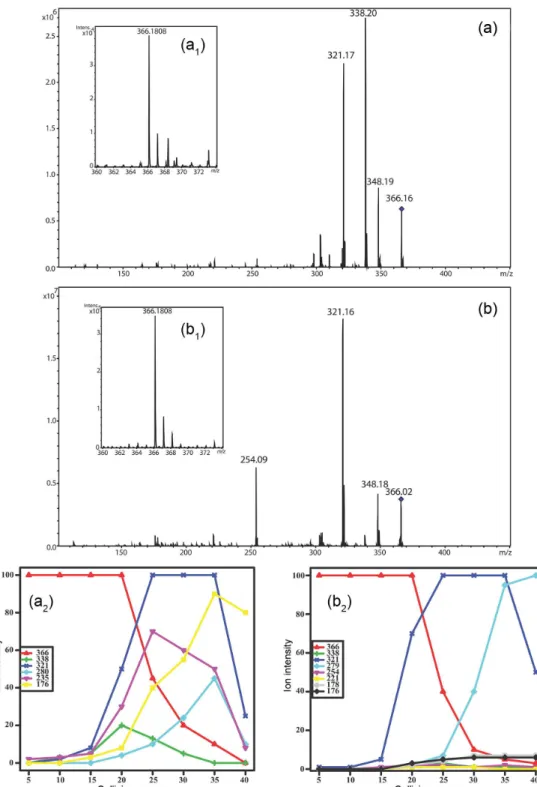
As expected, the initial fragmentation of brevianamide A was the loss of CO by anquimeric assistance followed by the loss of NH3 by an E2 mechanism. The [Brev. A–CO]+
ion of m/z 338 can be observed until a collision energy at 30 eV (see Figure 3 showing the Elab variation in QTOF) is
reached; however this ion was never observed as the base peak (see Figure 3).
The above observation can be easily explained by the sequential neutral elimination of NH3 affording the ion at
m/z 321 (Scheme 1), which has an additional conjugation step (in both proposed structures of Scheme 1) that can
help stabilize the charge. On the other hand, brevianamide C did not lose CO (absence of the ion at m/z 338) in QTOF experiments (see the Elab variation in QTOF), suggesting
divergent initial fragmentation mechanisms. In MS2 ion
trap experiments the ion at m/z 338 was observed for both compounds at the same energy (see experimental section), however for brevianamide A the ion at m/z 338 was the base peak while, C had a lower intensity (less than 30%). These data together suggest a strong influence of the structural moieties of A and C. Brevianamide C has the lactamic N linked to an allylic carbon, in which the ring can be opened from an opposite direction by the formation of a very stable allylic and tertiary carbocation (Scheme 1). This difference induces a heterolytic cleavage conserving the same mass charge ratio (m/z 366) as the protonated molecule. A sequential E2 mechanism yields a direct elimination of
45 mass units, while there was no observation of an initial CO elimination. These initial mechanistic differences allow the direct differentiation of brevianamides A and C which can be more easily observed using QTOF.
Scheme 2 shows the possible fragmentation pathway to obtain the ions at m/z 176 defined by MSn analyses (using an
ion trap machine) of the key ion at m/z 321 for brevianamide A and C. In both cases an internal rearrangement is necessary but for brevianamide C it is necessary to form a three membered ring structure before the neutral elimination. This restriction can be easily confirmed by the Elab experiments presented in Figure 3. The increase
of the collision energy over than 30 eV afforded the ion at m/z 176 in high intensity in breveniamide A spectra. On the other hand the same ion has less than 5% of the intensity in the breveniamide C spectra at all investigated collisions energies. Finally, in brevianamide C spectra, the key ion at m/z 321 afforded the ion at 279 by the propenyl neutral elimination. The long distance anquimeric assistance transfers the charge for the oxygen atom producing this ion as the base peak in the MS/MS analysis when applying high collision energies (Figure 3).
Conclusions
Analysis of two brevianamides isomers A and C showed the influence of the presence of a double bound between carbons 2-8 in brevianamide C instead of the additional ring contained in brevianamide A. The spectra acquired via the QTOF and ion trap spectrometers displayed the diagnostic m/z 338 fragment-ion (CO elimination). The intensity of the m/z 338 ion was higher for brevianamide A compared to C as shown by the ion trap, whereas QTOF did not detect this ion for brevianamide C. The m/z 279 ion also constituted a diagnostic fragment for brevianamide C which appeared
in the spectra recorded on both spectrometers. In the case of ion trap, the isolation of the key ion at m/z 321 makes a definitive verification of the difference for the ion at m/z 279 and provides a good strategy for differentiation between these compounds.
In conclusion, comparative ESI(+)MS/MS analysis between protonated brevianamide A and C molecules demonstrated that detection of the diagnostic m/z 338
and m/z 279 product ions helps to distinguish between them. The protonation site is crucial to understanding the different ways in which these compounds fragment. The use of ESI(+)MS/MS can be an alternative tool to nuclear magnetic resonance (NMR) when analyzing these natural compounds, especially when only small amounts or complex mixtures of the target material are available.
Figure 3. (a) Elab variation (QTOF spectrometer) of brevianamide A; (b) brevianamide C. Conditions: between 0 and 40 eV using CID mode and N2 as
H O
O
C NH2
N O N O O NH2 N H O N -CO H O N O N NH2 H -NH3 H O N O N H O N O N NH2 -NH3 H H O N O N H O O
C NH2
N O N O N H O N O NH2
-HCONH2 O N H O N O NH2
H O N H O N O NH2
H O N H O N O N H O N -HCONH2
Ring opening reaction
A
C
m/ z= 338
m/ z= 321
m/ z= 366
m/ z= 321
m/ z= 321
m/ z= 321
m/ z= 366
m/ z= 366 m/ z= 366
m/ z= 338
Scheme 1. Proposed initial fragmentation mechanisms for the protonated species of brevianamide A and C in ESI(+)MS/MS.
H O
N
O
N
m/ z= 321
N O H O N O N N O N O H H
m/ z= 176
O N H O N O N H O N H
m/ z= 321
N O N O N O H H
m/ z= 176
- 145 u - 145 u
A C O N H O N
m/ z= 321
O N H HO N H
m/ z= 279
C
Scheme 2. Proposed fragmentation mechanisms of the ions at m/z 176 observed in MSn analyses of the key ion m/z 321 for brevianamide A and C and the
Acknowledgements
The authors thank FAPESP (Fundação de Amparo à Pesquisa do Estado de São Paulo, grant No. 2009/51812-0), CAPES/PNPD (Coordenação de Aperfeiçoamento de Pessoal de Nível Superior) and CNPq (Conselho Nacional de Desenvolvimento Científico e Tecnológico) and INCT-if for fellowships and financial support. Also thanks Ellis O’Neill (Scripps Institution of Oceanography, San Diego, CA) and Cecilia Zappa (Albany College of Pharmacy and Health Sciences) for the language review.
References
1. Huang, R. M.; Zhou, X.; Xu, T.; Yang, X.; Liu, Y.; Chem. Biodivers. 2010, 7, 2809.
2. Wang, Y.; Wang, P.; Ma, H.; Zhu, W.; Expert Opin. Ther. Pat.
2013, 23, 1415.
3. Williams, R. M.; Cox; R. J.; Acc. Chem. Res. 2003, 36, 127. 4. Song, F. H.; Liu, X. R.; Guo, H.; Ren, B.; Chen, C. X.; Piggott,
A. M.; Yu, K.; Gao, H.; Wang, Q.; Liu, M.; Liu, X. T.; Dai, H. Q.; Zhang, L. X.; Capon, R. J.; Org. Lett. 2012, 14, 4770. 5. Torres-Garcia, C.; Diaz, M.; Blasi, D.; Farras, I.; Fernandez, I.;
Ariza, X.; Farras, J.; Lloyd-Williams, P.; Royo, M.; Nicolas, E.;
Int. J. Pept. Res. Ther. 2012, 18, 7.
6. Zhao, L.; May, J. P.; Huang, L.; Perrin, D. M.; Org. Lett. 2012,
14, 90.
7. Winnikoff, J. R.; Glukhov, E.; Dorrestein, P. C.; Gerwick, W. H.;
J. Antibiot. 2014, 67, 105.
8. Ernst, M.; Silva, D. B.; Silva, R. R.; Vêncio, R. Z. N.; Lopes, N. P.; Nat. Prod. Rep. 2014, 31, 784.
9. Crotti, A. E. M.; Vessechi, R.; Lopes, J. L. C.; Lopes, N. P.;
Quim. Nova 2006, 29, 287.
10. McLuckey, S. A.; J. Am. Soc. Mass Spectr. 1992, 3, 599. 11. Cheng, C.; Gross, M. L.; Mass Spectrom. Rev. 2000, 19, 398. 12. Furtado, N. A. J. C.; Vessechi, R.; Tomaz, J. C.; Galembeck,
S. E.; Bastos, J. K.; Lopes, N. P.; Crotti, A. E. M.; J. Mass Spectr.
2007, 42, 1279.
13. Williams, R. M.; Sanz-Cervera, J. F.; Sancenón, F.; Marcob, J. A.; Halligana, K. M.; Bioorg. Med. Chem. 1998, 6, 1233.
14. Birch, A. J.; Russel, R. A.; Tetrahedron 1972, 28, 2999. 15. Crotti, A. E. M.; Lopes, J. L. C.; Lopes, M. J.; J. Mass Spectrom.
2005, 40, 1030.
16. Frisch, M. J.; Trucks, G. W.; Schlegel, H. B.; Scuseria, G. E.; Robb, M. A.; Cheeseman, J. R.; Montgomery Jr., J. A.; Vreven, T.; Kudin, K. N.; Burant, J. C.; Millam, J. M.; Iyengar, S. S.; Tomasi, J.; Barone, V.; Mennucci, B.; Cossi, M.; Scalmani, G.; Rega, N.; Petersson, G. A.; Nakatsuji, H.; Hada, M.; Ehara, M.; Toyota, K.; Fukuda, R.; Hasegawa, J.; Ishida, M.; Nakajima, T.; Honda, Y.; Kitao, O.; Nakai, H.; Klene, M.; Li, X.; Knox, J. E.; Hratchian, H. P.; Cross, J. B.; Bakken, V.; Adamo, C.; Jaramillo, J.; Gomperts, R.; Stratmann, R. E.; Yazyev, O.; Austin, A. J.; Cammi, R.; Pomelli, C.; Ochterski, J. W.; Ayala, P. Y.; Morokuma, K.; Voth, G. A.; Salvador, P.; Dannenberg, J. J.; Zakrzewski, V. G.; Dapprich, S.; Daniels, A. D.; Strain, M. C.; Farkas, O.; Malick, D. K.; Rabuck, A. D.; Raghavachari, K.; Foresman, J. B.; Ortiz, J. V.; Cui, Q.; Baboul, A. G.; Clifford, S.; Cioslowski, J.; Stefanov, B. B.; Liu, G.; Liashenko, A.; Piskorz, P.; Komaromi, I.; Martin, R. L.; Fox, D. J.; Keith, T.; Al-Laham, M. A.; Peng, C. Y.; Nanayakkara, A.; Challacombe, M.; Gill, P. M. W.; Johnson, B.; Chen, W.; Wong, M. W.; Gonzalez, C.; Pople, J. A.; Gaussian 03, Gaussian, Inc., Wallingford CT, 2004. 17. Lee, C. T.; Yang, W. T.; Parr, R. G.; Phys. Rev. B. 1988, 37, 785. 18. Becke, A. D.; J. Chem. Phys. 1993, 98, 5648.
19. Kearney, G. C.; Gates, P. J.; Leadlay, P. F.; Staunton, J.; Jones, R.; Rapid Commun. Mass Spectrom. 1999, 13, 1650.
20. Alcamí, M.; Mó, O.; Yáñez, M.; Mass Spectrom. Rev. 2001, 20, 195.
21. Vessecchi, R.; Emery, F. S.; Lopes, N. P.; Galembeck, S. E.;
Rapid Commun. Mass Spectrom. 2013, 27, 816.
22. Vessecchi, R.; Emery, F. S.; Galembeck, S. E.; Lopes, N. P.;
J. Mass Spectrom. 2012, 47, 1648.
23. Singh, U. C., Kollman, P. A.; J. Comp. Chem., 1984, 5, 129.
Submitted: August 4, 2014
Published online: February 6, 2015
![Figure 1. Structures of brevianamides A (C 21 H 22 N 3 O 3, [M+H] + 366,1810) and C (C 21 H 22 N 3 O 3, [M+H] + 366,1809).](https://thumb-eu.123doks.com/thumbv2/123dok_br/18999481.463277/1.892.541.713.681.849/figure-structures-brevianamides-c-h-n-o-m.webp)