Received: November 26, 2011 Accepted: January 31, 2012
Conflict of Interests: The authors state that there are no financial and personal conflicts of interest that could have inappropriately influenced their work.
Copyright: © 2011 Drago et al.; licensee EDIPUCRS. This is an Open Access article distributed under the terms of the Creative Commons Attribution-Noncommercial-No Derivative Works 3.0 Unported License.
Evaluation of the lengths and angles of the tips
of rotatory files in root canal preparations
Avaliação do comprimento e do ângulo das pontas de
instrumentos rotatórios no preparo de canais radiculares
Mariana Aleluia Drago a
Rosana de Souza Pereira a
Miguel Ângelo Schettino Junior b
a Department of Endodontics, Federal University of
Espirito Santo (UFES), Vitória, ES, Brazil
b Department of Physics, Federal University of
Espirito Santo (UFES), Vitória, ES, Brazil
Correspondence:
Mariana Aleluia Drago
Rua Luis Fernandes Reis, 530/101 – Praia da Costa Vila Velha, ES – Brasil
29101120
E-mail: [email protected]
Abstract
Purpose: To detect changes in the angles and lengths of the tips of rotary nickel-titanium files after use.
Methods: Forty human teeth were prepared with eight sets of rotary ProTaper® Universal
nickel-titanium files with a length of 25 mm and rotation of 350 rpm. The files were evaluated in a scanning electron microscope (SEM) at three different times: Group A – before use; Group B – after using each file in the preparation of three molars; and Group C – after using each file in the preparation of five molars. The length of the tip was determined by measuring the length of a straight line drawn parallel to the file axis, from the apex of the tip to its posterior border. The SEM software provided the angle measurements. Data were analyzed by using paired t-tests and Wilcoxon tests.
Results: There were no differences between the groups for the S1 file. There was a decrease in length when the F2 file was used to prepare three and five molars, whereas decreases in length were seen when F5 files were used to prepare three or five molars.
Conclusion: The results suggest that the tips of the rotary instruments showed significant changes in length and angle even with relatively low use.
Key words: Dental files; Endodontics; molar
Resumo
Objetivo: Comparar as alterações no ângulo e comprimento da ponta dos alargadores rotatórios após sua utilização em relação à sua configuração original.
Metodologia: Quarenta dentes humanos foram preparados com instrumentos rotatórios de níquel-titânio ProTaper® Universal, 25 mm e rotação de 350 rpm. Os alargadores foram
avaliados em um microscópio eletrônico de varredura (SEM) em três grupos: Grupo A – antes do uso dos instrumentos; Grupo B – após o uso de cada instrumento na preparação de três molares; e Grupo C – após o uso de cada instrumento na preparação de cinco molares. O comprimento da ponta foi determinado medindo o comprimento de uma linha reta traçado paralelamente ao eixo do instrumento, desde o vértice da ponta até seu limite posterior. O software do SEM forneceu a medição do ângulo delimitado por dois segmentos de retas. Os dados foram analisados estatisticamente por teste t pareado teste de Wilcoxon.
Resultados: Para o instrumento S1 não houve diferença entre os grupos. Para o alargador F2 houve uma diminuição no comprimento entre os grupos B e C e uma diferença entre os grupos A com B e C foi observada no alargador F5.
Conclusão: Os resultados sugerem que a ponta dos instrumentos rotatórios sofreu alterações significativas de comprimento e ângulo com relativo pouco uso.
Introduction
Nickel-titanium alloys have been introduced for the
manufacture of endodontic instruments with the aim of
replacing the stiffness,
i.e
., the modulus of elasticity, of
stainless steel materials (1-3). These instruments are two
to three times more lexible than stainless steel iles (4).
The alloy used for manufacturing endodontic instruments
consists of approximately 55% nickel and 45% titanium,
and it is generically called 55-Nitinol (5).
Several instrument systems were introduced because of
the growing popularity of hand iles and nickel titanium
rotatory iles in endodontic practice, with various features
such as design, tip sizing, pitch, cross-sectional helix angle
and taper (6,7). In 2006, Dentsply Maillefer introduced the
ProTaper
®Universal System with modiied characteristics
of the original ProTaper
®. The changes included the new
rounded tip and removal of the angle of transition to reduce
canal transportation and provide greater security. Minor
changes were made in the S2 instrument to improve the
balance between S1, S2, and F1. The cross-sectional blades
of the F2 and F3 instruments were changed to a triangular
concave shape with a shallow U-shaped groove at the sides
to make them more lexible and homogeneous. The
cross-section of the F3 instrument also became lighter with grooves
to reduce the abrasion of the metal section (8-10). New iles,
F4 and F5, were introduced to accomplish a non-brushing
action inside the root canals (10,11).
However, these endodontic instruments may present
defects on their surface such as slots, microcavities and
a pronounced variation between the actual and nominal
dimensions originating in the manufacturing process. Due
to this lack of manufacturing precision in the shape and
size of these instruments, iatrogenic problems may occur
in the inal coniguration of the endodontic treatment of
root canals (3,10). Therefore, standardization has become
increasingly important due to demands for safety, quality
and uniformity in the production of materials (10). The aim
of this study was to detect possible changes in the angle and
in the lengths of the tips of the iles resulting from their use
in preparing root canals.
Material and methods
The study protocol was approved by the Federal
University of Espiríto Santo (UFES) Ethical Committee
and all the procedures followed were in accordance with
Resolution 196/96 of Health State Department, Brazil. Forty
human maxillary and mandibular molars, extracted with
completely formed apices, were obtained from the Human
Teeth Bank in the School of Dentistry of the University
of São Paulo (FOUSP), São Paulo, Brazil. The teeth were
prepared with the aid of eight sets of the rotary
nickel-titanium ProTaper
®Universal rotary system (Dentsply
Maillefer, Ballaigues, Switzerland) that were 25 mm in
length, and were driven by an electric motor with a constant
rotation of 350 rpm. The following ProTaper
®Universal iles
were tested: SX, S1, S2, F1, F2, F3, F4 and F5. According
to the manufacturer’s recommendations, the “crown-down”
technique was used. Sixty-four instruments were evaluated
in a scanning electron microscope (SEM, Superscan SS-550,
Shimadzu, Japan) before and after their use to measure
the lengths and the angles of their tips. Observations were
made during three different times and grouped as follows:
Group A – before the use of the instruments; Group B – after
using each instrument in the preparation of three molar teeth,
making a total of twenty four molars, and Group C – after
using each instrument in the preparation of ive molars each,
for a total of 16 molar teeth.
All canals were prepared by the same operator who
had been previously calibrated. The time taken for the
preparation of root canals by each instrument was recorded.
The use of a nickel-titanium ProTaper
®Universal rotary
system kit (Dentsply Maillefer, Ballaigues, Switzerland)
(SX, S1, S2, F1, F2, F3, F4 and F5) has been reported to be
safe in preventing the fracture of these instruments when
used to prepare up to ive molar teeth.
The analysis of these surfaces was performed using
scanning electron microscopy (SEM, Superscan SS-550,
Shimadzu, Japan) by measuring the angles and the lengths
of the tips at the Laboratory of Carbonaceous Materials and
Ceramic of the Department of Physics, Federal University
of Espirito Santo (CCE-UFES), according to Speciication
No. 101 of ANSI/ADA (12).
All iles of the ProTaper
®Universal System (Dentsply
Maillefer, Ballaigues, Switzerland) were disinfected
to remove the surface residues originating from the
manufacturing process and instrumentation by brushing
with enzymatic detergent, and sterilized in an autoclave in
200 mL of distilled water in a complete cycle for 30 minutes
at 121 °C and 15 psi.
The instruments were attached to the SEM specimen
holder with carbon double-sided tape, and the instrument cable
slot was positioned upward to standardize the measurements.
The long axis of each ile was placed parallel to a horizontal
reference. To measure the tip length and angle, the image of
the tip of the instrument was positioned in the center of the
screen at a magniication of 300X to 400X. The tip length was
determined by measuring the length of a straight line drawn
parallel to the axis of the instrument, from the apex to the
posterior border of the tip, according to ISO 3630-1 (Fig. 1)
(12,13). The value was recorded in nanometers (nm) and then
converted into millimeters (mm). The posterior limit of the
tip of the instrument was delimited by a line perpendicular
to the long axis of the instrument, which was tangential to
the initial limit of the irst helical channel of the active part.
The SEM software provided the measurements of the angle
enclosed by the two line segments (Fig. 2) (12).
Results
Table 1 shows the descriptive statistical analysis of the
angles of the tips measured with the help of the instruments.
Table 2 presents the comparisons of tip angle measures
between groups (Group A × Group B; Group B × Group C;
Group A × Group C). There was an increase in the tip angles
at all times (
P
<0.05).
Fig. 1. SEM image of the tip angle geometry of an F2 ProTaper
Universal instrument.
Fig. 2. SEM image of the length of the tip of an F2 ProTaper
Universal instrument.
Files Groups Minimum Maximum Median Mean Standard deviation
SX
A 41.000 56.000 46.000 47.125 4.853
B 42.000 57.000 48.000 48.813 4.735
C 43.000 58.500 49.000 49.813 5.000
S1
A 40.000 47.000 41.500 42.250 2.605
B 41.000 48.000 43.500 43.813 2.419
C 42.500 49.500 44.750 45.125 2.446
S2
A 42.000 50.000 45.000 45.375 3.068
B 42.000 52.000 46.000 46.688 3.535
C 43.000 53.000 47.250 47.938 3.438
F1
A 93.000 124.000 110.000 109.750 9.377
B 95.000 125.000 112.500 111.500 9.040
C 97.000 127.000 113.750 112.813 9.134
F2
A 87.000 105.000 98.500 97.500 6.234
B 89.000 107.000 99.500 99.250 6.065
C 90.000 109.000 101.000 100.500 6.425
F3
A 51.000 97.000 84.500 79.375 14.918
B 52.000 97.800 86.500 80.550 15.054
C 53.500 99.000 86.750 81.688 14.856
F4
A 100.000 109.000 103.000 103.625 2.774
B 102.000 110.000 105.000 105.375 2.774
C 102.500 111.000 105.750 106.500 2.752
F5
A 91.000 105.000 99.000 99.000 4.870
B 92.000 105.000 99.500 99.750 4.773
C 93.500 107.000 100.750 100.937 4.686
Table 1. Descriptive statistics of the
angle (in degrees) of the tips from different experimental groups (n=8).
Table 3 shows the descriptive statistical analysis of
the lengths of the tips (in mm) measured with the help of
the instruments. The data were asymmetric, as shown by
differences between the mean and median values. Table 4
shows the results of the Wilcoxon tests for the comparisons
of the tip lengths between groups (Group A × Group B;
Group B × Group C; Group A × Group C). Some groups
Files P-value*
Group A × Group B Group B × Group C Group A × Group C
SX 0.000 0.000 0.000
S1 0.000 0.000 0.000
S2 0.015 0.000 0.001
F1 0.000 0.002 0.000
F2 0.000 0.001 0.000
F3 0.000 0.001 0.000
F4 0.001 0.000 0.000
F5 0.048 0.002 0.000
* all comparisons are statistically significant at the significance level of 0.05
Files Groups Minimum Maximum Median Mean Standard deviation
SX
A 0.060 0.107 0.087 0.086 0.015
B 0.054 0.105 0.078 0.077 0.016
C 0.053 0.102 0.075 0.075 0.015
S1
A 0.071 0.128 0.114 0.103 0.024
B 0.064 0.119 0.107 0.098 0.023
C 0.061 0.116 0.108 0.096 0.024
S2
A 0.079 0.128 0.108 0.108 0.018
B 0.069 0.120 0.097 0.094 0.019
C 0.064 0.118 0.096 0.090 0.019
F1
A 0.030 0.057 0.039 0.039 0.008
B 0.027 0.052 0.030 0.033 0.009
C 0.025 0.050 0.028 0.031 0.009
F2
A 0.055 0.092 0.075 0.072 0.013
B 0.050 0.090 0.070 0.069 0.012
C 0.048 0.087 0.066 0.066 0.012
F3
A 0.060 0.120 0.079 0.085 0.022
B 0.011 0.099 0.061 0.056 0.029
C 0.014 0.740 0.082 0.207 0.269
F4
A 0.063 0.080 0.069 0.070 0.006
B 0.058 0.068 0.066 0.064 0.003
C 0.055 0.067 0.064 0.063 0.004
F5
A 0.065 0.102 0.085 0.087 0.012
B 0.060 0.092 0.079 0.079 0.011
C 0.057 0.090 0.079 0.078 0.010
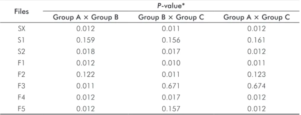
Files P-value*
Group A × Group B Group B × Group C Group A × Group C
SX 0.012 0.011 0.012
S1 0.159 0.156 0.161
S2 0.018 0.017 0.012
F1 0.012 0.010 0.011
F2 0.122 0.011 0.123
F3 0.011 0.671 0.674
F4 0.012 0.017 0.012
F5 0.012 0.157 0.012
* P-value in bold is statistically significant at the significance level of 0.05.
Table 2. Results of the paired t-test
for comparisons of angle measures between groups.
Table 3. Descriptive statistics of the
length (in mm) of the tips from different experimental groups (n=8).
Table 4. Results of the Wilcoxon test
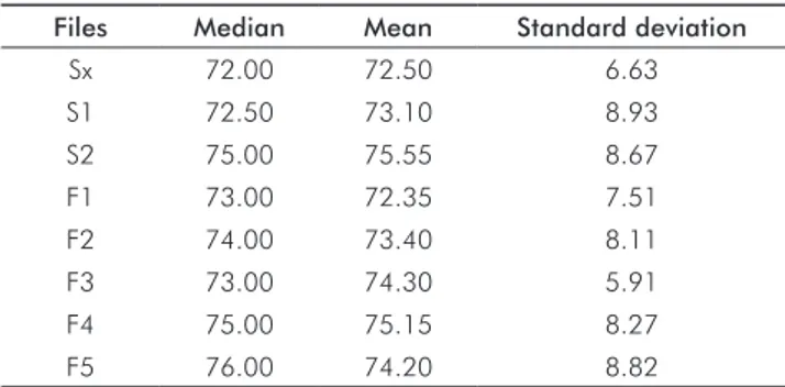
Table 5. Descriptive statistics of the instrumentation time (in seconds).
Files Median Mean Standard deviation
Sx 72.00 72.50 6.63
S1 72.50 73.10 8.93
S2 75.00 75.55 8.67
F1 73.00 72.35 7.51
F2 74.00 73.40 8.11
F3 73.00 74.30 5.91
F4 75.00 75.15 8.27
F5 76.00 74.20 8.82
Differences were observed among the instruments, Sx,
S2, F1 and F4, but there was always a decrease in length.
There were no differences between the groups for the S1
instrument. There was a decrease in length between B and
C groups for the F2 ile and between A and B groups for the
F3 ile. For the F5 ile, there was a decrease in length from A
to B groups, but not between B and C groups. Table 5 shows
the instrumentation time for all groups.
Discussion
Speciic design parameters of the tip, such as the angle,
length, cross-section and geometry can signiicantly inluence
the clinical eficacy of endodontic instruments (11). The tip
angle is formed by the contour of the instrument tip and its
vertex. The tip length is the distance between the vertex and
the base. The tip angle is related to its length,
i.e
., the lower
the angle of the tip of the ile, the greater is its length (14).
According to the manufacturer, all ProTaper Universal
instruments recently underwent a modiication of their tip
orientation and geometry.
The results of the present study show that the mean
values of the lengths of the tips ranged from 0.075 to
0.108 mm for Sx, S1 and S2 shaping instruments, and from
0.031 to 0.027 mm for F1, F2, F3, F4, and F5 inishing
instruments. The comparison of A, B, and C groups reveals
a decrease in the tip length for all groups of the shaping (Sx
and S2) and inishing instruments (F1 and F4).
Miserendino et al. (15) studied the dimensional aspects
of seven types of instrument tips as they inluence the cutting
eficiency. Tip length and angle were two of the dimensional
variables analyzed according to Speciication #28 ANSI/
ADA (12). Tips of iles with lengths lower than 0.5 mm and
1.0 to 1.5 mm had better cutting eficiency. Tip angles with
values between 60° and 69° showed better performance in
atresic root canals than the angles between 40° and 49°. In
root canals with a wider diameter, the tips having smaller
angles were the most eficient.
A previous comparison of dimensional and geometric
tip changes was reported by Câmara et al. for ProTaper and
ProTaper
®Universal instruments (17); their mean values for
initial tip lengths were greater than the ones reported in the
present study. The use of iles from different lots in this study
may have accounted for some of the confounding effects
on the outcome values. The angles of the tips for S1 and S2
ProTaper
®Universal iles decreased when compared with
the ProTaper system, but they increased for the inishing
instruments, F1, F2 and F3. The mean values for the tip
lengths ranged from 0.094 to 0.117 mm for the S1 and S2
shaping iles, and from 0.082 to 0.130 mm for the F1, F2,
and F3 inishing iles.
Some studies (11,17) reported that speciic features such
as the design of the tip, tip angle, length, cross-section and
tip geometry may signiicantly affect the penetration, cutting
and shaping by endodontic instruments inside the root canal.
For ProTaper
®Universal instruments, the reduction of the
tip angle from 66° to 39° is said to favor the centralization
of the iles inside the root canal walls, thus reducing the risk
of transportation. However, in the case of new inishing iles,
the increase in tip angle from 66° to 95° would lead to an
opposite effect.
In the present study, the tip angle values in all groups
of ProTaper
®Universal shaping iles ranged from 40°
to 50°, and from 87° to 124° in group A of the inishing
iles. There was an increase in tip angle of all groups of
ProTaper
®Universal instruments with use, which may
provide a safer cutting at the apical third, possibly reducing
the risk of fracture and deviation of these iles inside the root
canal.
Conclusions
Within the limitations of this study, the results suggest
that the tips of rotary instruments showed signiicant changes
in length and angle, even with relatively low use. There was
no fracture of shaping and inishing ProTaper
®Universal
iles during this experiment.
Acknowledgments
Kim HC, Yum J, Hur B, Cheung GS. Cyclic fatigue and fracture characteristics of ground 1.
and twisted nickel-titanium rotary files. J Endod 2010;36:147-51.
Bonetti Filho I, Miranda ER, De Toledo LR, Del Rio CE. Microscopic evaluation of three 2.
endodontic files pre- and posinstrumentation. J Endod 1998;24:461-4.
Alapati SB, Brantley WA, Svec TA, Powers JM, Mitchell JC. Scanning electron microscope 3.
observations of new and used nickel-titanium rotary files. J Endod 2003;29:667-9. Vahid A, Roohi N, Zayeri F. A comparative study of four rotary NiTi instruments in 4.
preserving canal curvature, preparation time and change of working length. Aust Endod J 2009;35:93-7.
Vaudt J, Bitter K, Kielbassa AM. Evaluation of rotary root canal instruments in vitro: a 5.
review. Endod 2007;1:189-203.
Bergmans L, Van Cleynenbreugel J, Beullens M, Wevers M, Van Meerbeek B, Lambrechts 6.
P. Progressive versus constant tapered shaft design using NiTi rotary instruments. Int Endod J 2003;26:288-95.
Inan U, Aydin C, Uzun O, Topuz O, Alcam T. Evaluation of the surface characteristics 7.
of used and new ProTaper instruments: an atomic force microscopy study. J Endod 2007;33:1334-7.
Ruddle CJ. The ProTaper technique: shaping the future of endodontics. Endod Topics 8.
2005;10:187-90.
Unal GC, Maden M, Savgat A, Orhan E. Comparative investigation of two rotary nickel-9.
titanium instruments: protaper universal versus protaper. Oral Surg Oral Med Oral Pathol Oral Radiol Endod 2009;107:886-92.
Vaudt J, Bitter K, Neumann K, Kielbassa AM. Ex vivo study on root canal instrumentation 10.
of two rotary nickel-titanium systems in comparison to stainless steel hand instruments. Int Endod J 2009;42:22-33.
West J. Progressive taper technology: rationale and clinical technique for the new ProTaper 11.
universal system. Dent Today 2006;25:66-9.
American National Standard/American Dental Association. Specification No. 101-2001 12.
[Internet]. Root Canal Instruments: General Requirements. [cited 2010 Jan 20]. Chicago: ANSI/ADA, 2001. Available from: http://webstore.ansi.org/RecordDetail.aspx?sku=ANSI/ ADA+Specification+No.+101-2001.
International Organization for Standardization. International Standard ISO 3630-1: 13.
2008 [Internet]. Dentistry - Root-canal instruments. Part 1: general requirements and test methods. [cited 2012 Jun 3]. Geneve: ISO, 2008. Avaliable from: http://webstore.ansi. org/RecordDetail.aspx?sku=ISO+3630-1%3a2008.
Lopes HP, Siqueira Jr JF. Endodontia: biologia e técnica. 3ª ed. Rio de Janeiro: Guanabara 14.
Koogan; 2010.
Miserendino LJ. Moser JB, Heuer MA, Osetek EM. Cutting efficiency of endodontic 15.
instruments. Part II: analysis of tip design. J Endod 1986;12:8-12.
American National Standard/American Dental Association. Specification No. 28-2008 16.
[Internet]. Root canal files and reamers, type K; 2008. [cited 2010 Jan 20]. Chicago: ANSI/ADA, 2008. Available from: http://webstore.ansi.org/RecordDetail.aspx?sku=ANS I%2fADA+Specification+No.+28-2008.
Câmara AS, De Castro MR, Viana AC, De Toledo LR, Buono VT, De Azevedo BMG. 17.
Flexibility and torsional strength of ProTaper and ProTaper Universal rotary instruments assessed by mechanical tests. J Endod 2009;35:113-6.