Short Report
0103 - 5053 $6.00+0.00
*e-mail: [email protected]
Conformational Analysis of Toxogonine, TMB-4 and HI-6 using
PM6 and RM1 Methods
Arlan da Silva Gonçalves,a Tanos C. C. França,b José D. Figueroa-Villarb and Pedro G. Pascutti*,a
aInstituto de Biofísica Carlos Chagas Filho, Universidade Federal do Rio de Janeiro, 21941-902 Rio de Janeiro-RJ, Brazil
bDivisão de Ensino e Pesquisa, Seção de Engenharia Química, Instituto Militar de Engenharia,
22290-270 Rio de Janeiro-RJ, Brazil
Neste trabalho, após validação por comparação com resultados obtidos por DFT, os métodos semi-empíricos PM6 e RM1 foram utilizados para análise conformacional de três oximas usadas em defesa química. Os resultados sugerem conformações de menor energia destes compostos que poderão ser utilizadas em futuras parametrizações para estudos por modelagem molecular.
In this work, after validation by comparison with results obtained by DFT, the semi-empirical methods RM1 and PM6 were employed to perform conformational analysis of three oximes employed in chemical defense. The results suggested low energy conformations for those compounds that could be useful in further parameterizations for molecular modeling studies.
Keywords: acetylcholinesterase, conformational analysis, oximes, neurotoxic agents
Introduction
The intensive use of neurotoxic organophosphorous compounds (OPCs) as pesticides in agriculture, as well as their potential use as chemical warfare agents, has attracted the attention of the scientiic community to develop eficient antidotes for this class of poisons.
One particularly important family of chemicals used in tactical warfare is the group broadly deined as nerve agents. They are closely related in chemical structure and biological action to many commonly used organophosphorous insecticides, but they are much more lethal.
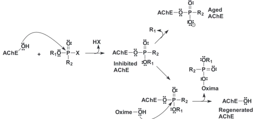
Nerve agents are esters of phosphoric acid that act as potent inhibitors of acetylcholinesterase (AChE), a fundamental enzyme for terminating neurotransmissions in all living animals. These compounds inhibit all AChEs, including the human enzyme (HuAChE), by phosphylating a serine hydroxyl group (Ser203 in HuAChE), which is directly responsible for the hydrolysis of the neurotransmitter acetylcholine. This reaction occurs very rapidly and can lead to irreversible inhibition by a process called aging.1 Before aging, the inhibition of AChE
can be reversed through dephosphylation of the serine residue by a nucleophile, usually an oxime (Figure 1).
Despite the existence of many different oximes in use today as antidotes against intoxication by neurotoxic agents, the literature has yet to report a universal oxime that is able to act eficiently against all existing neurotoxic agents. What has been observed experimentally is that oximes eficient in the treatment of intoxication by one speciic nerve agent can be completely ineffective against another.2-5 This probably happens because the mechanism of these compounds inside the AChE active site has not been fully elucidated yet. Some relevant factors required for obtaining a better understanding of this mechanism, like the orientation of the phosphoryl bond inside the active site, the proper charge and conformation of the oxime and the optimal angle for attacking the phosphylated serine, remain unknown.6 Theoretical studies have shown, for example, that the appropriate conformation is a fundamental requirement for the oxime to enter into the AChE active site’s gorge,6-10 reinforcing the importance of conformational analysis studies of these compounds.
pralidoxime (2-PAM), one of the oximes most commonly
used as an antidote against neurotoxic agents, in its lowest energy conformation, E-anti-anti, remained in a possible reactivation region in the AChE active site. This observation was conirmed by molecular dynamics simulations, where the E-anti-anti conformation and the positive charge of 2-PAM
(+1) were shown to be fundamental for its permanence inside HuAChE´s active site.12
In the present work, we performed conformational analysis studies using semi-empirical methods for the determination of possible global and local minima in each of the three different oximes: Toxogonine,13 TMB-414,15 and HI-6.16 The results obtained will be useful in parameterizing these molecules for further molecular modeling studies of their potential as AChE reactivators.
Methodology
The structures of Toxogonine, TMB-4 and HI-6 were built manually with GHEMICAL 2.1017 using the TRIPOS 5.2 force ield.18 Classical optimizations were carried out followed by a random conformational search using GHEMICAL 2.10,17 in order to ind structures near possible global minima. Then, the semi-empirical methods RM119 and PM620 implemented in MOPAC 200720 were used in two principal dihedral angles in those molecules, denoted Φ1 and Φ2 in Figure 2, which could causes further steric effects between two or more groups (i.e., aromatic rings) after optimizing the other degrees of freedom in the molecules.
The validations of RM1 and PM6 for conformational analysis were performed by calculations using the GAUSSIAN 2003 software package.21
Both dihedral angles, Φ1 and Φ2, were scanned from 0 to 360 degrees in 5-degree steps. This was performed with the MOPAC 200720 software package using the following
keywords in the input ile: PRECISE CHARGE=+2 RM1 (or PM6) STEP=5 POINT=72 EF NOXYZ COMPFG GEO-OK GRADIENTS, where CHARGE is the total charge of the system and the STEP and POINT keywords describe a coordinate as a dynamical coordinate, i.e., bond, angle and/or dihedral variation. In our case, the number of steps multiplied by the number of points gave us the total number of scanned angles (5 × 72 = 360). For each compound, we created two input iles, one using the RM1 method and the other using the PM6 method. Each output ile was parsed by a custom FORTRAN program (see supplementary information) to extract the XY coordinates in order to facilitate the comparison of the dihedral angle and ∆Hf.
Results and Discussion
It is important to mention here that the decision to only perform the conformational analysis on the dihedrals Φ1 and
Φ2 described in this work was supported by the following reasons: (i) Energetically, Φ1 and Φ2 are the dihedrals related to the highest energy points in Toxogonine, TMB-4 and HI-6 and, consequently, to the highest barriers of energy; (ii) Variations of these dihedrals intensify repulsions between two or more groups with the same charge, like aromatic rings; (iii) In order to avoid poor parameterizations and, consequently, trapping the molecule in local minima, it
Figure 1. Inhibition, disinhibition and aging of acetylcholinesterase. X is the leaving group.
is of fundamental importance to obtain information about putative minima obtained by the conformational analysis of those dihedrals; (iv) Variations in the other dihedrals in Toxogonine, TMB-4 and HI-6 do not cause enough steric effects to cause problems in further parameterizations.
In order to validate the RM1 and PM6 methods for the conformational search, we have performed the Φ2 dihedral variation of HI-6 from 0 to 180 degrees using DFT,22,23
with B3LYP24,25 and 6-31 G (d,p) basis sets and the RM1
and PM6 methods and compared the energy values and the energetic barriers found with each.26 In this case, despite
small differences in the minimal and maximal energies, the RM1, PM6 and DFT methods presented similar proiles, as can be seen in Figure 3. The highest and lowest energy points were located, respectively, at 20 and 140 degrees with DFT, 60 and 170 degrees with RM1 and at 40 and 160 degrees with PM6.
The energetic barriers computed from each method are presented in Table 1. The difference in the barriers calculated by DFT and RM1 was 1.41 kcal mol-1
(11.38-9.97 in Table 1), and that between DFT and PM6 was 1.78 kcal mol-1 (11.38-9.60 in Table 1). These deviations
are consistent with data already reported in the literature.19,20
After searching the points of maximal energy, it was also necessary to perform a vibrational analysis of the
stationary points found in the conformational analysis, in order to unequivocally characterize them as true energy maxima.26 This was done forΦ
2 = 140o using DFT/B3LYP
6-31G(d,p), Φ2 = 170o using RM1 and Φ2 = 160o using PM6. The single negative frequency vibrations found for DFT, RM1 and PM6 were 62.94i cm-1, 55.70i cm-1
and 42.2i cm-1, respectively. The occurrence of a single
imaginary frequency indicated that a good transition state (TS) had been located, and the vibrational analysis conirmed it as a true saddle point.
For a inal validation of the RM1 and PM6 methods for use in our conformational analysis, we calculated the root mean square deviations (RMSD) of the three TS geometries of HI-6 acquired by the RM1, PM6 and DFT methods. As shown in Figure 4, the three geometries presented a very good superimposition, with an RMSD of 0.269 Å
Figure 3. φ2 dihedral variation of HI-6, from 0 to 180 degrees, using DFT, B3LYP and the 6-31 G (d,p) basis set for the RM1 and PM6 methods.
Table 1. Energetic barriers acquired from the methods DFT, RM1 and
PM6 for the dihedral φ2 in HI-6
Method Energetic barriers*
DFT/B3LYP 6-31G** 11.38
RM1 9.97
PM6 9.60
*kcalmol-1.
between DFT and RM1, 0.394 Å between DFT and PM6 and 0.194 Å between DFT and PM6.
After the validation of these methods, conformational analyses of Toxogonine, TMB-4 and HI-6 were performed using the RM1 and PM6 methods by varying the dihedrals
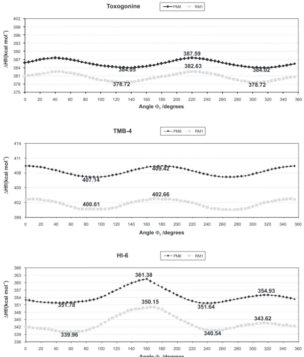
Φ1 and Φ2 from 0 to 360 degrees following the procedure already described in the methodology section. As can be seen in Figure 5, our results showed that, for the dihedral
Φ1 of Toxogonine, the lowest energy conformation was the one that presented torsion between 260 and 280 degrees. For dihedral Φ2 (Figure 6), two conformations were found, one between 120 and 140 degrees and the other between 300 and 320 degrees. The energetic barriers for Φ1 were
about 14.00 kcal mol-1 when using the PM6 method and 16.31 kcal mol-1 when using the RM1 method. For the dihedral Φ2,the barriers determined with the PM6 and RM1 methods were 3.5 and 3.9 kcal mol-1, respectively, suggesting that it is easier to turn Φ2 than Φ1.
When rotating the TMB-4 dihedral Φ1, even though the energy needed to transpose the barrier between the maximal and minimal energies was shown in this study to be about 5.81 kcal mol-1 with PM6 and 7.2 kcal mol-1 with RM1, intermediate conformational states were observed between the angles 60 and 100 and 200 and 300. The barriers to rotating dihedral Φ2 were 2.28 kcal mol-1 with PM6 and 2.05 kcal mol-1 with RM1 (Figure 6).
Figure 6.Plots of∆Hf×φ2for the oximes Toxogonine, TMB-4 and HI-6.
The results of the conformational analysis for HI-6 showed that, when varying both dihedral angles, Φ1 and Φ2, there were found, respectively, values with low (less than 3.0 kcal mol–1) and considerable (about 10.0 kcal mol–1) magnitudes, in terms of energetic barriers. As can be seen in Figure 5, the difference between the minima between 80 and 120 degrees and, for example, one of the maxima, located between 240 and 280 degrees, for the dihedral Φ1 was about 11.30 kcal mol-1 using PM6 and about 13.65 kcal mol-1 using RM1. This result suggests that, despite the existence of a local minimum between 280 and 320 degrees, it is dificult to cross the barrier with a magnitude between 11.00 and 14.00 kcal mol-1 and that the most stable conformation
for Φ1 in HI-6 is the one valued at 351.40 kcal mol-1 with PM6 and at 339.55 kcal mol–1 with RM1. For the analysis of the variation of the HI-6 angle Φ2 (Figure 6), it was shown that, for both PM6 and RM1, the major energetic barrier was about 10.00 kcal mol-1, suggesting that the most stable conformations are localized between the dihedral angles of 40 and 80 degrees and 240 and 260 degrees.
Conclusions
RM1, by comparison with the energetic barriers obtained for the Φ2 dihedral angle of the oxime HI-6 calculated using DFT, B3LYP and the 6-31 G (d,p) basis sets. The energetic
proiles obtained with the semi-empirical methods were very similar to the one obtained by DFT. Additionally, the superimposition of TSs obtained with the three methods did not present signiicant variations.
Further application of the RM1 and PM6 methods to the conformational analysis of Toxogonine, TMB-4 and HI-6 suggested that the ideal values for the dihedral angles
Φ1 and Φ2 could lead to the reduction of steric effects in future parameterizations in posterior molecular modeling
studies. Using RM1 and PM6 methods for conformational analysis provides more accuracy when compared to the other semi-empirical methods like AM119,20 and PM319,20
and, at the same time, is much less time consuming when compared with DFT.
Acknowledgments
The Authors wish to thank the Brazilian agencies CNPq, FAPERJ and CAPES/PRODEFESA for inancial support.
Supplementary Information
Supplementary data are available free of charge at http:// jbcs.sbq.org.br, as PDF ile.
References
1. Kryger, G.; Harel, M.; Giles, K.; Toker, L.; Velan, B.; Lazar, A.; Kronman, C.; Barak, D.; Ariel, N.; Shafferman, A.; Silman, I.; Sussman, J. L. Acta Crystallogr., Sect. D: Biol. Crystallogr. 2000, 56, 1385.
2. Somani, S. M.; Solana, R. P.; Dube, S. N.; Chemical Warfare Agents, Academic Press: San Diego, 1992.
3. Black, R. M.; Harrison, J. M. In The Chemistry of Organophosphorus Compounds, Volume 4, Ter- and
Quinque-Valent Phosphorus Acids and Their Derivatives; Hartley, F. R.
ed., John Wiley & Sons: New York, 1996.
4. Sidell, F. R.; Takafuji, E. T.; Franz, D. R. In Textbook of Military Medicine; Ofice of the Surgeon General, US Army, Washington
D.C., 1997, p. 129.
5. Marrs, T. C.; Maynard, R. L.; Sidell, F. R. In Chemical Warfare Agents: Toxicology and Treatment, 2nd ed., John Wiley & Sons:
New York, 2007.
6. Wong, L.; Radic, Z.; Brüggemann, R. J. M.; Hosea, N.; Berman, H. A.; Taylor, P. Biochemistry2000, 39, 5750.
7. Ekström, F.; Yuan-Ping, P.; Boman, M.; Artursson, E.; Akfur, C.; Börjegren, S.; Biochem. Pharmacol.2006, 72, 597.
8. Ashani, Y.; Radic, Z.; Tsigelny, I.; Vellom, D. C.; Pickering, N. A.; Quinn, D. M.; Doctor, B. P.; Taylor, P.; J. Biol. Chem.1995, 11, 6370.
9. Ekström, F. J.; Astot, C.; Pang, Y. P.; Clin. Pharmacol. Ther. 2007, 82, 282.
10. Worek, F.; Aurbek, N.; Koller, M.; Becker, C.; Eyer, P.; Thiermann, H. Biochem. Pharmacol.2007, 73, 1807. 11. Castro, A. T.; Figueroa-Villar, J. D.; Int. J. Quantum Chem.
2002, 89, 135.
12. Goncalves, A. D.; Franca, T. C. C.; Wilter, A.; Figueroa-Villar, J. D.; J. Braz. Chem. Soc.2006, 17, 968.
13. Smirnova, O. I.; Gurina, E. I.; Zhigalova, L. V.; Arestova, L. S.; Farmakologiya I Toksikologiya1975, 38, 467.
14. Bay, E.; Federation Proceedings1959, 18, 366.
15. Bay, E.; Krop, S.; Yates, L. F.; Proceedings of the Society for Experimental Biology and Medicine1958, 98, 107.
16. Bartosova, L.; Kuca, K.; Jun, D.; Kunesova, G.; Int. J. Toxicol. 2005, 24, 399.
17. Hassinen, T.; Peräkylä, M.; J. Comput. Chem.2001, 22, 1229. 18. Shih, J. H.; Chen, C. L.; Macromolecules1995, 28, 4509. 19. Rocha, G. B.; Freire, R. O.; Simas, A. M.; Stewart, J. J.;
J. Comput. Chem.2006, 27, 1101.
20. Stewart, J. J.; J. Mol. Model.2007, 13, 1173.
21. Frisch, M. J.; Trucks, G. W.; Schlegel, H. B.; Scuseria, G. E.; Robb, M. A.; Cheeseman, J. R.; Montgomery, J. A.; Vreven, T.; Kudin, K. N.; Burant, J. C.; Millam, J. M.; Iyengar, S. S.; Tomasi, J.; Barone, V.; Mennucci, B.; Cossi, M.; Scalmani, G.; Rega, N.; Petersson, G. A.; Nakatsuji, H.; Hada, M.; Ehara, M.; Toyota, K.; Fukuda, R.; Hasegawa, J.; Ishida, M.; Nakajima, T.; Honda, Y.; Kitao, O.; Nakai, H.; Klene, M.; Li, X.; Knox, J. E.; Hratchian, H. P.; Cross, J. B.; Bakken, V.; Adamo, C.; Jaramillo, J.; Gomperts, R.; Stratmann, R. E.; Yazyev, O.; Austin, A. J.; Cammi, R.; Pomelli, C.; Ochterski, J. W.; Ayala, P. Y.; Morokuma, K.; Voth, G. A.; Salvador, P.; Dannenberg, J. J.; Zakrzewski, V. G.; Dapprich, S.; Daniels, A. D.; Strain, M. C.; Farkas, O.; Malick, D. K.; Rabuck, A. D.; Raghavachari, K.; Foresman, J. B.; Ortiz, J. V.; Cui, Q.; Baboul, A. G.; Clifford, S.; Cioslowski, J.; Stefanov, B. B.; Liu, G.; Liashenko, A.; Piskorz, P.; Komaromi, I.; Martin, R. L.; Fox, D. J.; Keith, T.; Al-Laham, M. A.; Peng, C. Y.; Nanayakkara, A.; Challacombe, M.; Gill, P. M. W.; Johnson, B.; Chen, W.; Wong, M. W.; Gonzalez, C.; Pople, J. A.; Gaussian, Gaussian Inc.: Wallingford CT,2004. 22. Hohenberg, P.; Kohn, W.; Phys. Rev.1964, 136, B864. 23. Kohn, W.; Sham, L. J.; Phys. Rev.1965, 140, 4A. 24. Becke, A. D.; J. Chem. Phys.1993, 98, 7.
25. Lee, C.; Yang, W.; Parr, R. G.; Phys. Rev. B: Condens. Matter Mater. Phys. 1988, 37, 2.
26. We thank the referee for this suggestion.
Received: January 17, 2009
Supplementary Information
0103 - 5053 $6.00+0.00
*e-mail: [email protected]
Conformational Analysis of Toxogonine, TMB-4 and HI-6 using
PM6 and RM1 Methods
Arlan da Silva Gonçalves,a Tanos C. C. França,b José D. Figueroa-Villarb and Pedro G. Pascutti*,a
aInstituto de Biofísica Carlos Chagas Filho, Universidade Federal do Rio de Janeiro, 21941-902 Rio de Janeiro-RJ, Brazil
bDivisão de Ensino e Pesquisa, Seção de Engenharia Química, Instituto Militar de Engenharia,
22290-270 Rio de Janeiro-RJ, Brazil
cc Fortran code of program: Make Coordinate cc **************************************** cc Program Make Coordinate
cc Program for Linux Platform
cc Data the MOPAC program output ile (*.arc), this program make an output cc which may be imported
cc by programs as Microsoft Excel, GNUMERIC, etc cc By: Goncalves, A.S. (2008)
cc Note1: This program only is correctly executed if we make conformational cc search from zero to 360 degree with a step of 5 degrees.
cc
Program makecoord
character arc*30, nomeout*30 dimension x(1000),y(1000) a=system(‘clear’)
a=system(‘ls -ltrh *.arc’) Print *,’Give the name of arc ile’ Read *,arc
Print *,’Give the name of output ile’ Read *,nameout
OPEN(1,FILE=arc) OPEN(7,FILE=nameout) do m=1,9
read(1,*) enddo l=1 do n=1,9
read(1,’(8f7.2)’) x(l),x(l+1),x(l+2),x(l+3),x(l+4),x(l+5) & ,x(l+6),x(l+7)
read(1,’(8f7.2)’) y(l),y(l+1),y(l+2),y(l+3),y(l+4),y(l+5) & ,y(l+6),y(l+7)
read(1,*) l=l+8 enddo do n=1,72
write(7,’(f7.2,2x,f7.2)’) x(n),y(n) enddo