Platelet Activation and Thrombus Formation
over IgG Immune Complexes Requires
Integrin
α
IIb
β
3 and Lyn Kinase
Huiying Zhi1, Jing Dai2, Junling Liu3, Jieqing Zhu1,4, Debra K. Newman1,5,6, Cunji Gao1,7, Peter J. Newman1,6,8*
1Blood Research Institute, BloodCenter of Wisconsin, Milwaukee, Wisconsin, United States of America, 2Ruijing Hospital Affiliated Shanghai Jiao Tong University School of Medicine, Shanghai, People’s Republic of China,3Department of Biochemistry and Molecular Cell Biology, Shanghai Key Laboratory of Tumor Microenvironment and Inflammation, Shanghai JiaoTong University School of Medicine, Shanghai, China, 4Department of Biochemistry, Medical College of Wisconsin, Milwaukee, Wisconsin, United States of America,5Department of Microbiology, Medical College of Wisconsin, Milwaukee, Wisconsin, United States of America,6Department of Pharmacology and Toxicology, Medical College of Wisconsin, Milwaukee, Wisconsin, United States of America,7Chronic Disease Research Institute, Department of Nutrition and Food Hygiene, Zhejiang University School of Public Health, Hangzhou, China,8Department of Cell biology, Medical College of Wisconsin, Milwaukee, Wisconsin, United States of America
Abstract
IgG immune complexes contribute to the etiology and pathogenesis of numerous autoim-mune disorders, including heparin-induced thrombocytopenia, systemic lupus erythemato-sus, rheumatoid- and collagen-induced arthritis, and chronic glomerulonephritis. Patients suffering from immune complex-related disorders are known to be susceptible to platelet-mediated thrombotic events. Though the role of the Fc receptor, FcγRIIa, in initiating platelet activation is well understood, the role of the major platelet adhesion receptor, integrin
αIIbβ3, in amplifying platelet activation and mediating adhesion and aggregation down-stream of encountering IgG immune complexes is poorly understood. The goal of this inves-tigation was to gain a better understanding of the relative roles of these two receptor systems in immune complex-mediated thrombotic complications. Human platelets, and mouse platelets genetically engineered to differentially express FcγRIIa andαIIbβ3, were allowed to interact with IgG-coated surfaces under both static and flow conditions, and their ability to spread and form thrombi evaluated in the presence and absence of clinically-used fibrinogen receptor antagonists. Although binding of IgG immune complexes to FcγRIIa was sufficient for platelet adhesion and initial signal transduction events, platelet spreading and thrombus formation over IgG-coated surfaces showed an absolute requirement for
αIIbβ3 and its ligands. Tyrosine kinases Lyn and Syk were found to play key roles in IgG-induced platelet activation events. Taken together, our data suggest a complex functional interplay between FcγRIIa, Lyn, andαIIbβ3 in immune complex-induced platelet activation. Future studies may be warranted to determine whether patients suffering from immune complex disorders might benefit from treatment with anti-αIIbβ3-directed therapeutics.
a11111
OPEN ACCESS
Citation:Zhi H, Dai J, Liu J, Zhu J, Newman DK, Gao C, et al. (2015) Platelet Activation and Thrombus Formation over IgG Immune Complexes Requires IntegrinαIIbβ3 and Lyn Kinase. PLoS ONE 10(8): e0135738. doi:10.1371/journal.pone.0135738
Editor:Kathleen Freson, University of Leuven, BELGIUM
Received:June 2, 2015
Accepted:July 25, 2015
Published:August 20, 2015
Copyright:© 2015 Zhi et al. This is an open access article distributed under the terms of theCreative Commons Attribution License, which permits unrestricted use, distribution, and reproduction in any medium, provided the original author and source are credited.
Data Availability Statement:All relevant data are within the paper and its Supporting Information files.
Funding:This work was supported by National Natural Science Foundation of China #81270586 (to CG) and by the Heart, Lung, and Blood Institute of the National Institutes of Health #HL-44612 (to DKN and PJN). The funders had no role in study design, data collection and analysis, decision to publish, or preparation of the manuscript.
Introduction
IgG immune complexes contribute to the etiology and pathogenesis of a number of autoim-mune disorders, including heparin-induced thrombocytopenia [1], systemic lupus erythemato-sus [2,3], and collagen-induced/rheumatoid arthritis [4]. Patients with immune complex-related disorders are known to be hypercoagulable [5], and susceptible to both thrombocytope-nia [6,7] and thrombosis [8,9]. These disorders are thought to be precipitated, at least in part, by platelets that have become activated via their interaction with autoimmune antibody/anti-gen complexes—an event that was shown almost 50 years ago to induce secretion of platelet granule constituents [10], and that is now known to be mediated by the binding of the Fc region of IgG-containing immune complexes to the platelet cell surface Fc receptor, FcγRIIa.
FcγRIIa is a member of the immunoglobulin gene superfamily comprised of an extracellular domain that binds the Fc region of IgG, a single pass transmembrane domain, and a cyto-plasmic tail that contains two YxxL immune receptor tyrosine-based activation motifs (ITAMs) [11,12]. While FcγRIIa exhibits only low-affinity for monomeric IgG, it binds with high affinity to the Fc region of antigen-bound IgG immune complexes [11,13]. FcγRIIa is the only Fc receptor on human platelets, and is not expressed in mice [14]. Its cross-linking results in activation of associated Src-family kinases that phosphorylate the ITAM tyrosines, which act as a docking site for the SH2 domain-containing tyrosine kinase, Syk [15]. Activation of Syk, in turn, promotes an intracellular signaling cascade that eventually leads to phosphoryla-tion and activaphosphoryla-tion of phospholipase C (PLC)γ2 [16], resulting in calcium mobilization, gran-ule secretion, integrin activation, platelet aggregation, and thrombus formation.
In addition to its role as a receptor for IgG-containing immune complexes, FcγRIIa appears to be capable of promoting a number of other functions in platelets, most notably as an ampli-fier of integrinαIIbβ3-mediated platelet activation [17,18], and in cooperating with this integ-rin to mediate platelet activation by tumor cells [19] and certain strains of bacteria [20]. Interestingly, although FcγRIIa was found to mediate the initial attachment of Fcγ RIIa-trans-fected HEK293 to immobilized immune complexes, sustained signaling downstream of attach-ment required co-expression of the integrinαMβ2(Mac-1) [21]. Thus, at least in transfected cell lines, the ability of FcγRIIa to send productive activation signals into a cell requires integrin signaling as well. The purpose of the present investigation was to determine whether there is functional coupling between FcγRIIa andαIIbβ3 when platelets encounter immobilized IgG. Our results help define the molecular requirements for platelet activation and thrombus forma-tion in patients suffering from IgG immune complex disorders, and have potential therapeutic implications for treating and/or preventing the thrombotic complications associated with immune complex disorders.
Materials and Methods
Reagents and antibodies
Human fibrinogen was from Enzyme Research Laboratories Inc. Lyn and Fyn were obtained from Life Technologies. Src was purchased from Enzo Life Sciences.
Mice
Mice were maintained in the Biological Resource Center at the Medical College of Wisconsin (MCW). All animal protocols were approved by the MCW Institutional Animal Care and Use Committee. FcγRIIa transgenic mice [14] were littermates on a C57BL/6J background. Lyn -/-mice (C57BL/6 background) were from the Jackson Laboratories,β3-/-mice (C57BL/6 back-ground) were a gift of David A. Wilcox (MCW). Fyn-/-mice (129 background) were from Roy L. Silverstein (MCW). FcγRIIa mice were bred with Lyn-deficient mice (Lyn-/-), Fyn-deficient mice (Fyn-/-) andβ3 deficient mice (β3-/-) to obtain double-heterozygous mice that were used to establish Lyn-/-/FcγRIIapos, Lyn+/+/FcγRIIapos, Fyn-/-/FcγRIIapos, Fyn+/+/FcγRIIapos,β3-/-/ FcγRIIapos,β3+/+/FcγRIIaposcolonies. Mouse genotypes were verified by PCR amplification of genomic DNA. Expression of FcγRIIa, and lack of Lyn, Fyn, andβ3 were confirmed by West-ern blot analysis of platelet lysates. Platelet counts of the Lyn-/-/FcγRIIapos, Fyn-/-/FcγRIIapos mice were similar to that of wild-type controls.
Blood collection
Ethical approval was obtained from the institutional review board of the BloodCenter of Wis-consin in accordance with the Declaration of Helsinki. All study members provided informed, written consent to participate. Blood samples from a Type I Glanzmann thrombasthenic (GT) patient carrying an L924A point mutation in integrinαIIb, and from healthy volunteers free from medication for two weeks was collected into 90 mM PPACK (D-phenylalanyl-L-prolyl-L-arginine chloromethyl ketone). Mouse blood was drawn from the inferior vena cava of anesthe-tized mice into 3.8% sodium citrate (1/10 volume) or PPACK/heparin.
Platelet spreading assays
Platelets were obtained from 3.8% sodium citrate anticoagulated whole blood and added to 8-chamber glass tissue-culture slides (Becton Dickinson) that had been coated with human IgG (25μg/ml) or human fibrinogen (25μg/mL). Fibrinogen and BSA were precleared using
protein G Sepharose (GE Healthcare Bio-Sciences) to remove any traces of contaminating IgG. Spreading assays were performed as previously described [18]. Briefly, 200μl of washed
plate-lets at a concentration of 2.5x10⁷/mL were allowed to adhere to either immobilized fibrinogen or IgG for 30 minutes. Adherent platelets were fixed with 2% paraformaldehyde, permeabilized with 0.1% Triton X-100, and stained with phalloidin tetramethylrhodamine isothiocyanate. Images analyzed using Metamorph software (Universal Imaging). Statistical analysis of the area occupied by spread platelets was performed using a 2-tailed Student t-test for unpaired samples. For biochemical analysis, platelets were incubated at 37°C for 30 minutes on 10 cm tissue-culture dishes and lysed with 30 mM HEPES [pH 7.4], 300 mM NaCl, 20 mM EGTA, 0.2 mM MgCl2, 2% Triton X-100) containing protease and phosphatase inhibitor cocktails,
and subjected to immunoblot analysis.
In vitro thrombus formation under flow conditions
αIIbβ3 antagonists. Epifluorescence microscopic images of platelet adhesion and thrombus for-mation were acquired by in real time at one frame per second. Thrombus forfor-mation was deter-mined as the mean percentage of total area covered by thrombi and as the mean integrated fluorescence intensity perμm2. Image analysis was performed using Metamorph.
PF4 release assay
Washed platelets fromαIIbβ3-/-/FcγRIIaposandαIIbβ3+/+/FcγRIIaposmice were allowed to spread on glass slides that had been coated with BSA, 25μg/ml fibrinogen, or 25μg/mL IgG for
30 minutes. After spreading, the platelets were removed by centrifugation and the secreted PF4 quantified using a Quantikine ELISA kit (R&D systems).
CHO cell transfection and spreading assay
CHO-K1 cells stably expressing full-length humanαIIb andβ3 [22] were additionally trans-fected with pCMV FcγRIIa IRES neo (Addgene) using Lipofectamine LTX and PLUS Reagent. The expression levels ofαIIb,β3, and the FcγRIIa were confirmed by Western-blot. Spreading assays were performed by adding cells to glass slides that had been coated with either 25μg/ml
IgG or fibrinogen in the presence or absence of 250μg/ml soluble fibrinogen. Phase contrast
images of CHO cells were taken 45 minutes later.
Mass spectrometric analysis of phosphorylated Fc
γ
RIIa cytoplasmic
domain constructs
The region encoding amino acid residues 206–282 of the FcγRIIa cytoplasmic domain was PCR-amplified from pCMV FcγRIIa IRES neo and cloned into the bacterial expression vector pQE30-GB1 (Qiagen) in front of a histidine tag. The resulting construct was transduced into E. coli BL21 cells, induced with IPTG and purified from bacterial lysates using a nickel-Sepharose column (GE Healthcare Life Sciences). For kinase assays, recombinant FcγRIIa cytoplasmic domain proteins (1 mM) were incubated with Src, Lyn or Fyn in kinase assay buffer (250μM
ATP, 1 mM EGTA, 10 mM MgCl2, 0.01% Brij 35, and 250μM Na3VO4) for 60 min at 30°C
and then boiled in the presence of an equal volume of 2× SDS-PAGE sample reducing buffer. The resulting products were separated on a 12% SDS−polyacrylamide gel and stained with
Coomassie blue. Target bands were cut out, digested with trypsin, and subjected to mass spec-trometric analysis following a previously described protocol [23].
Statistical analysis
Statistically significant differences were identified by performing a one-way ANOVA followed by a two-tailed unpaired Student’s t test.
Results
α
IIb
β
3 and fibrinogen are required for platelet spreading, signal
amplification, and thrombus formation over immobilized IgG
We employed two complementary strategies to examine the potential contribution ofαIIbβ3 in amplifying platelet responses downstream of their binding to immobilized IgG. In the first, human platelets were incubated in IgG-coated chambers under either static or flow conditions in the presence versus absence of the fibrinogen receptor antagonist, abciximab. As shown in
αIIbβ3-fibrinogen interactions like eptifibatide and tirofiban similarly blocked platelet spread-ing on immobilized IgG (S1A–S1B Fig). Tirofiban also markedly inhibited the spreading of FcγRIIa-positive transgenicmouseplatelets on immobilized IgG (S2A–S2B Fig). Binding to
immobilized IgG resulted in strong phosphorylation of FcγRIIa ITAM tyrosines, and concomi-tant recruitment or activation of the tyrosine kinase Syk in both human (Fig 1C) and mouse (S2C Fig) platelets. Both FcγRIIa ITAM phosphorylation and Syk recruitment were suppressed by abciximab (Fig 1C), consistent with the known amplification of platelet activation responses via ligand binding-induced outside-in signaling throughαIIbβ3 [24,25]. Notably, pp125Fak, a reporter of integrin clustering downstream ofαIIbβ3/fibrinogen interactions [26], also became phosphorylated (Fig 1C), suggesting that platelet/IgG interactions had stimulated secretion of fibrinogen from plateletα-granules, leading to ligand binding-dependent clustering ofαIIbβ3
–a well-known inducer of FAK phosphorylation [26–29]. Consistent with the premise that αIIbβ3 requires fibrinogen to support cell spreading on immobilized IgG, CHO cells stably expressing both FcγRIIa andαIIbβ3 failed to spread on IgG-coated glass slides unless soluble fibrinogen was also present (S3 Fig). Finally, abciximab effectively blocked thrombus formation of whole blood, which contains ~3 mg/ml fibrinogen, that was passed over immobilized
IgG-Fig 1. BlockingαIIbβ3-fibrinogen interactions prevents spreading of human platelets and thrombus formation over immobilized IgG. (A)Washed human platelets spread on BSA- or IgG-coated coverslips for 30 minutes in the presence or absence of the integrinαIIbβ3 antagonist abciximab (6.7μg/ml) or Fab fragments of the FcγRIIa blocking antibody mAb IV.3 (10μg/ml). Spread platelets were fixed, permeabilized and stained with rhodamine-phalloidin. Scale bar, 5μm. Images are representative of three independent experiments.(B)Platelet spreading was quantified using Metamorph software and shown as the meanμm2±SEM of at least 200 platelets/group from one of 3 representative experiments. (
*P<0.05). Statistically significant differences were
identified by performing a one-way ANOVA followed by a two-tailed Student’s t test. Note that abciximab or IV.3 Fab significantly inhibited platelet spreading on immobilized IgG.(C)mAb IV.3 immunoprecipitates of lysed spread platelets were analyzed by Western blot with the indicated antibodies. The blots for P-FAK and FAK were performed using whole cell lysates. Note that platelet binding to immobilized IgG elicits strong activation of FcγRIIa and Fak, as well as enhanced recruitment of Syk, and that both abciximab and mAb IV.3 inhibit platelet spreading-induced phosphorylation.(D)Mepacrine-labeled whole blood was perfused at a shear rate 5 dynes/cm2over 100μg/ml IgG-coated microchannels in the presence or absence of 10μg/ml mAb IV.3 Fabs, 6.7μg/ml
abciximab, or an isotype-matched control Fab. Epifluorescence microscopic images of platelet adhesion and thrombus formation shown are representative of three separate experiments. Note that abciximab inhibits thrombus formation over immobilized IgG under conditions of flow.
coated chamber slides under conditions of venous flow (Fig 1D)–conditions likely to be present when platelets encounter IgG immune complexesin vivo.
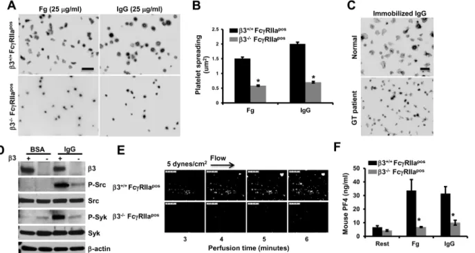
To confirm the importance ofαIIbβ3 in platelet reactions downstream of IgG-induced acti-vation, the effects ofαIIbβ3 deficiency on platelet reactivity to immobilized IgG were exam-ined. As shown inFig 2, both mouse (Fig 2A and 2B) and human (Fig 2C)αIIbβ3-deficient platelets spread poorly on immobilized IgG. Phosphorylation of Src and Syk induced by plate-let spreading on immobilized IgG was also greatly diminished in plateplate-lets missingαIIbβ3 (Fig 2D), as was thrombus formation (Fig 2E) andα-granule secretion (Fig 2F).
Involvement of Src- and Syk-family kinases in immobilized IgG-induced
platelet activation
The involvement of Src and Syk family kinases in cellular responses downstream from platelet-IgG interactions was examined using a series of kinase-specific inhibitors. As shown inFig 3A and 3B, spreading of human platelets on IgG-coated microtiter wells was abolished in the pres-ence of either the Src family kinase inhibitor, PP2, or the Syk kinase inhibitor PRT-060318 (PRT318) [30]. Spreading of FcγRIIa-positive transgenic mouse platelets on immobilized IgG was similarly affected by these two inhibitors (S4 Fig). In contrast, PP3, the inactive analogue of PP2, had no effect. PP2 and PRT318 also blocked tyrosine phosphorylation of multiple cellu-lar tyrosine kinase substrates, including FcγRIIa itself (Fig 3C).
To determine whether Src-family kinases were capable of phosphorylatingbothITAM
tyro-sine residues, a recombinant protein comprised of the entire FcγRIIa ITAM cytoplasmic domain (FcγRIIacyto) was subjected to anin vitrokinase assay, its products separated by
SDS-PAGE and then visualized by staining with Coomassie blue. As shown inFig 3D, both mono- and di-phosphorylated FcγRIIacytospecies were generated by Src. Kinase assays
employing either Lyn or Fyn showed identical results (data not shown). To determine the iden-tity of the ITAM tyrosine that became phosphorylated first, the lower bands from each of the three Src-family kinase reactions, thought to represent the mono-phosphorylated species, were cut out and subjected to trypsinization/mass spectrometry analysis. As shown inTable 1, pep-tides phosphorylated on either ITAM tyrosine residue—Y253or Y269—were found to be derived
from the lower MW band. That these two tyrosines are able to be phosphorylated independent of the phosphorylation state of the other was further shown by the ability of Fyn, Lyn, and Src to phosphorylate recombinant FcγRIIa cytoplasmic constructs in which either Y253or Y269had
been mutated to phenylalanine (Fig 3E).
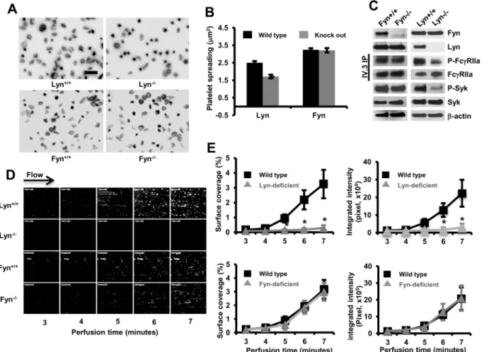
To determine the tyrosine kinase responsible for activation of intact platelets downstream of encountering immobilized IgG, we crossed FcγRIIaposmice with Lyn- or Fyn-deficient mice. The expression levels of FcγRIIa were comparable among different groups (flow-cytometry data not shown). We compared the ability of platelets to spread and form thrombi over immo-bilized IgG. As shown inFig 4A and 4B, whereas Fyn-/-/FcγRIIaposplatelets spread normally, spreading of Lyn-/-/FcγRIIaposplatelets was markedly impaired, despite normal expression of Fyn (Fig 4C) and Src (not shown). Tyrosine phosphorylation of FcγRIIa and Syk in Fyn-/-/ FcγRIIaposplatelets was also comparable to that observed in Fyn+/+/FcγRIIaposplatelets; how-ever, compared with Lyn+/+/FcγRIIaposplatelets, platelets from Lyn-/-/FcγRIIaposexhibited sig-nificantly reduced tyrosine phosphorylation of FcγRIIa and Syk, again despite normal
expression of Fyn and Src. Finally, when whole blood from Fyn+/+/FcγRIIapos, Fyn-/-
/FcγRIIa-pos, Lyn-/-/FcγRIIapos, or Lyn-/-/FcγRIIaposwas subjected to microfluidic flow conditions, only
Discussion
IgG immune complexes contribute to the etiology and pathogenesis of numerous autoimmune disorders, including heparin-induced thrombocytopenia, systemic lupus erythematosus, rheu-matoid- and collagen-induced arthritis, and chronic glomerulonephritis. Patients suffering from immune complex-related disorders are known to be susceptible to platelet-mediated thrombotic events. Though platelet activation and signal transduction pathways initiated by the binding of IgG immune complexes to its platelet receptor, FcγRIIa, are well understood, the role of the major platelet adhesion receptor, integrinαIIbβ3, in amplifying platelet activation and mediating adhesion and aggregation downstream of encountering IgG immune complexes is not known.
The purpose of the present investigation was to gain further insight into the molecular requirements for activation, spreading and thrombus formation in platelets encountering IgG immune complexes. We used small molecule antagonists ofαIIbβ3, as well as human and mouse platelets that selectively expressαIIbβ3 and FcγRIIa, to determine whether the integrin αIIbβ3 is required for platelet spreading and thrombus formation over immobilized IgG. We
Fig 2.αIIbβ3-deficient platelets fail to spread, form thrombi, or efficiently secrete granule contents over immobilized IgG. (A)Washed platelets from αIIbβ3-/-orαIIbβ3+/+FcγRIIaposmice were allowed to spread and analyzed as described in the legend forFig 1. Representative images of three independent experiments are shown. Scale bar, 5μm. Note thatαIIbβ3-deficient platelets failed to spread on fibrinogen, as expected, but also failed to spread on immobilized IgG.(B)Platelet spreading was quantified using Metamorph software and shown as the meanμm2±SEM of at least 200 platelets /group from
one of 3 representative experiments (*P<0.01). Statistically significant differences were identified by performing a one-way ANOVA followed by a two-tailed Student’s t test. (C) Washed human platelets from a Type 1 Glanzmann thrombasthenic (GT) and a healthy volunteer were allowed to spread on IgG for 30 minutes at 37°C. Note that the control platelets formed filopodia and large lamellipodia, while GT platelets failed to spread on immobilized IgG.(D)Lysates of spread murine platelets were analyzed by Western blotting with the indicated antibodies. Note thatαIIbβ3-/-/FcγRIIaposplatelets show decreased activation of
Src and Syk compared withαIIbβ3+/+/FcγRIIaposplatelets.(E)Anticoagulated, mepacrine-labeled whole blood fromαIIbβ3+/+/FcγRIIaposandαIIbβ3-/-/
FcγRIIaposmice was perfused over 100μg/mL IgG-coated flow chambers at a shear rate of 5 dynes/cm2and images acquired using epifluorescence microscopy. Data shown are representative of three separate experiments. Note thatαIIbβ3-deficient murine platelets exhibited dramatically-reduced thrombus formation compared to their wild-type counterparts. (F) PF4 secretion from washed murine platelets fromαIIbβ3-/-/FcγRIIaposandαIIbβ3+/ +/FcγRIIaposmice after 30 minute spreading BSA-, 25μg/ml fibrinogen-, or 25μg/mL IgG-coated glass slides. PF4 secreted into the culture supernatant was
determined by ELISA. Note that secretion was markedly reduced inαIIbβ3-/-/FcγRIIaposplatelets (*P<0.01). Statistically significant differences were
identified by performing two-tailed Student’s t test.
found that while FcγRIIa-IgG interactions are sufficient to support initial adhesion and stimu-late limited pstimu-latelet activation and secretion, events downstream of encountering immobilized IgG, including amplification of the release reaction, cell spreading and thrombus formation, require fibrinogen binding to itsαIIbβ3 receptor.
Previous studies have shown crosslinking FcγRIIa leads to activation of the Src-family kinases Src, Lyn, and to a lesser extent Fyn [31], as well as the protein tyrosine kinase Syk [15]. The involvement of these families of tyrosine kinases in FcγRIIa signaling is strikingly similar to the molecular requirements for platelets spreading on immobilized fibrinogen in that both Src and Syk family kinases have been known for nearly 20 years to play a significant role in out-side-in signaling and platelet spreading [32–34]. Two members of the Src family, Src and Fyn,
Fig 3. Role of Src- and Syk-family kinases in platelet activation by immobilized IgG. (A)Washed human platelets were incubated in IgG-coated microtiter chamber slides in the presence of DMSO (Control), PP3, PP2, or PRT318 for 30 minutes. Representative platelet spreading images of three independent experiments are shown. Scale bar, 5μm.(B)Quantitation shown is the meanμm2±SEM of at least 200 platelets /group
from one of three representative experiments. Statistically significant differences were identified by performing a one-way ANOVA followed by a two-tailed Student’s t test. Note that both Src and Syk family kinases appear to be involved compared with DMSO vehicle- or PP3-treated platelets (*P<0.01).(C) Washed human platelets were incubated in IgG-coated plates in the presence of DMSO (Control), PP3, PP2, or PRT318 for 30 minutes. IV.3 immunoprecipitation and Western blot reveals that inhibitors of Src and Syk kinase had pronounced effects on early tyrosine phosphorylation events, including phosphorylation of FcγRIIa ITAM tyrosines.(D)Src-mediated phosphorylation of purified, recombinant FcγRIIa cytoplasmic domain (FcγRIIacyto). Phosphorylated FcγRIIacytowas incubated for 60 minutes in the presence of purified
Src + ATP. Coomassie blue staining of SDS-PAGE gels of the resulting products reveal both mono- and di-phosphorylated FcγRIIacytospecies. Results are representative of two independent experiments.(E)Mutant
forms of FcγRIIacytocontaining only one of two ITAM tyrosines were incubated with Src, Fyn, or Lyn for 60
minutes, separated by SDS-PAGE and stained with Coomassie blue. Note that Src family kinases are able to phosphorylate either tyrosine residue independent of the phosphorylation state of the other ITAM tyrosine. Results are representative of two independent experiments.
are directly associated with distinct regions of the integrinβ3 cytoplasmic domain [35,36], and clustering ofαIIbβ3 has been shown to induce direct activation of Src [37]. Notably, while defi-ciency of Src leads to profound defects inαIIbβ3-mediated platelet spreading on immobilized fibrinogen, absence of Lyn actually promotes platelet spreading [38,39]. The roles of Src, Fyn, and Lyn on platelet spreading on immobilized IgG, on the other hand, remain to be defined.
Dual ITAM and ITIM-containing proteins have the capacity to become processively phos-phorylated—i.e. the first tyrosine residue, once phosphorylated, promotes high-affinity recruit-ment of the same, or a different, SH2 domain-containing kinase that then goes on to carry out efficient phosphorylation of the second tyrosine [40]. We have recently described such a mech-anism for phosphorylation of the two ITIM tyrosines of the inhibitory receptor, PECAM-1, in which phosphorylation of Y686by the Src-family kinase, Lyn, is a necessary prerequisite for
recruitment of Csk (C-terminal Src kinase) and its subsequent phosphorylation of Y663
[23,41,42]. Both Lyn and Syk have been shown inin vitrokinase assays to be capable of
tyro-sine phosphorylating a recombinant FcγRIIa cytoplasmic domain [43], and therefore, repre-sent the most likely candidate FcγRIIa ITAM kinases. Whether they exhibit similar sequence specificity and/or are reliant on sequential phosphorylation, however, has not been previously described. We found that inhibitors of Src and Syk kinase had pronounced effects on phos-phorylation of FcγRIIa ITAM tyrosines, however, unlike the ITIMs of PECAM-1, the Src-fam-ily kinases involved in FcγRIIa tyrosine phosphorylation exhibit no sequence specificity, and FcγRIIa ITAM tyrosines are independently, rather than sequentially, phosphorylated, at least
in vitro(Fig 3). Moreover, Lyn, but not Fyn or Src, appears to be required for initial platelet
activation over immobilized IgG. These findings are in stark contrast to the kinase require-ments for platelet spreading on immobilized fibrinogen, which requires Src, but not Lyn [38,39].
Since Lyn has an inhibitory role inαIIbβ3-mediated platelet spreading on immobilized fibrinogen [38,39], what might explain its essential role in supporting platelet spreading and thrombus formation on immobilized IgG? The answer, at least in part, may lie in the recent observation of Li et al., who found that Lyn is required forα-granule secretion [44]. Taken together with (1) the finding that fibrinogen is a necessary substrate for cells to spread on
IgG-Table 1. Mass spectrometry analysis of FcγRIIa cytoplasmic domain phosphopeptides generated fromin vitrokinase reactions.
Kinase used Phosphopeptides detected
Src QLEETNNDFETADGGpY253MTLNPR
APTDDDKNIpY269LTLPPNDHVNSNN NIpY269LTLPPNDHVNSNN
Lyn QLEETNNDFETADGGpY253MTLNPR
APTDDDKNIpY269LTLPPNDHVNSNN NIpY269LTLPPNDHVNSNN
Fyn QLEETNNDFETADGGpY253MTLNPR
APTDDDKNIpY269LTLPPNDHVNSNN NIpY269LTLPPNDHVNSNN
Recombinant full-length FcγRIIa cytoplasmic domain:
CRKKRISANSTDPVKAAQFEPPGRQQMIAIRKRQLEETNNDYETADGGYMTLNPRAPTDDDKNIYLTLPPNDHVNSNN was subjected to anin vitrokinase reaction, digested with trypsin, and subjected to mass spec analysis. The above phosphopeptides were detected. Note that both ITAM tyrosines 253 and 269 are targets for SFKs, at least using peptides as a substrate. The YxxL ITAM motifs are delimited with red bold letters, while the tryptic cleavage sites are underlined and italicized. The naturally-occurring non-ITAM tyrosine at residue 246 was mutated in the recombinant protein to a phenylalanine to prevent its phosphorylation.
coated surfaces (reference [45],Fig 1, andS3 Fig), (2) the ability of Lyn to carry out robust phosphorylation of both FcγRIIa ITAM tyrosines (Fig 3), and (3) the diminished recruitment of Syk to FcγRIIa ITAMs in Lyn-/-platelets (Fig 4C), it seems likely that Lyn is the physiologic FcγRIIa ITAM kinase responsible for initial platelet secretion and integrin activation following platelet/IgG interactions.
Platelet adhesion and aggregation at sites of vascular injury are essential for hemostasis, and ligand binding to integrins and immunoreceptor family members trigger signaling pathways that increasingly appear to share common components that converge to accomplish a common goal, namely that of integrin activation, granule release and controlled thrombus formation. In the present study, we have further defined the molecular requirements for platelet activation
Fig 4. Lyn, but not Fyn, is required for integrin-dependent platelet spreading and thrombus formation over immobilized IgG. (A)Washed FcγRIIa transgenic mouse platelets lacking the Src-family kinase Lyn or Fyn were plated on immobilized IgG-coated coverslips. After 30 minutes, spread platelets were fixed, permeabilized and stained. Images are representative of three independent experiments.(B)Quantification of platelet surface area of at least 200 platelets/group using Metamorph software as described above (*P<0.05). Results are reported as mean±S.E.M from one of three representative
experiments. Fyn- and Lyn-deficient mice are on different strains (seeMethods) and thus exhibit different degrees of spreading, even on wild-type backgrounds. Note that only Lyn-deficient platelets exhibit a spreading defect.(C)mAb IV.3 immunoprecipitates of lysed spread platelets were analyzed by Western blot with the indicated antibodies. The blots for (Fyn, Lyn, p-Syk, Syk,β-actin) were performed using whole cell lysates. Results were representative of two independent experiments. Note that tyrosine phosphorylation of FcγRIIa and Syk is reduced in Lyn-/-, but not Fyn-/-, FcγRIIaposplatelets during
spreading on immobilized IgG.(D)Whole blood from FcγRIIa transgenic mice lacking Lyn or Fyn was perfused at 5 dynes/cm2over IgG-coated coverslips and images acquired using epifluorescence microscopy.(E)Quantification of platelet thrombi expressed as the percentage of total area covered by thrombi (left panels) or total integrated fluorescence intensity (right panels) was performed using Metamorph program. Statistical analysis was performed using the Student’s t test, and data represented as mean±S.E.M (n = 3 per group). Note that thrombus formation was significantly inhibited (*P<0.05)in FcγRIIapos
platelets lacking Lyn (but not Fyn).
following their encounter with immune complexes. When cell surface FcγRIIa binds the Fc region of immobilized IgG, Lyn kinases located near FcγRIIa [46], perhaps due to their co-enrichment in lipid rafts [39,47] become activated and phosphorylate the ITAM tyrosines of FcγRIIa, initiating activation of PLCγ2 and generation of second messengers that result in Ca++mobilization, secretion ofα-granule fibrinogen, and activation ofαIIbβ3. Under the arti-ficialin vitroconditions used for platelet spreading assays, which take place in the absence of
extracellular fibrinogen, secretedα-granule-derived fibrinogen becomes the substrate that sup-portsαIIbβ3-mediated platelet spreading (Fig 1A and 1B,Fig 2A and 2C). BlockingαIIbβ3 with antagonists ofαIIbβ3/Fg interactions (Fig 1) or deficiency ofαIIbβ3 (Fig 2) totally abol-ishes the ability of platelets to spread on immobilized IgG. In the case of whole-blood,αIIbβ3, activated as a result of FcγRIIa/immune complex interactions, is absolutely required for throm-bus formation (Figs1Dand2E). These novel and somewhat unexpected observations extend previous notions about functionally-important integrin/ITAM connections, and provide com-pelling rationale for future clinical studies to determine whether anti-αIIbβ3-directed thera-peutics might benefit patients suffering from immune complex disorders in which thrombosis may be a complicating condition. In addition, the demonstrated requirement for specific tyro-sine kinases in these events (Figs3and4) suggests that Syk inhibitors already currently in clini-cal trials [48,49] may have the added benefit of suppressing not only the immune response responsible for immune complex formation, but also the confounding platelet activation events that occur downstream of platelet/immune complex interactions.
Supporting Information
S1 Fig. Small molecule antagonists of aIIbb3-fibrinogen interactions inhibit spreading of human platelets on immobilized IgG. (A)Washed platelets from human blood were incu-bated with BSA- or IgG-coated coverslips for 45 minutes in the presence or absence of the integrinαIIbβ3 antagonists Eptifibatide (6.7 mg/ml) or Tirofiban (10 mg/ml). After spreading, platelets were fixed, permeabilized and stained with rhodamine-phalloidin. Images are repre-sentative of three independent experiments. Scale bar, 5μm. (B) Platelet spreading was
quanti-fied using Metamorph software and shown as the meanμm2± SEM of at least 200 platelets/
group from one of 3 representative experiments. (P<0.01). Statistically significant differences
were identified by performing a two-tailed Student’s t test. Note that Eptifibatide or Tirofiban significantly inhibited platelet spreading on immobilized IgG.
(PDF)
S2 Fig. Small molecule antagonists ofαIIbβ3-fibrinogen interactions inhibit spreading of FcγRIIapostransgenic mouse platelets on immobilized IgG. (A)Washed platelets from FcγRIIaposmice were incubated over IgG-coated coverslips in the presence or absence of the integrinαIIbβ3 antagonist Tirofiban (10μg/ml) for 30 minutes at 37°C. Platelets were then
fixed, permeabilized and stained with rhodamine-phalloidin. Negative controls included spreading on BSA, or spreading in the presence of mAb IV.3 Fab fragments, which are known to block IgG/FcγRIIa interactions. Images are representative of three independent experiments. Scale bar, 5μm. (B) Platelet spreading was quantified using Metamorph software and shown as
the meanμm2± SEM of at least 200 platelets/group from one of 3 representative experiments.
(P<0.01). Statistically significant differences were identified by performing a two-tailed
independent experiments. (PDF)
S3 Fig. FcγRIIa binding to immobilized IgG is insufficient to support cell spreading. Chi-nese Hamster Ovary (CHO) cells stably expressing bothαIIbβ3 and FcγRIIa were incubated with glass slides that had been coated with 25μg/ml fibrinogen, 25μg/ml IgG, or 25μg/ml of
IgG to which 250μg/ml of soluble fibrinogen was added at the time of the assay. Images of cell
spreading shown are representative of three independent experiments. Note that cell spreading is dependent uponαIIbβ3 binding to either immobilized or co-added fibrinogen for spreading to occur.
(PDF)
S4 Fig. Src- and Syk-family kinase inhibitors block spreading of FcγRIIaposmouse platelets on immobilized IgG. (A)Washed FcγRIIaposplatelets were added to IgG-coated microtiter chamber slides in the presence of the indicated reagents, and allowed to adhere and spread for 30 minutes at 37°C. Representative platelet spreading images of three independent experiments are shown. Scale bar, 5μm. Platelet spreading was quantified(panel B)using Metamorph
soft-ware, with each bar representing the meanμm2± SEM of at least 200 platelets/group from one
of 3 representative experiments. Statistically significant differences were identified by perform-ing a one-way ANOVA followed by a two-tailed Student’s t test. (P<0.01, compared with
DMSO-treated control platelets.)Note that preincubation of murine platelets with SFK and Syk inhibitors significantly inhibited platelet spreading on immobilized IgG.
(PDF)
Acknowledgments
We are grateful to Portola Pharmaceuticals for supplying the Syk kinase inhibitor PRT-060318, to Dr. Steven McKenzie for supplying the FcγRIIa transgenic mice, and to Drs. Richard Hynes and David Wilcox for supplying the integrinβ3-deficient mice. Pu Liu, Nannan Wu, and Hu Hu from the Zhejiang University School of Medicine provided valuable technical assistance. Portions of this work were presented in abstract form at the 55thmeeting of the American Soci-ety of Hematology, December 8th, 2013.
Author Contributions
Conceived and designed the experiments: HZ CG PJN. Performed the experiments: HZ CG. Analyzed the data: HZ DKN CG PJN. Contributed reagents/materials/analysis tools: JD JL JZ DKN. Wrote the paper: HZ PJN.
References
1. Greinacher A (2009) Heparin-induced thrombocytopenia. J Thromb Haemost 7 Suppl 1: 9–12. doi:10. 1111/j.1538-7836.2009.03385.xPMID:19630757
2. Lewis JE, Fu SM, Gaskin F (2013) Autoimmunity, end organ damage, and the origin of autoantibodies and autoreactive T cells in systemic lupus erythematosus. Discov Med 15: 85–92. PMID:23449110
3. Aringer M, Vital E (2013) Lots of autoantibodies equal lupus? Arthritis Res Ther 15: 102. doi:10.1186/ ar4126PMID:23347779
4. Mewar D, Wilson AG (2006) Autoantibodies in rheumatoid arthritis: a review. Biomed Pharmacother 60: 648–655. PMID:17064873
6. Michel M, Lee K, Piette JC, Fromont P, Schaeffer A, et al. (2002) Platelet autoantibodies and lupus-associated thrombocytopenia. Br J Haematol 119: 354–358. PMID:12406068
7. Fernandez M, Alarcon GS, Apte M, Andrade RM, Vila LM, et al. (2007) Systemic lupus erythematosus in a multiethnic US cohort: XLIII. The significance of thrombocytopenia as a prognostic factor. Arthritis Rheum 56: 614–621. PMID:17265496
8. Mameli A, Barcellona D, Marongiu F (2009) Rheumatoid arthritis and thrombosis. Clin Exp Rheumatol 27: 846–855. PMID:19917173
9. Palatinus A, Adams M (2009) Thrombosis in systemic lupus erythematosus. Semin Thromb Hemost 35: 621–629. doi:10.1055/s-0029-1242716PMID:20013529
10. Movat HZ, Mustard JF, Taichman NS, Uriuhara T (1965) Platelet aggregation and release of ADP, sero-tonin and histamine associated with phagocytosis of antigen-antibody complexes. Proc Soc Exp Biol Med 120: 232–237. PMID:4159017
11. Rosenfeld SI, Looney RJ, Leddy JP, Phipps DC, Abraham GN, et al. (1985) Human platelet Fc receptor for immunoglobulin G. Identification as a 40,000-molecular-weight membrane protein shared by mono-cytes. J ClinInvest 76: 2317–2322.
12. Van den Herik-Oudijk IE, Capel PJ, van der Bruggen T, Van de Winkel JG (1995) Identification of sig-naling motifs within human FcgRIIa and FcgRIIb isoforms. Blood 85: 2202–2211. PMID:7718892
13. Karas SP, Rosse WF, Kurlander RJ (1982) Characterization of the IgG-Fc receptor on human platelets. Blood 60: 1277–1282. PMID:6215962
14. McKenzie SE, Taylor SM, Malladi P, Yuhan H, Cassel DL, et al. (1999) The role of the human Fc recep-tor FcγRIIA in the immune clearance of platelets: a transgenic mouse model. Journal of Immunology 162: 4311–4318.
15. Chacko GW, Duchemin A- M, Coggeshall KM, Osborne JM, Brandt JT, et al. (1994) Clustering of the platelet Fcg receptor induces noncovalent association with the tyrosine kinase p72syk. Journal of Bio-logical Chemistry 269: 32435–32440. PMID:7798242
16. Gratacap MP, Payrastre B, Viala C, Mauco G, Plantavid M, et al. (1998) Phosphatidylinositol 3,4,5-tri-sphosphate-dependent stimulation of phospholipase C-g2 is an early key event in FcγRIIa-mediated activation of human platelets. J Biol Chem 273: 24314–24321. PMID:9733717
17. Boylan B, Gao C, Rathore V, Gill JC, Newman DK, et al. (2008) Identification of FcgRIIa as the ITAM-bearing receptor mediatingαIIbβ3 outside-in integrin signaling in human platelets. Blood 112: 2780– 2786. doi:10.1182/blood-2008-02-142125PMID:18641368
18. Zhi H, Rauova L, Hayes V, Gao C, Boylan B, et al. (2013) Cooperative integrin/ITAM signaling in plate-lets enhances thrombus formation in vitro and in vivo. Blood 121: 1858–1867. doi: 10.1182/blood-2012-07-443325PMID:23264598
19. Mitrugno A, Williams D, Kerrigan SW, Moran N (2014) A novel and essential role for FcγRIIa in cancer cell-induced platelet activation. Blood 123: 249–260. doi:10.1182/blood-2013-03-492447PMID:
24258815
20. Arman M, Krauel K, Tilley DO, Weber C, Cox D, et al. (2014) Amplification of bacteria-induced platelet activation is triggered by FcgRIIA, integrinαIIbβ3 and platelet factor 4. Blood.
21. Xiong Y, Cao C, Makarova A, Hyman B, Zhang L (2006) Mac-1 promotes FcγRIIa-dependent cell spreading and migration on immune complexes. Biochemistry 45: 8721–8731. PMID:16846215
22. Zhu J, Boylan B, Luo BH, Newman PJ, Springer TA (2007) Tests of the extension and deadbolt models of integrin activation. J Biol Chem 282: 11914–11920. PMID:17301049
23. Paddock C, Lytle BL, Peterson FC, Holyst T, Newman PJ, et al. (2011) Residues within a lipid-associ-ated segment of the PECAM-1 cytoplasmic domain are susceptible to inducible, sequential phosphory-lation. Blood 117: 6012–6023. doi:10.1182/blood-2010-11-317867PMID:21464369
24. Pelletier AJ, Kunicki T, Ruggeri ZM, Quaranta V (1995) The activation state of the integrinαIIbβ3 affects outside-in signals leading to cell spreading and focal adhesion kinase phosphorylation. JBiolChem 270: 18133–18140.
25. Shattil SJ, Newman PJ (2004) Integrins: dynamic scaffolds for adhesion and signaling in platelets. Blood 104: 1606–1615. PMID:15205259
26. Kornberg L, Earp HS, Parsons JT, Schaller M, Juliano RL (1992) Cell adhesion or integrin clustering increases phosphorylation of a focal adhesion-associated tyrosine kinase. JBiolChem 267: 23439– 23442.
28. Hitchcock IS, Fox NE, Prevost N, Sear K, Shattil SJ, et al. (2008) Roles of focal adhesion kinase (FAK) in megakaryopoiesis and platelet function: studies using a megakaryocyte lineage specific FAK knock-out. Blood 111: 596–604. PMID:17925492
29. Hantgan RR, Lyles DS, Mallett TC, Rocco M, Nagaswami C, et al. (2003) Ligand binding promotes the entropy-driven oligomerization of integrinαIIbβ3. JBiolChem 278: 3417–3426.
30. Reilly MP, Sinha U, Andre P, Taylor SM, Pak Y, et al. (2011) PRT-060318, a novel Syk inhibitor, pre-vents heparin-induced thrombocytopenia and thrombosis in a transgenic mouse model. Blood 117: 2241–2246. doi:10.1182/blood-2010-03-274969PMID:21088136
31. Huang MM, Indik Z, Brass LF, Hoxie JA, Schreiber AD, et al. (1992) Activation of FcγRII induces tyro-sine phosphorylation of multiple proteins including FcγRII. J Biol Chem 267: 5467–5473. PMID:
1372004
32. Kaplan KB, Bibbins KB, Swedlow JR, Arnaud M, Morgan DO, et al. (1994) Association of the amino-ter-minal half of c-Src with focal adhesions alters their properties and is regulated by phosphorylation of tyrosine 527. EMBO J 13: 4745–4756. PMID:7525268
33. Clark EA, Shattil SJ, Ginsberg MH, Bolen J, Brugge JS (1994) Regulation of the protein tyrosine kinase pp72sykby platelet agonists and the integrinαIIbβ3. JBiolChem 269: 28859–28864. 34. Lowell CA, Fumagalli L, Berton G (1996) Deficiency of Src family kinases p59/61hck and p58c-fgr
results in defective adhesion-dependent neutrophil functions. J Cell Biol 133: 895–910. PMID:
8666673
35. Su X, Mi J, Yan J, Flevaris P, Lu Y, et al. (2008) RGT, a synthetic peptide corresponding to the integrin b3 cytoplasmic C-terminal sequence, selectively inhibits outside-in signaling in human platelets by dis-rupting the interaction of integrinαIIbβ3 with Src kinase. Blood 112: 592–602. doi: 10.1182/blood-2007-09-110437PMID:18398066
36. Reddy KB, Smith DM, Plow EF (2008) Analysis of Fyn function in hemostasis and aIIbβ3-integrin sig-naling. J Cell Sci 121: 1641–1648. doi:10.1242/jcs.014076PMID:18430780
37. Arias-Salgado EG, Lizano S, Sarkar S, Brugge JS, Ginsberg MH, et al. (2003) Src kinase activation by direct interaction with the integrinβcytoplasmic domain. Proc Natl Acad Sci U S A 100: 13298–13302. PMID:14593208
38. Obergfell A, Eto K, Mocsai A, Buensuceso C, Moores SL, et al. (2002) Coordinate interactions of Csk, Src, and Syk kinases with aIIbb3 initiate integrin signaling to the cytoskeleton. JCell Biol 157: 265–275. 39. Severin S, Nash CA, Mori J, Zhao Y, Abram C, et al. (2012) Distinct and overlapping functional roles of
Src family kinases in mouse platelets. J Thromb Haemost 10: 1631–1645. doi:10.1111/j.1538-7836. 2012.04814.xPMID:22694307
40. Ruzzene M, Brunati AM, Marin O, Donella-Deana A, Pinna LA (1996) SH2 domains mediate the sequential phosphorylation of HS1 protein by p72sykand Src-related protein tyrosine kinases.
Biochem-istry 35: 5327–5332. PMID:8611520
41. Ming Z, Hu Y, Xiang J, Polewski P, Newman PJ, et al. (2011) Lyn and PECAM-1 function as interdepen-dent inhibitors of platelet aggregation. Blood 117: 3903–3906. doi:10.1182/blood-2010-09-304816
PMID:21297004
42. Tourdot BE, Brenner MK, Keough KC, Holyst T, Newman PJ, et al. (2013) Immunoreceptor tyrosine-based inhibitory motif (ITIM)-mediated inhibitory signaling is regulated by sequential phosphorylation mediated by distinct nonreceptor tyrosine kinases: a case study involving PECAM-1. Biochemistry 52: 2597–2608. PMID:23418871
43. Ibarrola I, Vossebeld PJ, Homburg CH, Thelen M, Roos D, et al. (1997) Influence of tyrosine phosphor-ylation on protein interaction with FcγRIIa. BiochimBiophysActa 1357: 348–358.
44. Li Z, Zhang G, Liu J, Stojanovic A, Ruan C, et al. (2010) An important role of the Src family kinase Lyn in stimulating platelet granule secretion. JBiolChem 285: 12559–12570.
45. Legrand C, Dubernard V, Nurden AT (1989) Studies on the mechanism of expression of secreted fibrin-ogen on the surface of activated human platelets. Blood 73: 1226–1234. PMID:2539213
46. Ragab A, Severin S, Gratacap MP, Aguado E, Malissen M, et al. (2007) Roles of the C-terminal tyrosine residues of LAT in GPVI-induced platelet activation: insights into the mechanism of PLCγ2 activation. Blood 110: 2466–2474. PMID:17579183
47. Bodin S, Viala C, Ragab A, Payrastre B (2003) A critical role of lipid rafts in the organization of a key FcγRIIa-mediated signaling pathway in human platelets. ThrombHaemost 89: 318–330.
48. Gomez-Puerta JA, Bosch X (2011) Therapy: Spleen tyrosine kinase inhibitors—novel therapies for RA? Nat Rev Rheumatol 7: 134–136. doi:10.1038/nrrheum.2011.8PMID:21304505