INSTITUTO SUPERIOR DE CIÊNCIAS DA SAÚDE
EGAS MONIZ
MESTRADO INTEGRADO EM CIÊNCIAS FARMACÊUTICAS
FOLIC ACID, ONE-CARBON METABOLISM, MTHFR
POLYMORPHISMS AND PATHOLOGIES
Trabalho submetido por
Patrícia Muíla Bragança Sambo
para a obtenção do grau de Mestre em Ciências Farmacêuticas
INSTITUTO SUPERIOR DE CIÊNCIAS DA SAÚDE
EGAS MONIZ
MESTRADO INTEGRADO EM CIÊNCIAS FARMACÊUTICAS
FOLIC ACID, ONE-CARBON METABOLISM, MTHFR
POLYMORPHISMS AND PATHOLOGIES
Trabalho submetido por
Patrícia Muíla Bragança Sambo
para a obtenção do grau de Mestre em Ciências Farmacêuticas
Trabalho orientado por Doutora Alexandra Maia e Silva
D
EDICATION
I dedicate this thesis to my dearest husband, Anders, my partner
in life, for his remarkable patience, support and encouragement
during this challenging phase.
Additionally, I dedicate this thesis to my parents, Rosário and
Eduardo, for teaching me the value of hard work and for always
love me unconditionally, and to my sister, Indira, who always
Folic acid, one-carbon metabolism, MTHFR polymorphisms and pathologies
A
CKNOWLEDGEMENTS
My thanks and appreciation to Prof. Alexandra Maia e Silva, for
her leadership, guidance, attention and for persevering with me
as my supervisor during this period of scientific research.
Special thanks to my friends, colleagues and librarians who
supported and guided my research and writing.
Finally, I must acknowledge as well Instituto Superior de
Ciências da Saúde Egas Moniz, my second home, for providing
A
BSTRACT
The interaction between folate and methylenetetrahydrofolate reductase (MTHFR) gene is an example of a strong gene-nutrient interaction. MTHFR 677C→T
polymorphism may be associated with homocysteine in the modulation of the cardiovascular disease (CVD). Additionally, the interaction between the polymorphism and folate has been linked to a protective effect in individuals with colorectal cancer (CRC). The aim of this work is to assess the possible association between MTHFR
677C→T polymorphism, CVD and CRC, modified by folate and homocysteine. The predictive response of CRC patients carrying MTHFR 677C→T polymorphism, treated with 5-fluorouracil, is also briefly examined, along with the current strategies to inhibit tumours, involving MTHFR gene as a target. Studies were found by searches of electronic literature for papers up to October β014 using the terms “MTHFR 677C→T polymorphism” “folate,” “cardiovascular disease,” “homocysteine,” “colorectal cancer” and “chemotherapy”. Collected studies were mainly observational, randomised controlled trials, meta-analyses and systematic reviews, approximately from the last 15 years. The association between MTHFR 677C→T polymorphism and CVD was found, although results from folate supplementation trials demonstrated no benefit in CVD. High supply of folate and TT genotype carriers may have a lower risk to develop colon cancer in some populations. The type and amount of folate, along with its supplementation when carcinogenesis is already stablished may increase the risk for CRC. MTHFR 677C→T polymorphism seems to be associated with better prognosis and less toxicity in 5-fluorouracil monotherapy. MTHFR inhibition technique shows promising results as an anti-cancer therapy. Findings are inconsistent to recommend folate supplementation in TT genotype carriers with CVD and CRC. A genetic and environmental risk assessment for CRC risk in primary care, regarding folate and MTHFR 677C→T polymorphism is worth considering. Further research on combinatory MTHFR polymorphisms and riboflavin effect, could help clarify the association between MTHFR 677C→T polymorphism, folate and disease.
Folic acid, one-carbon metabolism, MTHFR polymorphisms and pathologies
R
ESUMO
Pensa-se que a variante do enzima codificado pelo polimorfismo MTHFR 677C→T possa estar associado à homocisteína e consequentemente, associado ao risco de doenças cardiovasculares (CVD); assim como a associação entre o folato e a mesma variante polimórfica possa proteger contra o cancro colorretal (CRC). O objetivo deste trabalho é correlacionar o polimorfismo MTHFR 677C→T com CVD e CRC, associado ao folato e à homocisteína. A resposta terapêutica ao 5-fluorouracil em pacientes com CRC e com o polimorfismo é também, brevemente analisada, juntamente com as atuais estratégias de inibição de tumores, envolvendo MTHFR. Os estudos foram identificados por pesquisa eletrónica de referências bibliográficas publicadas até outubro de 2014, usando as terminologias: “polimorfismo MTHFR 677C→T”, “folato”, “doença cardiovascular”, “homocisteína”, “cancro colorretal” e “quimioterapia”.
Compreenderam principalmente estudos observacionais, clínicos randomizados, meta-análises e revisões sistemáticas dos últimos 15 anos. A associação entre o polimorfismo e CVD foi encontrada, embora os resultados de estudos com suplementação de folato não demonstrem benefício, em CVD. O elevado consumo de folato e o genótipo TT parecem estar associados a menor risco de cancro do cólon. O tipo e quantidade de folato, juntamente com a fase da carcinogénese na qual se inicia a suplementação do mesmo podem estar relacionados com o aumento do risco de CRC. O polimorfismo parece estar associado a um melhor prognóstico e menor toxicidade em tratamentos com 5-fluorouracil. A técnica de inibição de MTHFR mostra resultados promissores como terapia anticancerígena. Os resultados são inconsistentes para recomendar suplementos de folato em indivíduos TT. Testes de avaliação da interação gene-ambiente, em relação ao folato e ao polimorfismo referido podem ser justificáveis, em indivíduos em risco de CRC. Mais estudos que combinem os polimorfismos do MTHFR e que analisem o efeito da riboflavina, ajudariam a compreender a associação entre o polimorfismo MTHFR 677C→T, o folato e a doença.
Palavras-chave:
R
ESUMÉ
Interaktionen mellem methylentetrahydrofolat reduktase (MTHFR) genet og folat er et eksempel på en stærk gen-næringsstof interaktion. MTHFR677C → T polymorfi kan
være associeret med homocystein i modulering af kardiovaskulær sygdom (KVS). Derudover er interaktionen mellem polymorfi og folat blevet associeret med en beskyttende effekt hos personer med kolorektal cancer (KRC). Målet med dette speciale er at vurdere den mulige sammenhæng mellem MTHFR 677C → T polymorfi, KVS og
KRC modificeret af folat og homocystein. Den prædiktive reaktion hos KRC- patienter, som bærer MTHFR 677C → T polymorfi behandlet med 5-fluorouracil-baseret
kemoterapi er også kortvarigt undersøgt sammen med de nuværende strategier til at hæmme tumorer, der involverer MTHFR som et mål. Studier blev fundet ved søgninger i elektronisk litteratur for artikler frem til oktober 2014 ved brug af søgetermerne "MTHFR 677C → T polymorfi", "folat", "hjertekarsygdom", "homocystein",
"kolorektal cancer" og "kemoterapi". Indsamlede studier var hovedsageligt observationsstudier, randomiserede kontrollerede studier, metaanalyser og systematiske reviews, alle hovedsageligt fra de sidste 15 år. Associationen mellem MTHFR677C →
T polymorfi og KVS blev fundet, selvom resultater fra folat-supplement studier ikke viste forbedring for KVS. Hypertension i TT-genotype bærere synes justerbare med riboflavin-tilskud. Høj forsyning af folat og TT-genotype bærere kan have en lavere risiko for at udvikle tyktarmskræft i visse populationer. Type og mængde af folater kan sammen med karcinogenes fase øge risikoen for KRC. MTHFR 677C → T polymorfi
synes at være associeret med bedre prognose og mindre toksicitet i 5-fluorouracil monokemoterapi. MTHFR-hæmning-teknik viser lovende resultater som en
anticancer-terapi. Resultaterne er inkonsistente i forhold til at anbefale folat-supplement i TT-genotype bærere med KVS og KRC. For visse populationer er gen-miljø vurderingstests vedrørende folat og MTHFR 677C → T polymorfi værd at overveje for KRC
risikoindivider. Yderligere forskning på kombinatoriske MTHFR-polymorfier og
riboflavin kunne bidrage til at afklare associationen mellem MTHFR 677C → T
polymorfi, folat og sygdom.
Folic acid, one-carbon metabolism, MTHFR polymorphisms and pathologies
C
ONTENTS
Dedication ... 2
Acknowledgements ... 3
Abstract ... 4
Resumo ... 5
Resumé ... 6
Contents ... 7
List of tables ... 8
List of figures ... 9
List of abbreviations ... 10
Introduction ... 12
Chapter I – Essential biochemistry of one-carbon metabolism ... 14
1.1. Overview ... 14
1.2. Folate cycle and metabolic pathway ... 15
1.3. Methionine and S-adenosylmethionine cycle ... 19
1.4. Homocysteine metabolism and transsulfuration pathway ... 21
Chapter II –MTHFR677C→T polymorphism: general aspects ... 23
2.1. Cytogenetic location ... 23
2.2. Population genetics ... 24
2.3. Relationship with plasma homocysteine ... 26
2.3.1. Molecular mechanisms of homocysteine ... 29
Chapter III –MTHFR677C→T polymorphism and cardiovascular risk ... 33
3.1. MTHFR 677C→T polymorphism, homocysteine and cardiovascular risk ... 33
3.2. Coronary heart disease ... 34
3.3. Venous thrombosis ... 36
3.4. Ischemic and haemorrhagic stroke ... 38
3.5. Can lowering homocysteine levels reduce cardiovascular risk?... 39
3.6. Possible explanations for discrepancies among studies ... 40
3.7. New perspectives: hypertension and riboflavin ... 44
Chapter IV –MTHFR677C→T polymorphism, folate and cancer ... 47
4.1. Gene-nutrition interaction and epigenetics on cancer ... 47
4.2. Folate and colorectal cancer ... 52
4.3. MTHFR 677C→T polymorphism and colorectal cancer ... 54
4.4. MTHFR 677C→T polymorphism and chemotherapy in colorectal cancer ... 58
4.5. MTHFR: an alternative in anti-tumour therapy. ... 60
Conclusions ... 61
L
IST OF TABLES
Table 1. Results from studies relating diseases and MTHFR677C→T polymorphism. ... 24
Table 2. Classification of hyperhomocysteinemia... 27
Table 3. Associations between medication use and homocysteine levels in an older population, and potential mediation by vitamin B12 and folate ... 28
Table 4. Results from studies relating coronary heart disease and MTHFR677C→T
polymorphism ... 35
Table 5. Results from studies relating venous thrombosis and MTHFR677C→T polymorphism ... 37
Table 6. Results from studies relating stroke and MTHFR677C→T polymorphism ... 38
Table 7. Results from studies relating folate supplementation and MTHFR677C→T
polymorphism ... 40
Table 8. Results from studies relating hypertension, riboflavin supplementation and MTHFR
677C→T polymorphism ... 45
Table 9. Results from studies relating cancer and MTHFR677C→T polymorphism... 50
Folic acid, one-carbon metabolism, MTHFR polymorphisms and pathologies
L
IST OF FIGURES
Figure 1. Inputs in one-carbon metabolism ... 14
Figure 2. Simplified representation of the one-carbon metabolism pathway ... 18
Figure 3. Structure of folic acid (a) and related compounds (b) ... 16
Figure 4. Methionine metabolism ... 20
Figure 5. Structure of S-adenosylmethionine ... 21
Figure 6. Transsulfuration pathway for homocysteine degradation and cysteine synthesis ... 22
Figure 7. Schematic representation of the MTHFR gene, located on chromosome 1p36.3 ... 23
Figure 8. Prevalence of homozygous TT genotype among newborns by area and ethnic background ... 25
Figure 9. Simplified homocysteine metabolism ... 29
Figure 10. Generation of reactive homocysteine thiolactone ... 31
Figure 11. Molecular mechanisms of homocysteine ... 30
Figure 12. Principle of Mendelian randomisation as applied to hyperhomocysteinemia ... 34
Figure 13.MTHFR677C→T frequencies of CC, CT and TT genotypes ... 36
Figure 14. Possible explanations for positive and negative association between MTHFR 677C→T polymorphism, homocysteine and cardiovascular disease among studies ... 41
Figure 15. Possible explanations for negative results from studies of folate supplementation, MTHFR 677C→T polymorphism and reduction of cardiovascular risk ... 43
Figure 16. Shared acquired capabilities that enable malignant growth in human cancers ... 47
Figure 17. Decreased dietary folate intake markedly perturbs both DNA methylation and biosynthesis ... 49
Figure 18. Steps in the development of sporadically occurring cancer in a normal colon epithelium ... 52
Figure 19. Dual effect of folate supplementation in the risk of colorectal cancer... 53
Figure 20. Interactions between high alcohol-low folate, TT genotype and risk of colorectal cancer ... 55
Figure 21. Interaction among folate, riboflavin, genotype, and colorectal cancer ... 57
L
IST OF ABBREVIATIONS
5-FU – 5-Fluorouracil
AdoHcy – S-Adenosyl-L-Homocysteine AdoMet – S-Adenosylmethionine
AHCY– Adenosylhomocysteinase ALL – Acute Lymphocytic Leukaemia APC - Adenomatous Polyposis Coli ASOs – Antisense Oligonucleotides
BHMT – Betaine-Homocysteine-Methyltransferase CBS – Cystathionine- -Synthase
CHD – Coronary Heart Disease CI - Confidence Interval
CRC – Colorectal Cancer CSE – Cystathionine- -Lyase CVD – Cardiovascular Disease DHF – Dihydrofolate
DHFR – Dihydrofolate Reductase DNA – Deoxyribonucleic Acid dNTP – Deoxynucleotide
dTMP – Deoxythymidine Monophosphate dUMP – Deoxyuridine Monophosphate dUTP – Deoxyuridine Triphosphate FAD – Flavin Adenine Dinucleotide
FdUMP – Fluorodeoxyuridine Monophosphate FOLFIRI – Folinic Acid, Fluorouracil, Irinotecan FOLFOX – Folinic Acid, Fluorouracil, Oxaliplatin HCC – Hepatocellular Carcinoma
Hcy – Homocysteine
Folic acid, one-carbon metabolism, MTHFR polymorphisms and pathologies
KRAS - Kirsten Rat Sarcoma Viral Oncogene Homolog LDL – Low-Density Lipoproteins
MAT – S-Adenosylmethionine Synthetase MI – Myocardial Infarction
MMIF – Macrophage Migration Inhibitory Factor miRNA – Micro Ribonucleic Acid
mRNA – Messenger RNA
MTHFD – Methylenetetrahydrofolate Dehydrogenase MTHFR – Methylenetetrahydrofolate Reductase
NADPH – Nicotinamide Adenine Dinucleotide Phosphate Hydrogen NO – Nitric Oxide
OR – Odds Ratio
PCFT – Proton-Coupled Folate Transporter PLP – Pyridoxal 5'-Phosphate
RNA – Ribonucleic Acid SAM – S-Adenosylmethionine tHcy – Plasma Homocysteine THF – Tetrahydrofolate
I
NTRODUCTION
Multifactorial diseases, such as cancer and cardiovascular disease are expanding globally in prevalence and mortality. Most of these conditions have a multifactorial inheritance due to genetics, but lifestyle and environmental factors, such as diet can prompt their start. Therefore, understand the interaction between genetics and diet is a major step in controlling multifactorial diseases (Lobo, 2008).
Extensive investigation upon the past 20 years has disclosed important aspects about one-carbon metabolism and its regulation. One-carbon metabolism plays a critical role in both DNA methylation and DNA synthesis, through the association of folate and methionine metabolism, along with homocysteine catabolism, being folate the central cofactor, ensuring the correct flow of carbon moieties (Ikeda et al., 2012).
The interconnection between folate and methionine cycle happens with the irreversibly reaction catalysed by methylenetetrahydrofolate reductase (MTHFR) that produces 5-methyltetrahydrofolate, a methyl donor in homocysteine remethylation to methionine, directing the folate pool towards methylation reactions, in detriment of DNA synthesis (Bailey & Gregory, 1999).
However, some polymorphisms from the MTHFR gene have been linked to the impairment of the enzyme function. MTHFR 677C→T polymorphism is the most significant, and accumulating evidences have shown that this variant of the MTHFR gene is associated with many disease outcomes, particularly in cardiovascular disease and colorectal cancer (Kennedy et al., 2012; Klerk et al., 2002). Individuals with the
677C→T homozygous variant (TT genotype) have lower MTHFR activity than heterozygotes (CT genotype) or homozygotes wild-type (CC genotype), and under conditions of low dietary folate, people with the TT genotype can have elevated plasma homocysteine concentrations because not enough folate is metabolized to 5-methyltetrahydrofolate (Trabetti, 2008).
Folic acid, one-carbon metabolism, MTHFR polymorphisms and pathologies
vascular injury (Brustolin, Giugliani, & Felix, 2010). Due to its impact on vascular tissues, homocysteine has been hypothesised as an independent risk factor for cardiovascular disease. However, homocysteine concentrations are positively correlated with many other cardiovascular risk factors, such as dyslipidemia, hypertension, sedentary lifestyle and unhealthy diet that can be potential confounders in epidemiological studies (Ueland & Loscalzo, 2012). Thus, many evidences suggest that homocysteine is more an epiphenomenon of vascular disease, rather than a casual factor (Brattström & Wilcken, 2000).
By contrast, several findings indicate that high intake of folate and the TT genotype are associated with reduced risk of colorectal cancer, compared to low intake of folate, heterozygous and wild-type genotypes (Kennedy et al., 2012). However, a dual effect of folate in colorectal cancer has been recently reported, suggesting a possible enhancement of carcinogenesis (Castillo-Lancellotti, Marí & Dagach, 2012). Consequently, folate supplementation effect has been vastly evaluated on lowering homocysteine concentrations in cardiovascular disease, through several large-scale interventional trials and in colorectal cancer patients, mainly through observational studies (Clarke et al., 2010; Hubner & Houlston, 2008).
The fact that MTHFR enzyme influences the concentrations of substrates involved in some antimetabolites chemotherapy supports the idea that MTHFR 677C→T polymorphism might have a pharmacogenetic role in predicting the efficacy and the toxicity of some cancer drugs (Chua et al., 2009). Additionally, emerging studies point to an alternative anti-cancer therapy targeting MTHFR enzyme (Stankova, Shang & Rozen, 2005).
Overall, in the present thesis, the possible association between MTHFR 677C→T
C
HAPTERI
–
E
SSENTIAL BIOCHEMISTRY OF ONE-
CARBON METABOLISM1.1. OVERVIEW
O
ne-carbon metabolism can be understood as a biochemical network that regulates nutrient physiologic status, centred on folate and methionine. This process involves three major pathways: folate metabolism, cycle of methionine and transsulfuration pathway of homocysteine (Hcy) [Fig. 1]. In addition to folate, methionine and Hcy, other nutrients are required to maintain the flow of carbon units, such as pyridoxal 5'-phosphate (PLP, vitamin B6), riboflavin (vitamin B2), cobalamin (vitamin B12), serineand betaine (Ikeda et al., 2012).
The terminology “one-carbon” refers to carbon moieties, such as methyl, methylene, methenyl and formyl units carried by folate coenzymes, essential for purine and thymidylate synthesis and, consequently, crucial for the preservation of genomic stability. Besides that, another function of one-carbon metabolism includes remethylation of Hcy in order to form methionine, the precursor of S-adenosylmethionine (SAM). SAM is a methyl donor for a wide range of methylation reactions involving lipids, hormones, DNA, proteins, etc. (Hazra et al., 2009). Therefore, one-carbon metabolism nutrients and metabolites participate in multiple
Serine Betaine
Choline Methionine cycle
Transsulfuration pathway of Hcy
Folate cycle
Riboflavin
PLP
Cobalamin
Figure 1. Inputs in one-carbon metabolism (adapted from: Locasale, 2013).
Chapter I – Essential biochemistry of one-carbon metabolism
biosynthesis. In addition to biosynthesis and methylation, one-carbon metabolism plays a role in redox homeostasis (Locasale, 2013).
The activity of enzymes involved in one-carbon metabolism is greater in the liver, followed by the pancreas and kidney. Moreover, one-carbon metabolism is highly compartmentalised in eukaryotic cells, with some studies suggesting that the compartmentalisation of folate coenzymes and one-carbon units are mechanisms of regulation for the entire process (Tibbetts & Appling, 2010).
1.2. FOLATE CYCLE AND METABOLIC PATHWAY
Folate is found mainly in leafy vegetables, justifying its name origin from the Latin
word “folium”, which means “leaf”. This nutrient is included in the vitamin B complex and cannot be produced by humans. However, folate differs from other B vitamins because it serves as a catalytic substrate for the transfer of one-carbon units, playing a major role ensuring the equilibrium between redox and methylation status in the body, in addition to the synthesis of purine and thymidylates (Czeizel, Dudás, Vereczkey & Bánhidy, 2013).
Using accurate terminology, folate refers to the family of substances containing a
pteridine ring conjugate to both ρ-aminobenzoic acid and (poly)-glutamate(s) found in dietary food. They can be found in a reduced form linked to a polyglutamyl chain containing different numbers of glutamic acids depending on the type of diet (Ikeda et al., 2012).
Polyglutamate residues are critical for the retention of folates in eukaryotic cells leading to efficient substrates for many folate-dependent enzymes in the one-carbon metabolism. Formyltetrahydrofolate synthetase is an example of that, the affinity of the enzyme for tetrahydrofolate (THF) with four or five glutamate residues is 150 times greater than for the monoglutamate form of THF (Fowler, 2001).
due to the fragile bond between the C-9 and N-10 leading to a substituted pteridine, and
ρ-aminobenzoylglutamate that carries no biologic activity. Even though the substitution of a carbon group at N-5 or N-10 lowers the tendency of breakage, the molecule is still vulnerable to oxidation, and, therefore, predisposed to inactivation (Brody & Shane, 2001).
Within the group of folate derivatives stands out folic acid, a synthetic compound not found in nature in significant quantities. Folic acid is fully oxidised, and used industrially as nutritional supplements in fortified foods, or for pharmaceutical purposes. In this form, the pteridine ring is not reduced, making the molecule more resistant to oxidative rearrangements (Hoffbrand & Weir, 2001).
Summing up, folate from food differs from folic acid, mostly in three aspects: in additional glutamate residues, in reduction to di- or tetrahydroforms, and in additional single carbon units attached to N-5 or N-10 nitrogen atoms [Fig. 2] (Brody & Shane, 2001; Ikeda et al., 2012).
Figure 2. Structure of folic acid (a) and related compounds (b) (adapted from: Fowler, 2001).
After ingestion, folic acid is rapidly absorbed, mainly in jejunum. Absorption occurs by active transport via the proton-coupled folate transporter (PCFT), and folic acid
a)
Chapter I – Essential biochemistry of one-carbon metabolism
THF by the enzyme dihydrofolate reductase (DHFR) in the presence of ascorbic acid. Folic acid appears to have a higher bioavailability than folate in food, about 80% or greater (Winkels et al., 2007).
In contrast, the polyglutamyl chain from natural folates is removed in the brush border of the mucosal cells by folate conjugase, reducing the bioavailability of folate as much as 25–50% because the reaction is not complete. After, folate monoglutamate is absorbed by both passive diffusion and active transport via PCFT. Both folate monoglutamates and folic acid, in the form of THF, are methylated to 5-methylTHF by one-carbon metabolism enzymes and release to circulation, being 5-methylTHF the principal circulating form of folate (Ikeda et al., 2012).
Peripheral cells take up 5-methylTHF via reduced folate carrier or folate receptor, and incorporate it into one-carbon metabolism, which then must be polyglutamated for cellular retention and one-carbon metabolism coenzyme function. Thus, the central folate acceptor molecule in the one-carbon cycle is a polyglutamyl form of THF (Bailey & Gregory, 1999). Therefore, 5-methylTHF must be converted to THF via methionine synthase, explained in more detail in the next section.
The beginning of one-carbon metabolism via folate cycle starts with the conversion of THF into 5, 10-methyleneTHF. This reaction is a decisive step in the cycle, using the carbon of serine as a main carbon source. Serine can be originated from 3-phosphoglycerate involved in glycolysis, or can also be directly imported from the extracellular environment by facilitated transport through amino acid transporters. The one-carbon unit is transferred from serine side-chain to THF via PLP-dependent serine hydroxymethyltransferase to form 5, 10-methylene-THF and glycine, in a reversible reaction (Locasale, 2013).
Figure 3. Simplified representation of the one-carbon metabolism pathway (Humpath.com - Human
pathology, 2003). FR – folate receptor; RFC – reduced folate carrier; MTHFR –
methylenetetrahydrofolate reductase; MTR – methionine synthase; MTRR – methyltetrahydrofolate-homocysteine methyltransferase reductase; SHMT – serine hydroxymethyltransferase; MTHFD - methylenetetrahydrofolate dehydrogenase; TS – thymidylate synthase; GARFT – glycinamide ribonucleotide formyltransferase; AICARFT – 5-aminoimidazole-4-carboxamide ribonucleotide formyltransferase; DHFR – dihydrofolate reductase; THF – tetrahydrofolate; AdoHcy – S-(5′ -adenosyl)-L-homocysteine; AdoMet – S-adenosylmethionine.
During the synthesis of thymidylates, the one-carbon group of 5, 10-methyleneTHF is donated to deoxyuridine monophosphate (dUMP), resulting in the formation of deoxythymidine monophosphate (dTMP) and dihydrofolate (DHF), reaction catalysed by the thymidylate synthase (TYMS). TYMS reaction is considered a limiting step in DNA synthesis, and, more importantly, reduces dUMP levels. Finally, DHFR catalyses the reduction of DHF back into THF (Forges et al., 2007).
Chapter I – Essential biochemistry of one-carbon metabolism
1.3. METHIONINE AND S-ADENOSYLMETHIONINE CYCLE
Methionine is an essential sulfur-containing proteinogenic amino acid in humans and serves as the initiating amino acid in eukaryotic protein synthesis. It is an overly consumed amino acid in developed countries at about 60% over that is required to the organism. Methionine cycle serves to regulate cellular methylation through adequate supply of SAM, and is a source of glutathione, the cell principle antioxidant, enabling the redox buffering. Methionine cycle could not accomplish its function without being interconnected with folate cycle in a network implying more than twenty enzymes (Joint, 1998).
The interconnection between folate and methionine cycle happens with the irreversibly conversion of 5, 10-methyleneTHF to 5-methylTHF, reaction catalysed by MTHFR, using NADPH or FAD, as reducing agents. MTHFR has a key role in one-carbon metabolism by irreversibly directing one-one-carbon moieties to Hcy remethylation, rather than DNA synthesis [Fig. 4] (Crider, Yang, Berry & Bailey, 2012). The enzyme is a dimer with each monomer constituted by a catalytic domain that binds the FAD cofactor and folate, and a regulatory domain that binds SAM (Ulvik et al., 2007).
After 5-methylTHF production, remethylation of Hcy to form methionine occurs via cobalamin–dependent methionine synthase, a ubiquitously expressed enzyme. Additionally, remethylation can happen as well via betaine-homocysteine-methyltransferase (BHMT), mainly expressed in the liver and kidneys, the main organs storing large amounts of betaine, required as a methyl group donor (Obeid, 2013).
Figure 4. Methionine metabolism (Lu & Mato, 2012). MAT – S-adenosylmethionine synthetase;
BHMT – betaine-homocysteine-methyltransferase; MS – methionine synthase; AHCY – S-adenosylhomocysteine hydrolase; MTHFR – methylenetetrahydrofolate reductase; MTs –
methyltransferases; Me2-glycine – dimethylglycine; AdoMet – S-adenosylmethionine; AdoHcy –
S-adenosyl-L-homocysteine; 5-MTHFR – 5-methyltetrahydrofolate; 5, 10-MTHFR – 5,10-methylenetetrahydrofolate.
Thus, 5-methylTHF is converted to THF closing the cycle of folate (Blom & Smulders, 2011). BHMT is an alternative pathway for the remethylation of Hcy, connecting the oxidative catabolism of choline to methionine metabolism. In this reaction, the methyl group is donated by betaine that is converted into dimethylglycine (Forges et al., 2007). Once remethylation of Hcy occurs, the resulting methionine is, either incorporated into proteins, or converted into SAM by S-adenosylmethionine synthetase (MAT) (Ikeda et al., 2012).
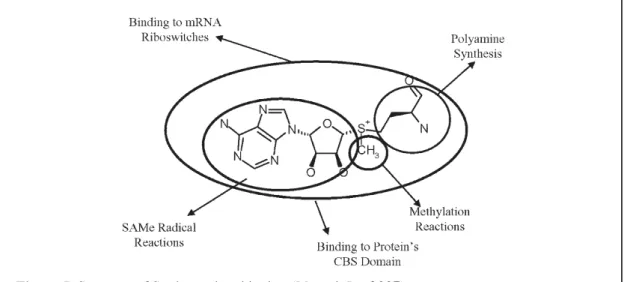
SAM is the major methyl donor for most methyltransferases and is involved in numerous cellular reactions, including methylation of DNA, histone, proteins, synthesis of polyamines, binding to mRNA, methylation and synthesis of lipids, and antioxidants reactions (such as glutathione and taurine) [Fig. 5] (Mato & Lu, 2007). Thus, SAM is determinant for a wide range of biological processes, from membrane fluidity, gene expression, cell growth, differentiation, to suppression of cell division and apoptosis.
Chapter I – Essential biochemistry of one-carbon metabolism
reverse situation can happen if adenosine and Hcy are being rapidly eliminated, which is crucial to avoid accumulation of SAH (Lu & Mato, 2012).
Figure 5. Structure of S-adenosylmethionine (Mato & Lu, 2007).
CBS – Cystathionine- -Synthase
The effectiveness of the cycle depends on the replenishment of methyl groups in order to keep a proper concentration of SAM. Therefore, the remethylation of Hcy back to methionine and thus to SAM is essential for the cell survival (Joint, 1998).
1.4. HOMOCYSTEINE METABOLISM AND TRANSSULFURATION PATHWAY
Hcy is a thiol-containing non-proteinogenic amino acid exclusively originated from the demethylation of methionine. In most mammalian cells, Hcy can suffer different metabolic fates: 1) remethylation, which is the conversion to methionine by methionine synthase or BHMT, as described before in last section; 2) catabolism via transsulfuration pathway, where it is irreversibly converted to cysteine [Fig. 6]; 3) release into the extracellular medium (Brustolin, Giugliani, & Felix, 2010). However, when the remethylation pathway is saturated, or when cysteine is required, Hcy follows the transsulfuration pathway (Blom & Smulders, 2011).
can be used for the generation of redox-controlling molecules, such as glutathione and taurine (Zhang et al., 2013).
Figure 6. Transsulfuration pathway for homocysteine degradation and cysteine synthesis (Lu &
Mato, 2012). CBS – cystathionine- -synthase; CSE - cystathionine- -lyase.
In the transsulfuration pathway, Hcy is condensed with serine in a -replacement irreversible reaction of the hydroxyl group of serine by cystathionine- -synthase (CBS) forming cystathionine, with release of a water molecule. CBS is a heme-containing enzyme that is subject to regulatory control, as it is the first and rate-limiting enzyme for the transsulfuration pathway (McBean, 2012). On the second step of the transsulfuration pathway, the resulting cystathionine, from CBS reaction, is then hydrolysed to cysteine, alpha-ketobutyrate (from the Hcy carbon chain), and ammonia (from the amino group of Hcy) by cystathionine- -lyase (CSE) [Fig. 6]. Both enzymes, CBS and CSE are dependent on PLP, the active form of vitamin B6. Alpha-ketobutyrate
can suffer oxidative decarboxylation catalysed by pyruvate dehydogenase complex producing propionylcoenzyme A that can enter the tricarboxylic acid cycle. Cysteine can be used for the glutathione biosynthesis, and originate metabolites in mammals such as taurine, pyruvate, and hydrogen sulfide (Stipanuk & Ueki, 2011).
Chapter II –MTHFR 677C→T polymorphism: general aspects
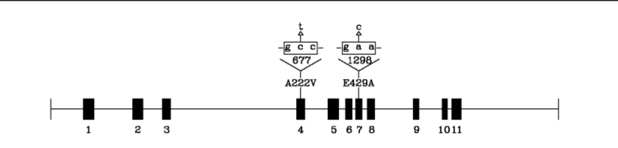
Figure 7. Schematic representation of the MTHFR gene, located on chromosome 1p36.3. Exons are
shown in black boxes. The two most investigated polymorphisms, 677C→T and 1β98A→C,
corresponding to the amino acid substitution A222V and E429A, respectively, are indicated above their corresponding exons (Trabetti, 2008).
C
HAPTERII
–
MTHFR
677C→T
POLYMORPHISM:
GENERAL ASPECTS2.1. CYTOGENETIC LOCATION
MTHFR gene is located on the short (p) arm of chromosome 1 at position 36.3. (Trabetti, 2008). The majority of polymorphisms reported for MTHFR gene have a minor impact on enzymatic activity, with the exception of 677C→T and 1298A→C. MTHFR 677C→T and 1298A→C polymorphisms seem to be in linkage disequilibrium, with findings pointing to the appearance of 677 variant later than the 1298C variant on a chromosome harbouring 1298A. However, only MTHFR 677C→T polymorphism has been established to lower considerably serum folate and higher plasma homocysteine (Ulvik et al., 2007).
MTHFR 677C→T polymorphism is transmitted in an autosomal recessive way and is characterized by a point mutation at base pair 677 (exon 4) that converts a cytosine (C) into a thymine (T) leading to an amino acid substitution (alanine to valine) at codon 222 in the MTHFR gene [Fig. 7] (Trabetti, 2008).
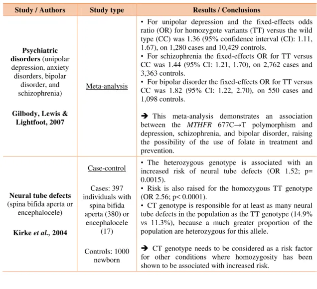
Many conditions have been associated with MTHFR 677C→T polymorphism, such as birth defects, psychiatric disorders [Table 1], vascular disease and cancer (both discussed in the following chapters); supporting the idea, that folate supplementation could have a role in the treatment and prevention of those diseases (Gilbody Lewis & Lightfoot, 2007). The relationship between the MTHFR 677C→T polymorphism and disease involves several mechanisms, such as plasma Hcy (tHcy), DNA methylation and DNA synthesis (Ueland et al., 2001).
Table 1. Results from studies relating diseases and MTHFR677C→T polymorphism.
Study / Authors Study type Results / Conclusions
Psychiatric disorders (unipolar depression, anxiety disorders, bipolar
disorder, and schizophrenia)
Gilbody, Lewis & Lightfoot, 2007
Meta-analysis
• For unipolar depression and the fixed-effects odds ratio (OR) for homozygote variants (TT) versus the wild type (CC) was 1.36 (95% confidence interval (CI): 1.11, 1.67), on 1,280 cases and 10,429 controls.
• For schizophrenia the fixed-effects OR for TT versus CC was 1.44 (95% CI: 1.21, 1.70), on 2,762 cases and 3,363 controls.
• For bipolar disorder the fixed-effects OR for TT versus CC was 1.82 (95% CI: 1.22, 2.70), on 550 cases and 1,098 controls.
This meta-analysis demonstrates an association between the MTHFR 677C→T polymorphism and
depression, schizophrenia, and bipolar disorder, raising the possibility of the use of folate in treatment and prevention.
Neural tube defects
(spina bifida aperta or encephalocele)
Kirke et al., 2004
Case-control
Cases: 397 individuals with
spina bifida aperta (380) or
encephalocele (17)
Controls: 1000 newborn
• The heterozygous genotype is associated with an increased risk of neural tube defects (OR 1.52; p= 0.0015).
• Risk is also raised for the homozygous TT genotype (OR 2.56; p< 0.0001).
• CT genotype is responsible for at least as many neural tube defects in the population as the TT genotype (14.9%
s 11.γ%), because a much greater proportion of the population are heterozygous for this allele.
CT genotype needs to be considered as a risk factor for other conditions where homozygosity has been shown to be associated with increased risk.
2.2. POPULATION GENETICS
Chapter II –MTHFR 677C→T polymorphism: general aspects
Northern Netherlands, and Russia), to intermediate values (8-10%) in France and Hungary, to higher values in Southern Europe (12-15% in Spain and Northern Italy), peaking in southern Italy (20-26% in Campania and Sicily). In North America, the frequency of TT homozygotes increase from Western Canada to Southeastern United States, reaching a peak in Mexico [Fig. 8] (Wilcken et al., 2003).
Figure 8. Prevalence of homozygous TT genotype among newborns by area and ethnic background
(Wilcken et al., 2003).
The prevalence of MTHFR 677C→T polymorphism varies considerably in China through a geographic gradient. For MTHFR 677C→T, the frequencies of the T allele and the TT genotype are notably higher in the north (63.1% and 40.8%, respectively) compared to the south (24.0% and 6.4%, respectively) (Yang et al., 2013).
frequency of the T allele was registered. Additionally, in West Africa, the genotype influence on tHcy was the most evident; the majority of individuals with the T allele had elevated tHcy. These data indicate a possible interaction between the environment and genetic pressure and suggest that TT genotype may confer a survival advantage in populations with adequate dietary folate consumption (Guéant-Rodriguez et al, 2006).
In fact, other studies indicate that heterozygous or homozygous mutant genotypes with adequate folic acid consumption may have, in certain circumstances, a selective advantage over wild-type genotype, which may explain the current observed variability in its frequency in different populations. Further facts that support this hypothesis are the beneficial effect to heterozygotes during times of starvation and the decreased risk of 677C→T homozygotes for colon cancer (Schneider, Rees, Liu & Clegg, 1998), which is going be examined in the last chapter.
2.3.RELATIONSHIP WITH PLASMA HOMOCYSTEINE
Hcy concentrations are regulated by many factors such as the cofactors cobalamin, PLP, folate and enzymes implicated in methionine metabolism. When discrepancies between Hcy production and catabolism take place, tHcy can increase. Impairments in Hcy metabolism can derive from various nutritional and/or congenital disorders, such as MTHFR 677C→T polymorphism (Medina, Urdiales, & Amores‐Sánchez, 2001).
Most of the tHcy is found in the oxidised form. Only 1–2% are present in its reduced form. Approximately 20% occurs as acid-soluble free Hcy in a form of homocysteine-cysteine mixed disulfide and homocystine (a dimer of Hcy). Approximately 70% to 80% circulate bounded to plasma proteins; the great majority travels linked to the cysteine of albumin, its main carrier in plasma. However, current data suggest that approximately 10%–30% of protein bound Hcy and cysteine are linked to globulins. The total tHcy is the sum of all protein-bound and free forms containing a thiol group (Amorim et al., 2011).
Chapter II –MTHFR 677C→T polymorphism: general aspects
and severity [Table 2]. HHcy is dependent on genetic, lifestyle, gender, being higher in men than in women and age, increasing from 10.8 µmol/L at age 40-42 up to 12.4 µmol/L between 65-67 years. Moreover, HHcy is observed in approximately 5% of the general population, and it is implicated in several morphological and physiological changes that are thought to increase the risk for many disorders, including vascular and neurodegenerative diseases, autoimmune disorders, birth defects, diabetes, renal disease, osteoporosis, neuropsychiatric disorders, and cancer (Brustolin, Giugliani, & Felix, 2010).
Table 2. Classification of hyperhomocysteinemia (Brustolin, Giugliani & Felix, 2010).
Severe hyperhomocysteinemia
tHcy levels at all times (31 to >100 µmol/L) caused, for example, by deficiencies in CBS, MTHFR, or in enzymes of cobalamin metabolism.
Mild hyperhomocysteinemia
Moderately high tHcy levels (15-30 µmol/L) under fasting conditions; reflects impaired Hcy methylation (folate, cobalamin or moderate enzyme defects, e.g., thermolabile MTHFR)
Post-methionine load
Abnormal increase in tHcy (>15 µmol/L) after a methionine load (100 mg/kg); reflects impaired Hcy transsulfuration (heterozygous CBS defects, PLP deficiency)
Apart from that, some findings reveal that elevated tHcy is associated with administration of certain medications, including antiepileptic drugs, methotrexate, antihypertensive, lipid-lowering and antidiabetic drugs (Malinow, Bostom & Krauss, 1999).
Ham et al (2014) conducted a study with 2,912 participants to assess the link between medication use, tHcy levels, and the potential mediation by serum vitamin B12
and folate [Table 3]. The only cases with higher mean Hcy levels were observed in users vs. non-users for diuretics, high-ceiling sulphonamide diuretics, medication acting via the renin-angiotensin system and metformin. Non-selective -blocker use was associated with lower mean Hcy levels and only this association was mediated by an underlying association with vitamin B12 and folate levels. Nonetheless, the associations
Table 3. Associations between medication use and homocysteine levels in an older population, and potential mediation by vitamin B12 and folate (Ham et al., 2014).
Medication group Results and possible mechanisms
•Diuretics in general
•High-ceiling sulphonamide diuretics
•Agents acting via the renin-angiotensin system
• Small but significant positive association with tHcy.
• Largely independent of vitamin B12 and folate levels.
• Diuretic use has previously been shown to be associated with higher tHcy, possibly through decreasing folate levels.
• High-ceiling sulphonamides are known as potent inhibitors of the reabsorption of electrolytes in the kidneys, resulting in reduced water reabsorption into the blood and thus increased water excretion. This alteration in fluid status, or mild dehydration, might cause the observed relative increase of the tHcy.
•Non-selective β-blocker
• Inverse significant association with tHcy was observed.
• Characterised by an underlying association with vitamin B12
and folate levels.
• How non-selective b-blockers may affect vitamin B12 and folate
levels is still unknown.
•Thiazides
•Selective β-blockers
•Statins
•Sulphonylurea derivatives
•No association was observed with tHcy, especially for metoprolol.
•Metformin
• Higher tHcy.
• Relationship between metformin and tHcy was independent of vitamin B12 and which became slightly stronger after including
folate levels.
•Metformin increases insulin sensitivity, and potentially insulin levels are involved. Nevertheless, the literature reports contradictory findings, as both insulin sensitivity as well as insulin resistance were associated with higher tHcy.
•Proton pump inhibitor
•Histamine H2 receptor antagonist
•Methotrexate
•Acetylsalicylic acid
•Theophylline
•L-dopa
• Lack of association with tHcy.
•Anticonvulsants
• Lack of association with tHcy.
• The association has been consistently reported in the literature.
• Anticonvulsant drugs may reduce folate levels and particularly in combination with the MTHFR TT genotype, this may result in
elevated tHcy.
• The discrepancy between these findings and other studies may be explained by the non-significant interaction with the MTHFR
polymorphism and the low frequency of folate deficiency in the population studied.
•Combination of medication groups significantly associated with higher homocysteine level
• No additive effect of the association was observed.
• There is no literature regarding a potential additive effect of a combination of medication use on tHcy.
Chapter II –MTHFR 677C→T polymorphism: general aspects
2.3.1.MOLECULAR MECHANISMS OF HOMOCYSTEINE
Endothelial cells participate in the regulation and preservation of vascular system and are extremely susceptible even to a mild increase in Hcy concentration. This sensitivity may be justified by the fact, that human endothelial cells do not express active form of CBS, and consequently, cannot initiate Hcy catabolism through transsulfuration pathway [Fig. 9]. Thus, a number of molecular mechanisms of Hcy have been suggested, and the underlying common pathogenic mechanisms are predominantly associated with vascular injury [Fig. 10] (Perła-Kaján, Twardowski & Jakubowski, 2007).
Figure 9. Simplified homocysteine metabolism (adapted from:Esse et al., 2012). AdoMet –
S-adenosylmethionine; AdoHcy – S-adenosyl-L-homocysteine; MAT – S-adenosylmethionine synthetase; Met – methionine; Cys – cysteine; Hcy – homocysteine.
Additionally, Hcy may affect glutathione peroxidase activity, changing the dissemination of reactive oxygen species. Endothelial glutathione peroxidase catalyses the reduction of hydrogen and lipid peroxides to the equivalent alcohol, avoiding the oxidative inactivation of NO. The mechanism by which Hcy may act is thought to involve the downregulation of glutathione peroxidase expression (Yilmaz, 2012).
Figure 10. Molecular mechanisms of homocysteine (adapted from: Brattström & Wilcken, 2000; Heneghan & Sultan, 2008; Medina, Urdiales & Amores‐Sánchez, 2001; Perna et al., 2003;
Stühlinger et al., 2001; Wang, Siow, & Karmin, 2001; Yilmaz, 2012; Zhang et al., 2012).
Hcy is also known as thrombogenic agent, boosting platelet aggregation, acting on the coagulation cascade, directly inducing a synergistic pathway with other risk factors. It activates coagulation factor V, X, and XII, inhibits protein C and cell-surface thrombomodulin, and modulates tissue plasminogen activator, through annexin II (an endothelial receptor) (Perna et al., 2003).
HHcy can have a direct impact on DNA methylation, leading to modifications in the gene expression, affecting both endothelial cells and smooth muscle cells. An increase in tHcy generates a growth in the intracellular concentration of SAH that can bind to methyltransferases with higher affinity than SAM. However, SAH is a potent inhibitor of DNA methyltransferases, affecting cell homeostasis (Castro et al., 2003). Several studies suggest that hypomethylation processes, due to of HHcy, induce proliferation of vascular smooth muscle cells, resulting in a decrease of the vessel calibre (Zhang et al.,
Hyperhomocysteinemia
Mechanisms
•Nitric oxide suppression
•Downregulation of glutathione peroxidase
•Protein thiolation
•Protein homocysteinylation
•Promotion of coagulation
•Oxidation of LDL
•Hypomethylation processes
•Promotion of inflammation and atherosclerosis
Vascular lesion
Chapter II –MTHFR 677C→T polymorphism: general aspects
Another molecular pathogenic mechanism of Hcy is believed to involve the formation of adducts with disulfide linkages with sulphydryl residues of proteins, in a reaction known as thiolation [Fig. 11]. Moreover, the detrimental effect of Hcy in proteins may be caused by the formation of Hcy thiolactone, leading to protein homocysteinylation, when transsulfuration or remethylation pathways are compromised. Both occurrences can lead to an irreversible damage of the enzymatic activity and denaturation of proteins, such as haemoglobin, low-density lipoproteins (LDL) and plasma proteins (Medina, Urdiales & Amores‐Sánchez, 2001).
Figure 11. Generation of reactive homocysteine thiolactone (adapted from: Medina, Urdiales &
Amores‐Sánchez, 2001).
As a result of the vascular injury effect, Hcy has been frequently referred as “the
cholesterol of the 21st century”. HHcy can produce vascular injury by increasing the oxidation of LDL within vascular cells and tissues. LDL aggregates of Hcy in the bloodstream can be taken up by macrophages, originating foam cells, the precursors of atherosclerotic plaques. Within these plaques, Hcy modifies the oxidative and synthetic processes of artery wall cells, propagating plaque formation (Heneghan & Sultan, 2008).
However, the origin HHcy is yet to be understood. Brattström and Wilcken (2000) suggest that, in reality, HHcy is more an epiphenomenon of vascular disease, having a minor role in the vascular damage. They hypothesised that the decline in renal function can be the actual cause of HHcy in vascular disease patients. Risk factors for vascular disease, such as atherogenesis and hypertension, frequently progress in a discrete way over the time before the appearance of clinically evident vascular episodes. Therefore, atherogenesis and hypertension can conduct to renal impairment, affecting the clearance of Hcy. As matter of fact, the existence of vascular illnesses would promote the raise of Hcy in plasma through the impairment of renal function.
Chapter III –MTHFR 677C→T polymorphism and cardiovascular risk
CHAPTER III –MTHFR 677C→T POLYMORPHISM AND CARDIOVASCULAR RISK
3.1.MTHFR677C→T POLYMORPHISM, HOMOCYSTEINE AND CARDIOVASCULAR RISK
Cardiovascular disease (CVD) or heart disease is a group of illnesses that involves the heart and/or the blood vessels, leading mostly to cardiac disease, vascular diseases of the brain and kidney, and peripheral arterial disease (Kelly, 2010). The concerns about CVD are reasonable due to its considerable high mortality rate, being the primarily worldwide cause of death. Additionally, the overall CVD is estimated to cost
the EU economy almost €196 billion a year (Nichols et al., 2012). The causes underlying CVD are diverse and multifactorial, but atherosclerosis and hypertension are the most common causes, defined by genetic and environmental aspects, such as gene-gene and gene-gene-environment interactions (Sabetisoofyani, Larson & Watson, 2010).
As discussed in the last chapter, HHcy is cogitated by many researchers to grant a mild vascular risk alone, and to increase the risk of vascular lesions in association with other factors. Therefore, the attention given to cardiovascular risk and mild increase of circulating Hcy has been motivating investigators to establish a connection between MTHFR 677C→T polymorphism and CVD risk (Clarke et al., 2012; Xuan et al., 2011).
The metabolic phenotype of low concentrations of folate and tHcy elevated over 25% in individuals with the TT genotype compared with those with the CC genotype has been vastly assessed in Mendelian randomisation studies (Brattström, Wilcken, Öhrvik & Brudin, 1998). If a gene variant is responsible for higher tHcy levels, random allocation of this gene during conception produces a randomised trial of low versus high Hcy from birth on [Fig. 12]. Many studies revealed increased cardiovascular risk in individuals with the TT genotype compared with those carrying the wild-type (Smulders & Blom, 2011).
recommendations concerning nutrient consumption, and avoidance of other cardiovascular risk factors.
Figure 12. Principle of Mendelian randomisation as applied to hyperhomocysteinemia.
Essential for the validity of the model is the absence of arrows between the gene and confounders, as well as the absence of an arrow (a direct association not dependent on the risk factor) between the gene and the disease (Smulders & Blom, 2011).
Several MTHFR 677C→T polymorphism studies, including retrospective and prospective studies, suggest that the association between the polymorphism and CVD is causal, stating HHcy as the underlying mechanism (Wald, Law & Morris, 2002). However, the debate about this topic has intensified in the beginning of the 21st century
with several studies showing discordant results. Many studies advocate that mild HHcy associated with MTHFR 677C→T polymorphism is not high enough to be an independent risk factor for the development of CVD. Thus, although the association between tHcy concentrations and CVD has been already demonstrated, the causality is not yet established (Brattström, Wilcken, Öhrvik & Brudin, 1998).
3.2. CORONARY HEART DISEASE
Chapter III –MTHFR 677C→T polymorphism and cardiovascular risk
However, results of CVD studies do not seem to be concordant for the overall population. In 2011, another meta-analysis on published studies, studied the association between the polymorphism and risk of myocardial infarction (MI), and found association only for Caucasians (Xuan et al., 2011).
Even though collected evidences have shown an association between HHcy and CHD, there is a strong divergence in results, with studies supporting the idea that HHcy might be just a consequence of CHD. Additionally, the divergence is magnified when the causality between MTHFR 677C→T polymorphism and risk of CHD is hypothesised. An updated meta-analysis of unpublished studies on MTHFR 677C→T
polymorphism and CHD performed in 2012 found that the additional CHD risk in TT compared to CC genotype carriers was only 2%, a negligible increase, certainly coincidental. The stratification of the study populations by folate status showed, as well, no evidence that T allele had an extra CHD risk compared to the wild-type [Table 4] (Clarke et al., 2012).
Table 4. Results from studies relating coronary heart disease and MTHFR677C→T polymorphism.
Study / Authors Study type Sample Results
Coronary heart disease
Klerk et al., 2002
Meta-analysis of 40 published and unpublished studies 11,162 cases 12,758 controls
Individuals with the TT genotype had a 16% (OR, 1.16; 95% CI, 1.05-1.28) higher odds of CHD than those with the CC genotype.
Significant heterogeneity between the results obtained in European populations (OR, 1.14; 95% CI, 1.01-1.28) compared with North American populations (OR, 0.87; 95% CI, 0.73-1.05).
Myocardial Infarction
Xuan et al., 2011
Meta-analysis of 30 published studies 8,140 cases 10,522 controls
Significant association was found between
MTHFR677C→T polymorphism and risk of MI
(OR, 1.183; 95% CI, 1.076–1.300).
The same association was found in overall Caucasians (OR, 1.139; 95% CI: 1.007–1.288) and young/middle-aged (<50 years) Caucasians (OR, 1.275; 95% CI, 1.077–1.509).
No associations were detected between MTHFR
677C→T polymorphism and the risk of MI in elderly male or female Caucasians, East Asians, South Asians, and African-Americans.
Coronary heart disease
Clarke et al., 2012
Meta-analysis of 19 unpublished studies 48,175 cases 67,961 controls
Figure 13. MTHFR 677C→T
frequencies of CC, CT and TT genotypes. Frequencies were 38.5, 35.7 and 25.5% in the VTE group and 41.4, 43.2 and 15.5% in the controls, respectively. In the two groups, the level of Hcy in subjects with the TT genotype or CT genotype was higher than that in subjects with the CC genotype. The plasma level of Hcy in the VTE group
(1γ.05±β.γ7 mol/l) was significantly
higher than that in the control group
3.3. VENOUS THROMBOSIS
The specific mechanism of susceptibility for venous thrombosis remains unclear. Interest in the genetic basis of venous thrombosis was enhanced by discover of genes with a possible effect in thrombophilia, such Leiden V factor, prothrombin G20210A and MTHFR 677C→T polymorphism (Yin et al., 2012).
Due to HHcy association with venous thrombosis, MTHFR 677C→T polymorphism has been proposed as a genetic risk factor candidate for venous thrombosis. Consequently, several studies have suggested that the effect of MTHFR 677C→T polymorphism on venous thrombosis is only perceptible in particular minor groups with other predisposing genetic or environmental factors or, in contrast, in minor groups in which conventional risk factors for venous thrombosis are missing. Based on this premise, one could suggest that in clinical practice, measurement of Hcy levels (and the assessment of folate levels) would be reasonable for individuals with unsolved idiopathic, persistent venous thrombosis, or venous thrombosis occurring at early age or at an uncommon site (Den Heijer, Lewington & Clarke, 2005).
In 2012, a study examined the possible relation between MTHFR 677C→T
Chapter III –MTHFR 677C→T polymorphism and cardiovascular risk
Joachim and colleagues (2013) conducted a study to assess the prevalence of HHcy and MTHFR variant in a pediatric population with VTE, and the association with thrombus outcome. Subjects were enlisted in an institution-based prospective cohort of children with VTE. The prevalence of HHcy or MTHFR variant was not amplified in comparison with the control group. Plasma Hcy did not change between those with CC genotype versus MTHFR 677C→T heterozygotes or homozygotes, and adverse thrombus outcomes were not associated either.
In concordance with the previous study, a meta-analysis evaluating risk factors for venous thrombosis showed the same lack of association. The study used the Multiple Environmental and Genetic Assessment including subjects with first venous thrombotic event, deep vein thrombosis of either the leg or pulmonary embolism [Table 5]. In this study, MTHFR 677C→T polymorphism was not associated with the risk of venous thrombosis. Stratification by risk factors did not provide evidence of an association in specific groups. Since genotype distributions did not differ between case and control groups, there was no excess risk of venous thrombosis associated with MTHFR
677C→T polymorphism. Consequently, based on this study, routine testing of MTHFR
677C→T genotype as part of a thrombophilia evaluation with venous thrombosis would not be justified (Bezemer et al., 2007).
Table 5. Results from studies relating venous thrombosis and MTHFR677C→T polymorphism.
Authors Study type Sample Results
Yin et al., 2012 Population -based case-control 440 cases 440 controls
The plasma levels of Hcy in the VTE group (13.05±2.37 µmol/l) were significantly higher compared with those in the control group (11.94±2.03 µmol/l, P<0.001).
Compared with the CC genotype, the TT genotype was significantly correlated with an increased risk of VTE (OR, 1.753; 95% CI, 1.215–2.529; P=0.003).
In the VTE group (F=106.051; P<0.001), the level of Hcy in subjects with the CT+TT genotype (14.49±2.51
mol/l) was significantly higher than that in subjects with the CC genotype (11.γ5±1.57 mol/l, P<0.001).
Bezemerl et al., 2007 Population -based case-control 4375 cases 4856 controls
The TT genotype was present in 440 patients (10%) and 517 control subjects (11%), and the CT genotype in 1891 patients (43%) and in 2094 control subjects (43%).
Stratification by known risk factors for venous
thrombosis, such as V Leiden, prothrombin β0β10G→A,
3.4. ISCHEMIC AND HAEMORRHAGIC STROKE
The MTHFR 677C→T polymorphism has been associated, as well, to ischemic and haemorrhagic stroke in many studies but not in others [Table 6]. In 2010, a study evaluated the MTHFR 677C→T polymorphism in ischemic and haemorrhagic stroke
patients in a Northern Indian population. Despite the elevated tHcy found in TT genotype ischemic stroke patients, MTHFR 677C→T gene polymorphism was
associated with neither haemorrhagic nor ischemic stroke (Somarajan, Kalita, Mittal & Misra, 2011). However, recent meta-analyses suggest that the T allele of the MTHFR
677C→T polymorphism is associated with increased risk of haemorrhagic stroke (HS) and ischemic stroke (IS) [Table 6]. These results, if confirmed, are significant for the genetic epidemiology, diagnosis, treatment and prevention of HS and ARE (Kang et al., 2013; Li & Qin, 2014).
Table 6. Results from studies relating stroke and MTHFR677C→T polymorphism.
Study /
Authors Study type Sample Results
Ischemic stroke and Intracerebral haemorrhage
(ICH)
Somarajan et al., 2007
Case–control
Cases: 207 IS 215 ICH
188 controls
The frequency of the CC genotype in controls was 68.6%, CT in 28.7% and TT in 2.7%. It was 75.3%, 20.5% and 4.2% in ICH and 66.2%, 39.4% and 2.4% respectively in IS.
The frequency of these genotypes as well as allele frequency was not different in IS, ICH as compared to controls
Allele was more frequent in IS compared to ICH. Hcy level was higher in IS patients with variant genotype.
Haemorrhagic stroke
Kang et al., 2013
Meta-analysis of 15 case-control studies
2034 cases
4485 controls
Significant associations between the MTHFR
677C→T polymorphism and the risk of HS were observed in dominant (OR, 1.611, 95% CI, 1.336–
1.942), codominant (OR, 1.500, 95% CI, 1.330–
1.692), and recessive (OR, 1.695, 95% CI, 1.409–
2.038) models.
Ischemic stroke
Li & Qin, 2014
Meta-analysis of 19 case-control studies
2223 cases
2936 controls
Statistically significant association with IS was identified for allele T (OR = 1.28; 95% CI, 1.17–
1.40, P < 0.00001).
Marginally significant association was detected with genotype CT (OR = 1.13; 95% CI: 1.01–127; P = 0.04) and genotype (OR = 1.43; 95% CI: 1.20–
Chapter III –MTHFR 677C→T polymorphism and cardiovascular risk
A possible mechanism linking MTHFR 677C→T polymorphism and stroke events would be through elevated tHcy inducing endothelial dysfunction, mostly through coagulative effect in IS and rupture of microaneurysms in HS (Li et al., 2003). It was also postulated that elevated tHcy could cause, initially, IS and later, HS (Sazci et al., 2006).
3.5. CAN LOWERING HOMOCYSTEINE LEVELS REDUCE CARDIOVASCULAR RISK?
An approach that could disclose the uncertainty around TT genotype, HHcy and CVD triangle would be to carry out several trials that evaluate the decrease of cardiovascular risk or the protective effect in TT genotype when tHcy level is reduced by supplementation of folate. If folate supplementation were demonstrated to reduce cardiovascular risk, this would be of considerable importance for public health and clinical practice. However, the majority of follow-up studies on the assessment of therapeutic intervention with folic acid-based vitamin supplements has indicating no beneficial effect in CVD prevention (Schwammenthal & Tanne, 2004).
A daily dose of folic acid ranging 0.5 and 5 mg was associated with a 25% decrease in tHcy levels addressed in a meta-analysis of the short-term trials. This decrease in tHcy, accomplished with folic acid supplements, is similar to the variation in tHcy levels between TT and CC genotypes. Despite the positive results in some observational studies, a great part of interventional trials firmly contests any satisfactory result of lowering Hcy concentrations with folic acid treatment on CVD (Den Heijer, Lewington & Clarke, 2005).
Table 7. Results from studies relating folate supplementation and MTHFR677C→T polymorphism.
Authors Study type Sample Results
Clarke et al., 2010
Meta-analysis of 8 large, randomised, placebo-controlled trials 37 485 individuals at increased risk of cardiovascular disease
Folate supplementation led to an average of 25% reduction in Hcy levels
During a median follow-up of 5 years, folate supplementation had no significant effects on vascular outcomes, with an OR of 1.01 (95% CI, 0.97-1.05) for major vascular events; OR, 1.03 (95% CI, 0.97-1.10) for major coronary events, and 0.96 (95% CI, 0.87-1.06) for stroke.
Clarke et al., 2012
Meta-analysis of 10 large-scale placebo-controlled trials 50,378 individuals
Little or no effect on the 5 years incidence of CHD (OR, folate vs placebo,
1.02; 95% CI, 0.96–1.08).
3.6. POSSIBLE EXPLANATIONS FOR DISCREPANCIES AMONG STUDIES
Sample dimension, linkage disequilibrium between polymorphisms, nutrition status, setting, and study design could be responsible for the discrepancy [Fig. 14]. Population specificity might play a role, because different groups of patients may have various sets of genetic factors that predispose to the disease, stressing the importance of ethnicity and genetic background (Yin et al., 2012). Specific alleles of candidate genes can be deeply correlated with the disease in one population, while in another this connection can be low because of other genetic factors or particular interactions between genetic and non-genetic factors (Trabetti, 2008).