Duration-controlled swimming
exercise training induces cardiac
hypertrophy in mice
1Instituto do Coração, Faculdade de Medicina, and
2Escola de Educação Física e Esportes, Universidade de São Paulo,
São Paulo, SP, Brasil F.S. Evangelista1,
P.C. Brum2 and J.E. Krieger1
Abstract
Exercise training associated with robust conditioning can be useful for the study of molecular mechanisms underlying exercise-induced car-diac hypertrophy. A swimming apparatus is described to control training regimens in terms of duration, load, and frequency of exer-cise. Mice were submitted to 60- vs 90-min session/day, once vs twice a day, with 2 or 4% of the weight of the mouse or no workload attached to the tail, for 4 vs 6 weeks of exercise training. Blood pressure was unchanged in all groups while resting heart rate decreased in the trained groups (8-18%). Skeletal muscle citrate synthase activity, measured spectrophotometrically, increased (45-58%) only as a result of duration and frequency-controlled exercise training, indicating that endurance conditioning was obtained. In groups which received dura-tion and endurance condidura-tioning, cardiac weight (14-25%) and myo-cyte dimension (13-20%) increased. The best conditioning protocol to promote physiological hypertrophy, our primary goal in the present study, was 90 min, twice a day, 5 days a week for 4 weeks with no overload attached to the body. Thus, duration- and frequency-con-trolled exercise training in mice induces a significant conditioning response qualitatively similar to that observed in humans.
Correspondence
J.E. Krieger
Laboratório de Genética e Cardiologia Molecular, InCor Av. Dr. Enéas C. Aguiar, 44 Bloco B, 10º andar 05403-000 São Paulo, SP Brasil
Fax: +55-11-3069-5022 E-mail: [email protected] Research supported by FAPESP (No. 01/00009-0) and CNPq (No. 471219/01-0). F.S. Evangelista is the recipient of a doctoral fellowship from FAPESP (No. 99/01881-1).
Received February 21, 2003 Accepted October 21, 2003
Key words
•Exercise training •Swimming
•Cardiac hypertrophy •Mice
•Myocardium
Introduction
In response to a variety of mechanical, hemodynamic, hormonal and pathologic stimuli, the myocardium adapts to increased workloads through the hypertrophy of indi-vidual muscle cells (1). Cardiac hypertrophy can occur as an adaptive response to a physi-ological (exercise training) or pathphysi-ological (valvular disease, hypertension, or obesity) increase in cardiac work (2,3). Different forms of cardiac hypertrophy arise as a result of a combination of genetic, physiologic, and environmental factors. The molecular
di-lated (7) cardiomyopathies, but the lack of standardized and reproducible exercise train-ing protocols has limited the understandtrain-ing of this complex biological process.
Exercise training leads to improvement of cardiovascular capacity which is associated with lower resting and submaximal heart rates, increased ventricular weights and volume, and myocyte hypertrophy (8,9). The adaptation to exercise training is dependent on factors such as training load, duration and frequency. Swim-ming is recognized for its efficiency in induc-ing myocardial hypertrophy and a significant increase in left ventricular end-diastolic vol-ume in rats (10,11). In the present study, we developed a robust and reproducible exercise training protocol for the development of car-diac hypertrophy in mice. A swimming train-ing apparatus was built and used to test differ-ent swimming programs regarding duration, frequency and load of physical exercise. Us-ing this approach, we provided evidence that duration and frequency but not load-controlled swimming training regimens lead to signifi-cant endurance conditioning and myocardial hypertrophy in mice.
Material and Methods
Study population
Nine-week-old male C57/BL6 mice (N = 62) were maintained in a light- (12-h light
cycle) and temperature- (22ºC) controlled environment and were fed a pellet rodent diet (Nuvital Nutrientes S/A, Curitiba, PR,
Brazil) ad libitum and had freeaccess to
water. The training sessions were performed during the dark cycle of the mice which in the present study were kept from 7:00 to
19:00 h. Theanimals were randomly
as-signed to 3 sedentary (S1-S3, N = 19) and 7 exercise-trained (T1-T7, N = 43) groups ac-cording to the two protocols described be-low.
All animal experimental procedures fol-lowed Institutional guidelines and were ap-proved by the Ethics Committee of the Uni-versity of São Paulo Medical School.
Training protocols
Group assignment and number of mice used in each protocol are given in Table 1.
Protocol 1. The adaptation to exercise training depends on factors such as training load, duration and frequency. In protocol 1, duration, frequency and load of physical exercise were investigated. Training dura-tion was associated with changes in length of
exercise sessions (60 vs 90 min), while
exer-cise with the addition of different workloads (2 or 4% of body weight) modified the train-ing load. Traintrain-ing frequency was investi-gated by comparing the number of exercise
training sessions per day (once vs twice a
day). The aim was to optimize these param-eters to induce robust cardiovascular effects, such as resting bradycardia and cardiac hy-pertrophy. The mice were randomly assigned to a sedentary (S1, N = 13) and five trained (T1-5, N = 29) groups. The T1, T2 and T3 groups were exercised at the same load but with different duration or frequency of exercise, with T1 and T2 being used as duration groups and T3 as the frequency group. In contrast, T4 and T5 groups (load groups) were trained with the same duration and frequency of exercise but with different loads.
Table 1. Summary of swimming exercise training schedules used in the present study.
Protocol Training N Volume Intensity Frequency Duration group (min) (% bw) (sessions/day) (weeks)
1 T1 6 90 0 2 6
T2 5 60 0 2 6
T3 6 90 0 1 6
T4 5 60 2 2 6
T5 7 60 4 2 6
2 T6 7 90 0 2 4
T7 7 90 0 2 6
The T1 group trained 5 days a week, twice a day with a gradual progression to-ward a 90-min session for 6 weeks. The T2 and T3 groups performed a swimming train-ing protocol similar to that of the T1 group, except that exercise sessions were shorter (a 60-min session, twice a day). The training frequency of the T2 and T3 groups was also reduced (once a day, 90-min session). The T4 and T5 groups performed a swimming protocol similar to that of the T2 group (a 60-min session), except that they were submit-ted to swimming with a 2 or 4% body weight workload, respectively. All mice were weighed once a week and when necessary the workload (2 or 4% of body weight) was adjusted to body weight changes. Sedentary mice were placed in the swimming appara-tus for 5 min twice a week to mimic the water stress associated with the experimental pro-tocol.
Protocol 2. In this protocol the results obtained for group T1 were evaluated with
regard to the duration (4 vs 6 weeks) of
training. Mice were randomly assigned to 2
sedentary (S2 and S3, 4 vs 6 weeks,
respec-tively, N = 6) and 2 exercise-trained (T6 and
T7, 4 vs 6 weeks, respectively, N = 14)
groups.
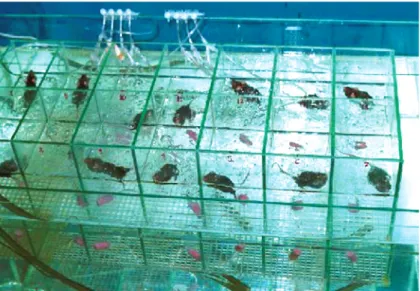
Swimming apparatus
We designed a swimming apparatus es-pecially planned for exercise training of mice. The system consists of two coupled 200-l water glass tanks of different dimensions. The outer tank measures 60 cm in diameter, 100 cm in width and 50 cm in height. The inner tank is divided into 14 lanes with a
surface area of 15 x 15 cmper lane and a
depth of 35 cm to allow individual training. To prevent floating during the swimming session, water bubbling was produced by tubes connected to an air pump system. A heating system kept the water temperature between 30 and 32ºC and a water filter with a flow capacity of 420 l/h was used to clean
the swimming apparatus (Figure 1).
Resting systolic blood pressure and heart rate measurements
Tail-cuff systolic blood pressure and heart rate (HR) were determined during the 4- or 6-week-period of study using a computer-ized tail-cuff system (BP 2000 Visitech Sys-tems, Apex, NC, USA) (12). Blood pressure values were determined for each animal by averaging blood pressure measurements ob-tained on two different days of the same week during the animal’s dark cycle.
Analysis of cardiac structure
Twenty-four hours after the last exercise training session, sedentary and exercise-trained mice were killed and tissues har-vested. The weights of the heart and of the dissected chamber atria, right ventricle and left ventricle were measured. The dissected chambers were then fixed by immersion in 4% buffered formalin and embedded in par-affin for routine histologic processing. Sec-tions (4 µm) were stained with hematoxylin and eosin for examination with a light micro-scope. Myocyte width was measured in the left ventricle free wall with a
assisted morphometric system (Leica Quan-timet 500, Cambridge, UK). For 10 myo-cytes containing a nucleus visible in the field, a single transverse measurement of width passing through the nucleus was de-termined. Myocyte diameter was determined for each animal by averaging the measured myocytes.
Skeletal muscle oxidative enzyme activity
Muscle samples were taken from the left and right soleus at the time of killing and
frozenin liquid nitrogen for later processing.
Citrate synthaseactivity was measured
spec-trophotometrically in whole muscle
homo-genates and the amount of the complex re-sulting from coenzyme A and oxaloacetate was determined (13).
Statistical analysis
Data are reported as means ± SEM. Data for the exercise-trained groups were compared to those for the sedentary groups using one-way ANOVA for repeated measures and
two-way ANOVA followed by the Tukey post
hoc test in Protocols 1 and 2, respectively.
Statistical significance was set at P ≤ 0.05.
Results
Resting systolic blood pressure and heart rate during the exercise-training period
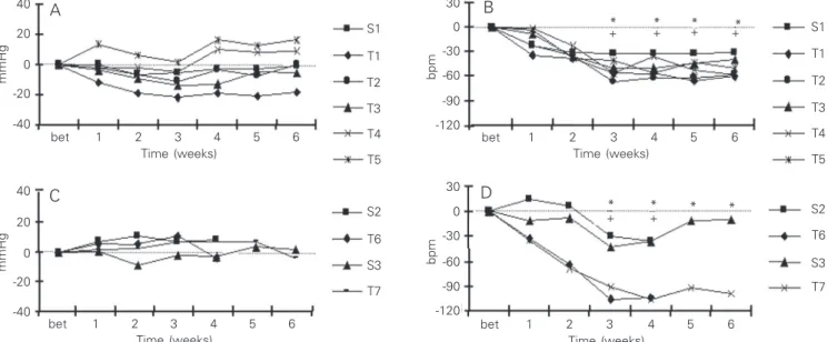
Baseline blood pressure did not differ
among the groups studied in Protocol 1
(Fig-ure 2A). In contrast, after 3 weeks of exer-cise training, HR decreased significantly in all trained groups compared to the pre-exer-cise period (T1, 12.3%; T2, 12%; T3, 8.3%; T4, 10%, and T5, 8%) as well as to sedentary littermates (Figure 2B). In contrast, HR was unchanged in the sedentary group through-out the study. The development of resting bradycardia in the exercising mice indicates that aerobic conditioning was achieved with these training regimens (14).
In Protocol 2, reduction of exercise train-ing from 6 to 4 weeks resulted in no change in blood pressure or HR responses (Figure 2C and D, respectively). Baseline blood pres-sure remained unchanged in all sedentary and exercise-trained groups, while
signifi-S1
T1
T2
T3
T4
T5
mmHg
40
20
0
-20
-40
S2
T6
S3
T7 S1
T1
T2
T3
T4
T5
bpm
30
0
-30
-120 -60
-90
mmHg
40
20
0
-20
-40
bpm
30
0
-30
-120 -60
-90 bet 1 2 3 4 5 6
Time (weeks)
bet 1 2 3 4 5 6 Time (weeks)
S2
T6
S3
T7
bet 1 2 3 4 5 6 Time (weeks)
bet 1 2 3 4 5 6 Time (weeks)
* * * *
* * * *
+ + + +
+ +
A
C
B
D
cant resting bradycardia was achieved at both T6 (18%) and T7 (16.4%) 4 and 6 weeks of exercise training, respectively, compared to sedentary littermates (Figure 2D).
Cardiac structure analysis
The body weights of exercise-trained mice from the T1 to T4 groups were similar to those of sedentary mice (Table 2). In con-trast, mice exercised at a higher load (T5) showed a significantly lower body weight than sedentary mice. In Protocol 2, the body weight of T6 mice did not differ from that of their sedentary littermates, while T7 mice had a significantly lower body weight than S3.
Cardiac hypertrophy was observed only in mice from duration (T1) and load groups (T4 and T5) in Protocol 1 (Table 2). The increased heart weight to body weight ratio was mainly due to an increase in left ven-tricle weight in the T1 and T4 groups. In the load group (T4), the normalized weight of the atria also contributed to the increased heart weight. In contrast, in the group exer-cised at a higher load (T5) the normalized increase in heart weight was associated
mainly with a smaller body weight.
The magnitude of cardiac hypertrophy was not influenced by reduction of exercise training from 6 to 4 weeks. Both the T6 and T7 groups presented increased heart weight to body weight ratios mainly due to an in-creased left ventricle weight to body weight ratio (Table 2).
Changes in myocyte width paralleled the changes observed in the left ventricle to body weight ratios. A significant increase in myocyte width was observed in the exercise-trained groups (T1, T4, T6 and T7) com-pared to sedentary littermates in both proto-cols (Figure 3A,C).
Skeletal muscle oxidative enzyme activity
An increased muscle oxidative activity concomitant with an increase in aerobic work capacity is one of the hallmarks of skeletal muscle adaptation to aerobic conditioning. We measured the maximal activity of citrate synthase (an enzyme involved in the citric acid cycle) in the soleus muscle of all exer-cise-trained and sedentary mice as a marker of muscle oxidative activity.
Interestingly, in Protocol 1, the maximal activity of citrate synthase was significantly
Table 2. Cardiac morphometric analysis following physical training in Protocols 1 and 2.
Protocol Group BW (g) H/BW (mg/g) LV/BW (mg/g) RV/BW (mg/g) AT/BW (mg/g)
1 S1 25.5 ± 0.50 4.02 ± 0.07 3.04 ± 0.06 0.78 ± 0.02 0.19 ± 0.01 T1 23.0 ± 0.66 4.51 ± 0.00* 3.47 ± 0.07* 0.82 ± 0.03 0.23 ± 0.01 T2 23.4 ± 0.12 4.35 ± 0.09 3.32 ± 0.07 0.82 ± 0.03 0.23 ± 0.01 T3 24.7 ± 0.94 4.20 ± 0.20 3.21 ± 0.17 0.78 ± 0.04 0.21 ± 0.01 T4 23.6 ± 0.44 4.62 ± 0.19* 3.48 ± 0.14* 0.87 ± 0.05 0.27 ± 0.01* T5 22.7 ± 0.60* 4.50 ± 0.09* 3.34 ± 0.07 0.89 ± 0.04 0.28 ± 0.01*
2 S2 23.17 ± 0.80 3.94 ± 0.10 2.92 ± 0.03 0.80 ± 0.03 0.22 ± 0.01 S3 25.90 ± 0.85 4.21 ± 0.01 3.09 ± 0.04 0.90 ± 0.07 0.22 ± 0.01 T6 21.34 ± 0.46 4.93 ± 0.04** 3.67 ± 0.04** 0.93 ± 0.01 0.34 ± 0.01** T7 22.20 ± 0.82+ 5.13 ± 0.13+ 3.80 ± 0.10+ 1.01 ± 0.04 0.29 ± 0.02
AT = atria, BW = body weight, H = heart, LV = left ventricle, RV = right ventricle, S = sedentary group, T = training group. Data are reported as mean ± SEM.
higher only in exercise-trained mice from the duration group when compared with sed-entary mice and other trained groups (Figure 3B). In Protocol 2, exercise-trained mice from the T6 and T7 groups showed a signifi-cant increase in citrate synthase activity when compared to their respective sedentary litter-mates (Figure 3D).
Discussion
In the current study we examined exer-cise conditioning, which represents one of the major cardiovascular adaptations to chronic cardiovascular stress, and provide evidence that duration- and frequency-con-trolled but not load-confrequency-con-trolled exercise train-ing regimens induce substantial endurance conditioning and myocardial hypertrophy in mice. These results suggest that the dura-tion- and frequency-controlled training regi-mens can be useful to unravel the role of particular genes and pathways in exercise-induced cardiac hypertrophy in the context of the whole animal.
Exercise training model
Swimming rather than running was cho-sen as a model because of its efficiency in inducing myocardial hypertrophy and greater
left ventricular end-diastolic volume in rats (10,11). Although most treadmill running studies have failed to show cardiac hypertro-phy in rats (15,16), some investigators (17,18) have observed cardiac hypertrophy in mice trained in voluntary running protocols or intensity-controlled treadmill running. How-ever, it is important to emphasize that condi-tioning associated with voluntary running in wheels is difficult to quantify because there is a wide variation in the amount of running among animals. Even though Kemi et al. (17) recently showed that graded running intensity results in major increments of ven-tricular mass than fixed running intensity, their protocol was probably restricted to healthy mice considering the intensity levels achieved during the exercise training period
(85-90% VO2max). Regarding swimming,
most training protocols (3,19-21) have adopted group swimming, which promotes a vigorous response but adds stress as an im-portant confounding variable. One excep-tion is the study by Kaplan et al. (19) show-ing a swimmshow-ing trainshow-ing-induced cardiac hypertrophy limited to female mice.
In the present study, we developed a swimming apparatus for mice with individual lanes and bubbling water to avoid animal floating, which facilitated the control of load, duration and frequency during the
swim-µm
30
µmol mg
-1 ml -1 400
300
200
100
0
µmol mg
-1 ml -1 400
300
200 100
0 25
20
15
10
30
25
20
15
10
S1 T1 T2 T3 T4 T5 S1 T1 T2 T3 T4 T5
µm
4 weeks 6 weeks 4 weeks 6 weeks S
T
S T
* *
* *
* *
*
Groups Groups
Groups Groups
A
C
B
D
Figure 3. Left cardiac myocyte dimensions and skeletal muscle maximal citrate synthase activ-ity following physical training in Protocols 1 (A and B) and 2 (C and D). Data are reported as means ± SEM. *P < 0.05 com-pared to the sedentary groups S1 (A and B; one-way ANOVA test) and S (C and D; two-way ANOVA test).
ming sessions. As previously described (19), minimal diving was observed in mice and their forelegs were relatively inactive during swimming, with almost all of the vigorous muscular activity being performed by the hindlegs.
The current study demonstrated that mice who are close to their limit of functional adaptation (very high peak oxygen uptake, intrinsic HR and cardiac sympathetic tone) develop cardiac hypertrophy and endurance conditioning by swimming.
Blood pressure and heart rate responses
Resting bradycardia is a useful and reli-able indicator of endurance conditioning (22). The exercise-trained groups showed a lower resting HR when compared to the sedentary groups (P < 0.05). However, the magnitude of resting bradycardia was higher in the du-ration-controlled trained groups (12-18%) than in the load- and frequency-controlled trained groups (8-10%). The bradycardic re-sponse occurred two to three weeks after training in all groups. Although the magni-tude of resting bradycardia did not differ significantly among the trained groups, mice from the T6 and T7 groups (duration-con-trolled training regimen, Protocol 2) tended to present a greater magnitude of resting bradycardia than T1 mice (duration-con-trolled training regimen, Protocol 1). This response might be related to the variability of resting HR values in this species. In a recent review, Bernstein (23) pointed out the wide variability in the definition of normal resting HR in mice, with reported values ranging from 212 to 690 bpm (24,25).
There was no difference in systolic blood pressure after all swimming training regi-mens when exercise-trained mice were com-pared with sedentary littermates. These re-sults are consistent with previous observa-tions by our group (26,27) and others (28) showing that arterial pressure remains un-changed in exercise-trained normotensive
animals and humans.
Skeletal muscle oxidative capacity
The adaptation of skeletal muscle oxida-tive capacity induced by exercise training is well established and is also considered to be a good marker for exercise training effi-ciency (29). However, few swimming stud-ies in rodents have documented an increased skeletal muscle oxidative capacity associ-ated with exercise training (30). One of the purposes of the present study was to deter-mine if skeletal muscle oxidative capacity would be affected by different swimming training regimens in mice. Interestingly, we observed a significant elevation in citrate synthase activity only in the duration-con-trolled exercise groups from Protocol 1 (T1, 58%), and Protocol 2 (T6, 45%, and T7, 54%) groups. In contrast, the lower duration (T2), frequency (T3), and load groups (T4 and T5) showed no significant changes in citrate synthase activity. These results sug-gest that duration-controlled exercise train-ing is the most efficient regimen for inductrain-ing skeletal muscle adaptation in mice. In fact, duration of training has been identified as the main factor affecting skeletal muscle oxidative capacity after exercise training in rodents (31). In contrast, the effect of load exercise training on oxidative enzyme activ-ity in rodents remains controversial (32,33).
Ventricular weights and left ventricular cardiac myocyte dimensions
voluntary wheel running (18) and treadmill running (17) have reported smaller cardiac hypertrophies (10 and 12.3%, respectively) compared to the present study (25%). The hypertrophic responses were accompanied by an increase in myocyte width. Interest-ingly, the time course of exercise training (4
vs 6 weeks) did not influence the magnitude
of cardiac hypertrophy (groups T6 vs T7).
Limitations of the present study
Swimming has been suggested to be the most efficient type of exercise for inducing conditioning and cardiac hypertrophy in ani-mals (36) even though it is associated with an important stress response (28). We placed the mice from the sedentary groups in the water for 5 min twice a week in an attempt to minimize the effect of stress when
compar-ing sedentary vs trained groups.
Further-more, although the corticosterone levels were not assessed in these animals, the adrenal gland weight was determined and did not differ among groups (data not shown).
Earlier studies have demonstrated that exercise training by swimming increases the heart weight-to-body weight ratio by 12 to 31% in rats (16) and by 16 to 29% in mice
(19,34). Cardiac hypertrophy is well known to occur in response to various stimuli, such as pressure and volume overload. Exercise training is mainly related to a volume over-load-induced cardiac eccentric hypertrophy with predominant longitudinal myocyte growth (1). Although myocyte length has not been determined, we were able to detect an increase in myocyte width which paralleled the changes observed in left ventricle to body weight ratios. This is consistent with the idea that exercise training-induced ec-centric hypertrophy can lead to proportional myocyte width and length growth (37).
In the present investigation, training regi-mens associating duration, load, and fre-quency of exercise were restricted to male mice. We cannot exclude a gender effect on this response although similar cardiovascu-lar and metabolic responses to physical train-ing have been shown in male and female rats (17).
In the present study we describe a swim-ming apparatus to produce exercise-induced cardiac hypertrophy in mice and provide evidence indicating that duration- and fre-quency-controlled but not load-controlled exercise is associated with physical condi-tioning in mice.
References
1. Lorell BH & Carabello BA (2000). Left ventricular hypertrophy: patho-genesis, detection, and prognosis. Circulation, 102: 470-479. 2. Hunter JJ & Chien KR (1999). Signaling pathways for cardiac
hyper-trophy and failure. New England Journal of Medicine, 341: 1276-1283.
3. Scheuer J, Malhotra A, Hirsch C, Capasso J & Schaible TF (1982). Physiologic cardiac hypertrophy corrects contractile protein abnor-malities associated with pathologic hypertrophy in rats. Journal of Clinical Investigation, 70: 1300-1305.
4. Bianciotti LG & de Bold AJ (2002). Natriuretic peptide gene expres-sion in DOCA-salt hypertenexpres-sion after blockade of type B endothelin receptor. American Journal of Physiology, 282: H1127-H1134. 5. Iemitsu M, Miyauchi T, Maeda S, Sakai S, Kobayashi T, Fujii N,
Miyazaki H, Matsuda M & Yamaguchi I (2001). Physiological and pathological cardiac hypertrophy induce different molecular pheno-types in the rat. American Journal of Physiology, 281: R2029-R2036. 6. Aronow BJ, Toyokawa T, Cannin A, Haghighi K, Delling U, Kranias E, Molkentin JD & Dorn GW (2001). Divergent transcriptional
re-sponses to independent genetic causes of cardiac hypertrophy. Physiological Genomics, 6: 19-28.
7. Hardt SE, Geng YJ, Montagne O et al. (2002). Accelerated cardio-myopathy in mice with overexpression of cardiac G(s)alpha and a missense mutation in the alpha-myosin heavy chain. Circulation, 105: 614-620.
8. Wisloff U, Helgerud J, Kemi OJ & Ellingsen O (2001). Intensity-controlled treadmill running in rats: VO(2 max) and cardiac hypertro-phy. American Journal of Physiology, 280: H1301-H1310. 9. Yamamoto K, Miyachi M, Saitoh T, Yoshioka A & Onodera S (2001).
Effects of endurance training on resting and post-exercise cardiac autonomic control. Medicine and Science in Sports and Exercise, 33: 1496-1502.
10. Geenen D, Buttrick P & Scheuer J (1988). Cardiovascular and hor-monal responses to swimming and running in the rat. Journal of Applied Physiology, 65: 116-123.
program. American Journal of Physiology, 220: 1238-1241. 12. Krege JH, Hodgin JB, Hagaman JR & Smithies O (1995). A
noninva-sive computerized tail-cuff system for measuring blood pressure in mice. Hypertension, 25: 1111-1115.
13. Srere PA (1969). Citrate synthase. In: Lowenstein JM (Editor), Meth-ods in Enzymology. Academic Press, New York, USA.
14. Tipton CM (1965). Training and bradycardia in rats. American Jour-nal of Physiology, 209: 1089-1094.
15. Fitzsimons DP, Bodell PW, Herrick RE & Baldwin KM (1990). Left ventricular functional capacity in the endurance-trained rodent. Jour-nal of Applied Physiology, 69: 305-312.
16. Schaible TF & Scheuer J (1981). Cardiac function in hypertrophied hearts from chronically exercised female rats. Journal of Applied Physiology, 50: 1140-1145.
17. Kemi OJ, Loennechen JP, Wisloff U & Ellingsen O (2002). Intensity-controlled treadmill running in mice: cardiac and skeletal muscle hypertrophy. Journal of Applied Physiology, 93: 1301-1309. 18. Allen DL, Harrison BC, Maass A, Bell ML, Byrnes WC & Leinwand
LA (2001). Cardiac and skeletal muscle adaptations to voluntary wheel running in the mouse. Journal of Applied Physiology, 90: 1900-1908.
19. Kaplan ML, Cheslow Y, Vikstrom K, Malhotra A, Geenen DL, Nakouzi A, Leinwand LA & Buttrick PM (1994). Cardiac adaptations to chronic exercise in mice. American Journal of Physiology, 267: H1167-H1173.
20. Nakao C, Yamada E, Fukaya M, Tayama K, Tsukamoto Y & Sato Y (2001). Effect of acetate on glycogen replenishment in liver and skeletal muscles after exhaustive swimming in rats. Scandinavian Journal of Medicine and Science in Sports, 11: 33-37.
21. Ueno N, Oh-ishi S, Kizaki T, Nishida M & Ohno H (1997). Effects of swimming training on brown-adipose-tissue activity in obese ob/ob mice: GDP binding and UCP m-RNA expression. Research Commu-nications in Molecular Pathology and Pharmacology, 95: 92-104. 22. Lewis S, Thompson P, Areskog NH, Vodak P, Marconyak M, DeBusk
R, Mellen S & Haskell W (1980). Transfer effects of endurance training to exercise with untrained limbs. European Journal of Ap-plied Physiology and Occupational Physiology, 44: 25-34. 23. Bernstein D (2003). Exercise assessment of transgenic models of
human cardiovascular disease. Physiological Genomics, 13: 217-226.
24. Iwase M, Bishop SP, Uechi M et al. (1996). Adverse effects of chronic endogenous sympathetic drive induced by cardiac Gsa overexpression. Circulation Research, 78: 517-524.
25. Gardin JM, Siri FM, Kitsis RN, Edwards JG & Leinwand LA (1995). Echocardiographic assessment of left ventricular mass and systolic
function in mice. Circulation Research, 76: 907-914.
26. Krieger EM, Brum PC & Negrão CE (1998). Role of arterial barore-ceptor function on cardiovascular adjustments to acute and chronic dynamic exercise. Biological Research, 31: 273-279.
27. Negrão CE, Irigoyen MC, Moreira ED, Brum PC, Freire PM & Krieger EM (1993). Effect of exercise training on RSNA, baroreflex control, and blood pressure responsiveness. American Journal of Physiolo-gy, 265: R365-R370.
28. Desai KH, Sato R, Schauble E, Barsh GS, Kobilka BK & Bernstein D (1997). Cardiovascular indexes in the mouse at rest and with exer-cise: new tools to study models of cardiac disease. American Journal of Physiology, 272: H1053-H1061.
29. Wibom R, Hultman E, Johansson M, Matherei K, Constantin-Teodosiu D & Schantz PG (1992). Adaptation of mitochondrial ATP production in human skeletal muscle to endurance training and detraining. Journal of Applied Physiology, 73: 2004-2010. 30. Pereira BCRLF, Safi DA, Medeiros MH, Curi R & Bechara EJ (1994).
Superoxide dismutase, catalase and glutathione peroxidase activi-ties in muscle and lymphoid organs of sedentary and exercise-trained rats. Physiology and Behavior, 56: 1095-1099.
31. Benzi G, Panceri P, de Bernardi M, Villa R, Arcelli E, D’Angelo L, Arrigoni E & Berte F (1975). Mitochondrial enzymatic adaptation of skeletal muscle to endurance training. Journal of Applied Physiolo-gy, 38: 565-569.
32. Dohm GL, Beecher GR, Stephenson TP & Womack M (1977). Adaptations to endurance training at three intensities of exercise. Journal of Applied Physiology, 42: 753-757.
33. Gillespie AC, Fox EL & Merola AJ (1982). Enzyme adaptations in rat skeletal muscle after two intensities of treadmill training. Medicine and Science in Sports and Exercise, 14: 461-466.
34. Rothermel BA, McKinsey TA, Veja RB et al. (2001). Myocyte-en-riched calcineurin-interacting protein, MCIP1, inhibits cardiac hyper-trophy in vivo. Proceedings of the National Academy of Sciences, USA, 98: 3328-3333.
35. Spencer FA, Meyer TE, Gore JM & Goldberg RJ (2002). Heteroge-neity in the management and outcomes of patients with acute myocardial infarction complicated by heart failure: the national reg-istry of myocardial infarction. Circulation, 105: 2605-2610. 36. Schaible TF & Scheuer J (1979). Effects of physical training by
running or swimming on ventricular performance of rat hearts. Journal of Applied Physiology, 46: 854-860.