Venous sinus thrombosis in a child with nephrotic
syndrome: a case report and literature review
INTRODUCTION
Nephrotic syndrome (NS) is associated with a hypercoagulable state due to the increase in the plasma levels of ibrinogen and coagulation factors V and VIII, urinary loss of antithrombin III, changes in the ibrinolytic system, thrombocytosis and increased platelet activation and aggregation. he incidence of thromboembolic complications in children with NS varies between 2 and 5% and is less than in adults. he age distribution of NS subtypes explains this diference; membranous nephropathy predominates among adults and is more associated with thromboembolic events; minimal lesion NS predominates among children and presents a lower risk of those outcomes. hrombosis can be venous or arterial, although arterial events are less commonly associated with NS. he most frequently afected vessels include the renal vein, the pulmonary artery, the deep veins of the lower limbs, the inferior vena cava and the femoral artery.(1-7)
In conjunction with an international literature review using deined search strategy in the PubMed, SciELO and Lilacs databases with the terms ‘nephrotic syndrome’ and ‘cerebral sinovenous thrombosis’, the discussion of this case emphasizes that early diagnosis of thromboembolytic syndrome in NS is crucial to the introduction of anticoagulant therapy and good prognosis.
Ronaldo Afonso Torres1,2, Bruna Ribeiro Torres3,
Alessandra Soares Rocha de Castilho4,
Ronaldo Honorato5
1. Neonatal and Pediatric Intensive Care Unit, Hospital Santa Isabel - Ubá (MG), Brazil. 2. Department of Medicine and Nursing, Universidade Federal de Viçosa - Viçosa (MG), Brazil.
3. Academic Course of Medicine, Faculdade de Ciências Médicas e da Saúde de Juiz de Fora - SUPREMA - Juiz de Fora (MG), Brazil. 4. Children’s Nephrology Service, Hospital Santa Isabel - Ubá (MG), Brazil. 5. Radiology Service, Hospital Santa Isabel - Ubá (MG), Brazil.
Nephrotic syndrome is associated with a hypercoagulable state and an increased risk of thromboembolic complications. Cerebral venous sinus thrombosis is a rare complication of nephrotic syndrome, with few cases described in the literature, although the disease may be under-diagnosis. he true incidence of cerebral venous sinus thrombosis may be underestimated because many events are asymptomatic or are not diagnosed in time. Here, we describe the case of a male child, 2 years and 10 months old, with nephrotic syndrome presenting with headache, epileptic seizures and sensory inhibition
Conflicts of interest: None. Submitted on June 30, 2014 Accepted on November 15, 2014
Corresponding author:
Ronaldo Afonso Torres
Rua Frei Cornélio, 200 - Laurindo de Castro Zip code: 36500-000 - Ubá (MG), Brazil E-mail: [email protected]
Responsible editor: Werther Brunow de Carvalho
Trombose de seios venosos em criança com síndrome nefrótica:
relato de caso e revisão da literatura
ABSTRACT
Keywords: Sinus thrombosis, intracranial/etiology; Venous thrombosis; Nephrotic syndrome/complications; Child; Case reports
who was diagnosed with superior sagittal and transverse sinuses thrombosis. An international literature review was performed with a deined search strategy in the PubMed, SciELO and Lilacs databases using the terms ‘nephrotic syndrome’ and ‘cerebral sinovenous thrombosis’. he diagnosis of venous thrombosis should be considered in any patient with nephrotic syndrome who presents with neurological signs and symptoms, as early clinical diagnosis promotes favorable outcomes.
CASE REPORT
Informed consent to present this case report was obtained from the responsible parties.
A 34-month-old Caucasian male child with a 1-month history of NS treated by corticotherapy with prednisolone associated with loop diuretics (furosemide) was admitted in the pediatric intensive care unit (PICU) of the Hospital Santa Isabel in Ubá (MG) from Emergency Services. Upon hospital admission, the patient exhibited headache, vomiting and liquid diarrhea of 48-hour duration with sensory loss. he day of admission, the child experience subintrant generalized tonic-clonic convulsive seizures. he mother reported recent use of azithromycin to treat an upper respiratory tract infection. On physical examination, the child exhibited pallor, borderline dehydration, arterial hypertension, photoreactive pupil, edema ++/4+, and no signs of meningeal irritation. Venous access was obtained, and diphenylhydantoin and continuous infusion of midazolam were administered. Laboratory exams
revealed leukocytosis (global leukocytes: 28,000/mm3),
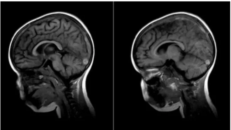
hypoalbuminemia (2.1g/dL), a normal ionogram and C-reactive protein (CRP) of 25.9mg/dL (reference value: up to 6mg/dL). We chose to collect cephalorachidian liquor, which revealed hyperproteinorachia (67mg/dL; reference values: 15 to 40mg/dL), cellularity, normal cytometry and glycorrhachia. Cerebral magnetic resonance imaging was conducted, which revealed an image compatible with superior sagittal sinus and transverse sinus thrombosis, largely restricted to the left hemisphere (Figure 1). Sagittal, axial and coronal planes at T1, T2, T2* and Flair sequences revealed the presence of a hypointense lesion on T1 and a hyperintense lesion on T2 inside the superior sagittal sinus and the left transverse sinus, suggesting thrombosis in both sinuses. here was no enhancement of pathologic area after the injection of contrast.
Once the radiologic diagnosis was established, treatment for cerebral venous thrombosis (CVT) began with a continuous infusion of intravenous heparin. he use of corticosteroid was maintained, and loop diuretics were stopped. he patient required a gradual increase of heparin up to 35UI/kg/hour because of the mild efect on the partial thromboplastin time. When midazolam was removed, we observed hazy mydriasis without focal signs or loss of muscle strength. Ophthalmic examination revealed optic papilla with physiologic excavation and mild to moderate blurred edges, which was consistent with mild to moderate papillitis. Routine exams reveal the persistence of hypoalbuminemia and massive proteinuria, and three cycles of methylprednisolone pulse therapy were administered. Reduced proteinuria and increased serum albumin were initially observed with pulse therapy but were accompanied by rapid relapse of the nephrotic framework. With the onset of fever, chills, incoercible vomiting and cyanosis of the extremities, new exams were conducted, revealing anemia (hemoglobin: 9.7g/dL; reference values: 11.5 to 13.5g/dL), leukocytosis with left
shift (19,600/mm3) and CRP of 23.1mg/L. Antimicrobial
therapy for hospital-acquired infection with ceftazidime (150mg/kg/day) and vancomycin (40mg/kg/day) began, with a gradual improvement in the infection response. Once improvements in nephrotic syndrome ceased, cyclophosphamide treatment was started, which improved the proteinuria and reduced the edema. Phenobarbital was also administered, and diphenylhydantoin was progressively reduced. he patient was discharged from intensive treatment after 45 days of hospitalization for ambulatory care in pediatric nephrology and ophthalmology because of poor visual acuity. he patient experienced no relapses of the nephrotic syndrome for 8 months after hospitalization but experienced three relapses after this period and recurrent proteinuria. Histopathology of the renal biopsy was compatible with minimal glomerular lesions. Currently, the patient exhibits good renal function without proteinuria. From the neurological point of view, he exhibits appropriate neuro-psychomotor development without focal changes. he patient experiences poor visual acuity and regularly uses glasses.
DISCUSSION
NS, a glomerular disease that mostly afects children, is classically deined by massive proteinuria (>40mg/m2/hour), hypoalbuminemia (<2.5g/dL), generalized edema and hyperlipidemia. NS complications can occur as part of the
course of disease or as a consequence of the pharmacological treatment. Among the complications associated with the disease, CVT is an unusual condition, with few reports and case series described in the literature, especially among children. However, the recent increase in the number of cases may indicate that NS has been underdiagnosed in the past.(1-10)
Considering all possible etiologies, CVT has an incidence of 0.67 cases per 100 thousand children per year, i.e., it is likely underestimated. he exact incidence of CVT associated with NS is unknown, but CVT is believed to represent 4.7 to 6% of all CVT cases in children, excluding newborns. CVT involves the thrombosis of the intracranial venous sinus and cerebral veins, which leads to impaired venous drainage and, thus, intracranial hypertension and/or venous infarction. Because of the highly variable clinical presentation and because the disease is relatively uncommon in pediatric patients, the diagnosis of CVT is diicult, delayed or missed in some cases. he low incidence contributes to our poor understanding of the source and physiopathology of CVT in pediatric patients. Risk factors for CVT vary by age and frequently diferent in number and quality between children and adults. Infections are the most common factor in newborns as well as in older children, followed by hypercoagulable states and dehydration. Most of the cases arise from a combination of prothrombotic risk factors with or without subjacent clinical condition.(2,10,11-17)
NS is a known risk factor for arterial or venous thromboembolism (TE), and patients with severe proteinuria exhibit a 3.4-fold increased risk of venous thrombosis. hrombosis risk is higher at the onset of NS and during relapses because of the loss of coagulation factors and acute intravascular volume depletion during this phase of the disease. he risk is also higher in corticoresistant NS compared with corticosensitive NS. hrombosis can occur in NS due to the loss of proteins involved in systemic hemostasis inhibition, the increase in the synthesis of prothrombotic factors, or the local activation of the glomerular hemostatic system. TE-predisposing factors in NS include the following: anomalies in platelet activation and aggregation; coagulation system activation; increased synthesis of coagulation factors V, VII, VIII, and X as well as von Willebrand factor, ibrinogen,
and accumulation of α2-macroglobulin; reduction of
endogenous anticoagulants (antithrombin III, C protein and S protein); decreased activity of the ibrinolytic system and imbalance of the plasmin formation system; changes in the glomerular hemostatic system; intravascular volume depletion; and the use of diuretics.(1-4,6,7,9,11-15)
Considering the factors involved in the genesis of the thromboembolic framework, caution should be taken in the use of diuretics in NS because the treatment enhances hypovolemia in nephrotic patients, thereby increasing the risk for TE. he clinical manifestations of CVT are nonspeciic and vary with age. Convulsive seizures (generalized or focal) are the most common presentation among newborns and children. Focal signs and neurological symptoms, such as decreased consciousness, nausea, vomiting, motor deicits (hemiplegia and ataxia), headache and visual changes (diplopia and papilledema), are more commonly found in children. Among the less frequent symptoms are vertigo, somnolence, confusion and neck pain. Because CVT symptoms are nonspeciic, the disease requires a high degree of clinical suspicion.(2-4,8,10-14)
In the case reported, the patient exhibited with seizure, sensory deicits, headache, vomiting and visual alterations. he child had been taking oral furosemide associated with corticotherapy. Special care must be taken when administering diuretics in NS; diuretics may be used in symptomatic treatment of edema in massive anasarca patients with diiculty breathing, cellulitis refractory to antimicrobial and corticoresistant children. Because the patient did not exhibit these conditions and because of the possible drug inluence on CVT genesis, the use of furosemide was discontinued.
headache and papilledema are typical manifestations in older children.(2-4,7-14,18,19)
Treatment of CVT is based on symptomatic and supportive measures. As in any emergency, airway, breathing and circulation management are imperative. Benzodiazepine therapy is the irst line of treatment of seizures in CVT. he treatment of choice is the use of heparin followed by oral anticoagulants. Diiculty in achieving anticoagulation is describe in NS-associated cases due to antithrombin III urinary loss.(11,14,20)
In the present case report, there was a need to gradually increase the heparin dose due to insuicient efects on the partial thromboplastin time, which was maintained during the acute phase, followed by oral anticoagulant throughout the remainder of the treatment and during relapses. Oral anticoagulants have similar results but exhibit less frequent adverse efects. he treatment of choice is hypocoagulation with non-fractional heparin or low-molecular weight heparin (LMWH) for 5 to 7 days, followed by LMWH or a vitamin K antagonist for 3 to 6 months or as long as the patient exhibits nephrotic proteinuria (albumin level <2g/dL) or both.
Prophylactic hypocoagulation is controversial; however, prophylactic hypocoagulation should be administered in a patient with a thromboembolic event and high risk of recurrence based on an albumin concentration <2g/dL, ibrinogen >6g/L or an antithrombin III level <70% of the normal value.(6,11)
Long-term neurological sequelae, in descending order of frequency, are as follows: motor deicits (hypotonia, hemiplegia and hemiparesis), seizures, cognitive dysfunction, developmental and/or speech delay, and visual disturbances. Children who develop CVT in the neonatal
period exhibit normal intellectual abilities. However, a considerable number exhibit a heterogeneous cognitive proile, with partial cognitive disorders. his inding may indicate that although intellectual functioning is generally normal, children with CVT may sufer from attention and perception disorders that are only evident during alphabetization. he occurrence of venous infarctions, seizures, Glasgow scale <12 at admission and the presence of cerebral parenchyma lesions are associated with a worse prognosis. A European cohort study published in 2007 associated a higher risk of CVT recurrence with four factors: age over 2 years; absence of secondary anticoagulant prophylaxis; absence of recanalization; and the presence of G20210A mutation in factor II. Two cohort studies described the recurrence of symptoms in 12 to 13% of 180 long-term survivors, occurring on average 12 to 18 months after initial presentation.(10,11,13,16,21,22)
he patient of this report had low visual acuity as the only sequelae. He did not experience other CVT events and was administered an oral anticoagulant during NS relapses. A multidisciplinary approach to CVT in NS is imperative and must include pediatric neurology monitoring, ophthalmology and, when indicated, rehabilitative therapy.
CONCLUSION
Cerebral venous thrombosis should be considered in any patient with nephrotic syndrome who presents with neurological signs or symptoms. When suspected, imaging exams should be promptly conducted. Upon conirmation of cerebral venous thrombosis, anticoagulant therapy should be started immediately. Importantly, early clinical suspicion is correlated with a more favorable outcomes.
A síndrome nefrótica associa-se a um estado de hipercoagulabilidade, apresentando risco aumentado de complicações tromboembólicas. A trombose dos seios venosos cerebrais é uma complicação rara da síndrome nefrótica, com poucos casos descritos na literatura, mas com diagnósticos cada vez mais frequentes. A verdadeira incidência pode estar subestimada, uma vez que muitos eventos são assintomáticos ou não são diagnosticados a tempo. Descrevemos aqui o caso de uma criança do sexo masculino, de 2 anos e 10 meses, com síndrome nefrótica, que apresentou, na evolução, cefaleia,
crises epilépticas e rebaixamento sensorial, com o diagnóstico de trombose do seio sagital superior e transverso. Foi realizada revisão da literatura internacional por meio de estratégia de busca deinida, nas bases de dados PubMed, SciELO e Lilacs, utilizando os termos “nephrotic syndrome” e “cerebral sinovenous
thrombosis”. O diagnóstico de trombose venosa deve ser
considerado em qualquer paciente com síndrome nefrótica que manifeste sinais e sintomas neurológicos, destacando que a suspeita clínica precoce tem relação com um desfecho favorável.
RESUMO
REFERENCES
1. Park SJ, Shin JI. Complications of nephrotic syndrome. Korean J Pediat. 2011;54(8):322-8. Erratum in: Korean J Pediatr. 2012;55(4):151. 2. Rodrigues MM, Zardini LR, de Andrade MC, Mangia CM, Carvalhaes JT,
Vilanova LC. Cerebral sinovenous thrombosis in a nephrotic child. Arq Neuropsiquiatr. 2003;61(4):1026-9.
3. Pillekamp F, Hoppe B, Roth B, Querfeld U. Vomiting, headache and seizures in a child with idiopathic nephrotic syndrome. Nephrol Dial Transplant. 1997;12(6):1280-1.
4. Bhoobun S, Jalloh AA, Jacobsen KH. Cerebral venous thrombosis in a child with nephrotic syndrome: case report. Pan Afr Med J. 2012;13:57. 5. Meirelles PZ, Watanabe A, Carneiro JD, Koch VH. Peculiaridades da
terapia trombolítica na síndrome nefrótica pediátrica: monitorização do fator anti-Xa. Rev Paul Pediatr. 2008;26(2):183-7.
6. Balona F, Ferreira G, Marques E, Vilarinho A. Trombose dos seios venosos cerebrais em criança com síndrome nefrótica: caso clínico. Nascer e Crescer. 2009;18(2):85-8.
7. Zaki SA, Shanbag P. Persistent headache in a child with the nephrotic syndrome. Saudi J Kidney Dis Transpl. 2010;21(5):951-3.
8. Menascu S, Lotan A, Ben Zeev B, Nowak-Goottl U, Kenet G. Cerebral venous thrombosis in the Mediterranean area in children. Mediterr J Hematol Infect Dis. 2011;3(1):e2011029.
9. Al Fakeeh KN, Al Rasheed SA. Cerebral venous thrombosis in the nephrotic syndrome. Saudi J Kidney Dis Transpl. 2000;11(1):59-63.
10. deVeber G, Andrew M, Adams C, Bjornson B, Booth F, Buckley DJ, Camfield CS, David M, Humphreys P, Langevin P, MacDonald EA, Gillett J, Meaney B, Shevell M, Sinclair DB, Yager J; Canadian Pediatric Ischemic Stroke Study Group. Cerebral sinovenous thrombosis in children. N Engl J Med. 2011;345(6):417-23.
11. Hashmi M, Wasay M. Caring for cerebral venous sinus thrombosis in children. J Emerg Trauma Shock. 2011;4(3):389-94.
12. Fluss J, Geary D, deVeber G. Cerebral sinovenous thrombosis and idiopathic nephrotic syndrome in childhood: report of four new cases and review of the literature. Eur J Pediat. 2006;165(10):709-16.
13. Jackson BF, Porcher FK, Zapton DT, Losek JD. Cerebral sinovenous thrombosis in children: diagnosis and treatment. Pediatr Emerg Care. 2011;27(9):874-80; quiz 881-3.
14. Sébire G, Tabarki B, Saunders DE, Leroy I, Liesner R, Saint-Martin C, et al. Cerebral venous sinus thrombosis in children: risk factors, presentation, diagnosis and outcome. Brain. 2005;128 (Pt 3):477-89.
15. Heller C, Heinecke A, Junker A, Knöfler R, Kosch A, Kurnik K, Schobess R, von Eckardstein A, Sträter R, Zieger B, Nowak-Göttl U; Childhood Stroke Study Group. Cerebral venous thrombosis in children: a multifactorial origin. Circulation. 2013;108(11):1362-7.
16. Grunt S, Wingeier K, Wehrli E, Boltshauser E, Capone A, Fluss J, Gubser-Mercati D, Jeannet PY, Keller E, Marcoz JP, Schmitt-Mechelke T, Weber P, Weissert M, Steinlin M; Swiss Neuropaediatric Stroke Registry. Cerebral sinus venous thrombosis in Swiss children. Dev Med Child Neurol. 2010;52(12):1145-50.
17. Kenet G, Waldman D, Lubetsky A, Kornbrut N, Khalil A, Koren A, et al. Paediatric cerebral sinus vein thrombosis. A multi-center, case-controlled study. Thromb Haemost. 2004;92(4):713-8.
18. Gasparetto EL. Trombose venosa cerebral. Radiol Bras. 2006;39(5):III. 19. Monteiro AM, Lima CM, Ribeiro EB, Lins MC, Miranda S, Miranda LE.
Diagnóstico por imagem e aspectos clínicos da trombose venosa cerebral em recém-natos a termo sem dano cerebral: revisão em 10 anos. Radiol Bras. 2010;43(3):149-53.
20. Dlamini N, Billinghurst L, Kirkham FJ. Cerebral venous sinus (sinovenous) thrombosis in children. Neurosurg Clin N Am. 2010;21(3):511-27. 21. Ichord R. Outcome in childhood cerebral venous thrombosis--new insights.
Nat Clin Pract Neurol. 2008;4(1):16-7.