*Correspondence: Unidade de Pesquisa em Genética e Biologia Molecular UPGEM
Faculdade de Medicina de São José do Rio Preto – FAMERP Av. Brigadeiro Faria Lima, 5416 - Bloco U-6 São José do Rio Preto - SP, 15090-000
Telefone: (17) 3201-5720 Fax: (17) 3201-5708 [email protected]
SUMMARY
Objective. Considering that studies about the frequencies of phenotypic features of Down syndrome
(DS) in the Brazilian population with large ethnic variability are scarce in literature, this study analyzed clinical and demographic characteristics of DS children from the Southeastern region of Brazil. MethOds. Sixty-two DS children with free trisomy 21 were evaluated by physical examination using
reference values that considered the children´s gender and age at their presentation. Data about clinical complications were collected by retrospective analysis of the children’s medical records and/ or information supplied by their mothers. Statistical analysis was performed using Likelihood Ratio Test, with signiicance level less or equal to 5%.
Results. Clinical features observed in more than 90% of the individuals were lat facial proile,
brachycephaly, slanted palpebral issures, hypotonia at birth and lat nasal bridge. Congenital heart disease was present in 56.5% of the cases, verbal language acquisition disorder in 87%, and global delayed development in 77.8%.
cOnclusiOn. The comparison between our data and related literature showed a great variability of the
phenotype features frequencies of DS among studies. Besides environmental factors, this can relect individual as well as population characteristics.
Keywords: Down syndrome. Phenotype. Physical Examination.
c
linical
pROfile
Of
childRen
with
dOwn
syndROMe
tReated
in
a
genetics
Outpatient
seRvice
in
the
s
Outheast
Of
b
Razil
ÉRika cRistina pavaRinO beRtelli1*, jOice MatOs biselli2, daiana bOnfiM3, eny MaRia gOlOni-beRtOllO1
Trabalho realizado na Unidade de Pesquisa em Genética e Biologia Molecular – UPGEM, Departamento de Biologia Molecular da Faculdade de Medicina de São José do Rio Preto – FAMERP, São José do Rio Preto, SP
1. Professoras Livre-docentes da Faculdade de Medicina de São José do Rio Preto – FAMERP, São José do Rio Preto, SP
2. Mestre em Ciências da Saúde - Doutoranda em Ciências da Saúde da Faculdade de Medicina de São José do Rio Preto – FAMERP, São José do Rio Preto, SP 3. Enfermeira da Faculdade de Medicina de São José do Rio Preto – FAMERP, São José do Rio Preto, SP
i
ntROductiOnDown syndrome (DS) is a common chromosomal abnormality, usually due to an extra copy of the 21. Its incidence is approximately 1:660 live births1, and it is
the most common genetic cause of mental disability.2,3
DS phenotype is complex and varies among individuals, who may present a combination of dysmorphic features,4-6
congenital heart disease,7,8,9 neurological abnormalities, such
as early manifestation of Alzheimer’s disease,10 immunological
deficiency,11 and elevated risk of specific types of leukemia,12
among other clinical complications. 13-15.
Diagnostic hypothesis of DS can be performed in the pre and postnatal period and conirmed by chromosome analysis (karyotype examination). Cytogenetic investigation is important to conirm clinical diagnosis and to determinate the recurrence risks of DS, helping the genetic counseling process.16,17
Considering that studies about the frequencies of phenotypic features of Down syndrome (DS) in the Brazilian population, that presents a great ethnic variability, are scarce in literature, the objective of this study was to analyze clinical and demographic
characteristics of DS children with free trisomy 21 from a teaching hospital linked to a medical school in the Southeastern region of Brazil.
M
ethOdsThis study was approved by the Research Ethics Committee of the São José do Rio Preto Medical School / SP (CEP-FAMERP), and by the National Research Ethics Commission (CONEP), Brazil. All individuals were included in this study only after the Informed Consent of their parents.
The retrospective study was carried out at a general genetics outpatient service of a teaching hospital in Sao José do Rio Preto, São Paulo, Brazil, which assists a region of about 100 municipalities in the Southeast of Brazil. Patients assisted in the general genetics outpatient service usually have a low socio-economic status.
features of DS which is not routinely used at the service. Data regarding brachydactyly, hypertelorism, length of the hands and microcephaly were determined using reference values for non-DS individuals that consider the children’s gender and age at their presentation.18
Evaluation of weight and height was carried out using speciic curves for DS individuals19, and these anthropometric data were
used to assess the nutritional status of the children by the body mass index for age (BMI-for-age, Kg/m2). The resulting data were
plotted in speciic charts of BMI for individuals DS19. Distribution
of indices was expressed in Z-scores terms, also referred to as standard deviation (SD) units, and the Z-score cutoff point to classify low anthropometric levels was 2 SD units below the reference mean.
Data about clinical complications were collected by retrospective analysis of the children’s medical records and/ or information provided by their mothers. When an individual was not evaluated for a speciic characteristic, he/she was excluded from the frequency analysis of this characteristic. Data concerning age were presented by mean and standard deviation (SD). For statistical analysis of differences between our study and the related literature, the Likelihood Ratio Test (Chi-square test) with a signiicance level > 5% was used.
R
esultsOf the 62 children included in the study, 27 were female (43.5 %) and 35 male (56.5%). The average age at presentation was 3.0 years (range: 0.12 to 12.7 years). In relation to weight and height, 12.3% and 7.0% of the individuals presented low and overweight, respectively, while 16.4% presented low height and 9.1% presented increased height. Regarding the BMI, 78.2% of the DS children presented BMI below 18.5 Kg/ m2 and 3.6% above 25 Kg/m2. Analysis of the nutritional status
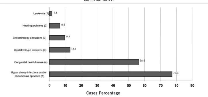
Figure 1. Clinical complications presented by DS patients. Distribution according to the number of patients evaluated: (1) 59; (2) 51; (3) 53; (4) 62; (5) 61.
Cases Percentage
(BMI-for-age) showed that 16.4% presented BMI below 2 SD and 10.9% above 2 SD.
All cases were diagnosed in postnatal period. The average of maternal age at birth of the affected child was 32.0 ± 8.6 years (range: 13.1 - 46.9 years). The majority of mothers (58.1%) was under 35 years old at birth of the affected child.
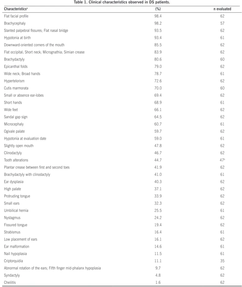
Frequencies of clinical features were presented in Table 1. The clinical characteristics presented in more than 90% of individuals were lat facial proile, brachycephaly, slanted palpebral issures, hypotonia at birth and lat nasal bridge. Verbal language acquisition disorder, and overall delayed development were referred by mothers in 87% and 77.8% of cases, respectively.
Clinical complications presented in DS individuals are shown in Figure 1. Congenital heart diseases were represented by interatrial communication (57.1%), patent ductus arteriosus (34.3%), interventricular communication (22.9%), defect of the atrioventricular septum (17.1%), tetralogy of Fallot (5.7%), and valve insuficiency (14.3%). In one case, the problem was not speciied. Forty-three percent of children presented more than one type of congenital heart disease.
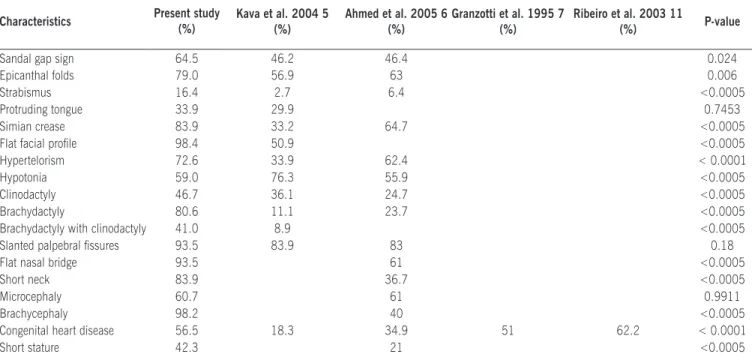
We performed a comparison among frequencies observed in our study and other studies previously published, and this analysis disclosed signiicant statistical differences for the majority of characteristics (Table 2).
d
iscussiOnThree types of chromosomal alterations may result in DS: free trisomy, translocation, and mosaicism.18 Advanced maternal
Table 1. Clinical characteristics observed in DS patients.
Characteristicsa (%) n evaluated
Flat facial proile 98.4 62
Brachycephaly 98.2 57
Slanted palpebral issures; Flat nasal bridge 93.5 62
Hypotonia at birth 93.4 61
Downward-oriented corners of the mouth 85.5 62
Flat occipital; Short neck; Micrognathia; Simian crease 83.9 62
Brachydactyly 80.6 60
Epicanthal folds 79.0 62
Wide neck; Broad hands 78.7 61
Hypertelorism 72.6 62
Cutis marmorata 70.0 60
Small or absence ear-lobes 69.4 62
Short hands 68.9 61
Wide feet 66.1 62
Sandal gap sign 64.5 62
Microcephaly 60.7 61
Ogivale palate 59.7 62
Hypotonia at evaluation date 59.0 61
Slightly open mouth 47.8 62
Clinodactyly 46.7 62
Tooth alterations 44.7 47b
Plantar crease between irst and second toes 41.9 62
Brachydactyly with clinodactyly 41.0 61
Ear dysplasia 40.3 62
High palate 37.1 62
Protruding tongue 33.9 62
Small ears 32.3 62
Umbilical hernia 25.5 61
Nystagmus 24.2 62
Fissured tongue 19.4 62
Strabismus 16.4 61
Low placement of ears 16.1 62
Ear malformation 14.6 61
Nail hypoplasia 11.5 61
Criptorquidia 11.1 35
Abnormal rotation of the ears; Fifth inger mid-phalanx hypoplasia 9.7 62
Syndactyly 4.8 62
Cheilitis 1.6 62
Table 2. Comparison of clinical characteristics between our study and related literature* Characteristics Present study
(%)
Kava et al. 2004 5 (%)
Ahmed et al. 2005 6 (%)
Granzotti et al. 1995 7 (%)
Ribeiro et al. 2003 11
(%) P-value
Sandal gap sign 64.5 46.2 46.4 0.024
Epicanthal folds 79.0 56.9 63 0.006
Strabismus 16.4 2.7 6.4 <0.0005
Protruding tongue 33.9 29.9 0.7453
Simian crease 83.9 33.2 64.7 <0.0005
Flat facial proile 98.4 50.9 <0.0005
Hypertelorism 72.6 33.9 62.4 < 0.0001
Hypotonia 59.0 76.3 55.9 <0.0005
Clinodactyly 46.7 36.1 24.7 <0.0005
Brachydactyly 80.6 11.1 23.7 <0.0005
Brachydactyly with clinodactyly 41.0 8.9 <0.0005
Slanted palpebral issures 93.5 83.9 83 0.18
Flat nasal bridge 93.5 61 <0.0005
Short neck 83.9 36.7 <0.0005
Microcephaly 60.7 61 0.9911
Brachycephaly 98.2 40 <0.0005
Congenital heart disease 56.5 18.3 34.9 51 62.2 < 0.0001
Short stature 42.3 21 <0.0005
* The comparison between the frequencies observed in this study and other studies was possible with those that present the number of individuals evaluated.
several studies have related the maternal risk for DS with genetic polymorphisms involved in the folate metabolism.17,23-28 DS is responsible for manifestation of a combination of clinical features that makes clinical diagnosis possible. However, some characteristics in frequency and severity are variable among the individuals. 5, 6,11,29. The knowledge of clinical manifestations of
DS by physicians and other health professionals is important to make an early postnatal diagnosis possible, since prenatal diagnosis is not frequently performed, as in our sample, where all cases were diagnosed in the postnatal period, with diagnostic hypothesis conirmed by cytogenetic analysis. Late DS diagnosis can result in delayed early intervention and appropriate therapy for some risk conditions, such as congenital heart diseases. These must be detected and treated, otherwise they contribute to morbidity and mortality of these children, in addition, resulting on an impact in the physical and psychological development.5,30
Frequencies of DS clinical features vary within the different studies, and some of them described only speciic characteristics. Bibliographic reviews on DS have shown a large variability of these frequencies.1,4-8,10-12,16-18,31. Comparison of frequencies
observed in our study and other studies disclosed signiicant statistical differences for most characteristics (Table 2). It is possible that the differences may relect the cytogenetic proile of the individuals included in the study, since in our study only individuals with free trisomy 21 were included while other studies used for comparison evaluated individuals with free trisomy, translocation and mosaicism, even though free trisomy is present in the majority (>90%) of DS cases,5,6,18,32 alongside individuals
with diagnostic hypothesis of DS without cytogenetic diagnosis. According to Devlin and Morrison´s study1, the accuracy of DS
clinical diagnosis varies according to the cytogenetic alteration
involved (90% for free trisomy, 100% for translocation and 37.5% for mosaicism), apparently relecting a clinical variability according to cytogenetic abnormality.
Several other factors may contribute to phenotype variability in DS, such as allelic heterogeneity for chromosome 21 genes present in three copies, the individual’s genetic constitution and environmental factors.29. Brazil is one of the countries with
the largest ethnic heterogeneity in the world. This is a result of miscegenation in the immigrant population for centuries, mainly Portuguese, African and Amerindians. This process of miscegenation has provided the gene propagation and contributed to the characteristics of the Brazilian population. Moreover, since Brazil is a country with a large territory, there is a great geographic heterogeneity.33
In relation to weight, a higher frequency of low weight (12.3%) as compared to overweight (7.0%) was observed in the present study. Regarding the BMI, a high frequency of
DS children presented BMI below 18.5 Kg/m2 (78.2%), while
only 3.6% presented BMI above 25 Kg/m2. Although obesity is
frequent in DS,16 the higher frequency of low weight and BMI
observed in our study may be explained by the fact that 50% of the evaluated children presented an age below 2 years old. DS neonates are often born below the usual weight and gain weight as age increases. Analysis of the nutritional status showed that 16.4% of the children presented undernutrition (BMI-for-age below -2 SD), probably due to the low socioeconomic status of the patients assisted by the general genetics outpatient service of the Hospital de Base de Sao José do Rio Preto. Regarding height, according to literature,34 low height is common in DS, and was
The incidence of congenital heart diseases in DS is about 60%.9,35 In this study, 56.5% of the children presented
congenital heart diseases, similar frequency in Brazilian studies, which ranges from 51 to 62.2%.7,11
The frequencies of types of congenital heart diseases were similar between our study and Ribeiro et al.11 with a greater
frequency of interatrial communication in both studies (62% and 46.4%, respectively). In Granzotti et al.´s study,7
frequencies of types of congenital heart diseases differ from those observed in our study.
Upper airway infections and/or pneumonia episodes presented a frequency of 77.4%, while in literature this was 50-60% and 40%, repectively.11 DS individuals presented
high risk for endocrinology alterations, hearing problems and development of leukemia.12,15,16,31 In our study, the mothers
reported endocrine alterations and hearing problems in 9.7 and 6.6%, respectively. One case had a diagnosis of acute myeloid leukemia.
Some DS characteristics can be less evident in the individual over the years, 15,36 , and environmental factors,
such as early stimulation, may also contribute to this change. An example is the difference in frequency of muscular hypotonia at birth (93.4%) and at evaluation date (59%), observed in this study. Kava et al.5 observed similar
frequencies of muscular hypotonia between neonates and non-neonates; however, distinct individuals comprised both groups.
The children included in this study were screened in the Outpatient Service of Genetics of a medical school hospital that offers early stimulation services, such as Phonoaudiology, Physiotherapy, and Occupational Therapy. Other characteristic, the protruding tongue, observed in 33.9% of the evaluated children, can be modified by
earlier stimulation, mainly by Phonoaudiology. Kava et al.5
described similar frequency (29.9%) in a sample of DS individuals in India. The performance of a multidisciplinary group of health professionals that assist the needs of DS persons and their families, the Ding-Down workgroup, in the medical school - FAMERP, can provide an effective care to DS individuals and family compliance to the programs of earlier stimulation, therefore improving the quality of life of these persons.
c
OnclusiOnThe comparison between our data and related literature showed a great variability of the phenotype features frequencies of DS among studies. In addition to environmental factors, this can also relect individual and population characteristics.
f
inancials
uppORtFundação de Amparo à Pesquisa do Estado de São Paulo – FAPESP (04/15944-5); Coordenação de Aperfeiçoamento de Pessoal de Nível Superior – CAPES (CGPP 046/2006); Conselho Nacional de Desenvolvimento Cientíico e Tecnológico – CNPq (3181/2005); Conselho Nacional de Desenvolvimento Cientíico e Tecnológico – CNPq (302157/2008-5).
Conlict of interest: none
R
esuMOpeRfilclínicOdecRiançascOMsíndROMede dOwnatendidaseM uMaMbulatóRiOdegenÉticanaRegiãO sudestedO bRasil
ObjetivO. Considerando que estudos relacionados às
frequências das características fenotípicas da síndrome de Down (SD) na população brasileira, que apresenta grande variabilidade étnica, são escassos na literatura, este estudo analisou características clínicas e demográficas de crianças com SD da região Sudeste do Brasil.
MétOdOs. Sessenta e duas crianças com SD com trissomia
livre do 21 foram avaliadas por meio de exame físico utilizando-se valores de referência que consideram o gênero e idade da criança na data da avaliação. Dados sobre complicações clínicas foram coletados por análise retrospectiva dos prontuários médicos e/ou informação das mães. A análise estatística foi realizada utilizando-se o teste da razão de máxima verossimilhança com nível de significância menor ou igual a 5%.
ResultadOs. As características clínicas observadas em
mais de 90% dos pacientes foram perfil facial achatado, braquicefalia, fenda palpebral oblíqua, hipotonia muscular ao nascimento e ponte nasal baixa. Doenças cardíacas congênitas estiveram presentes em 56,5% dos casos, distúrbio de aquisição de linguagem em 87% e atraso do desenvolvimento global em 77,8%.
COnClusãO. A comparação entre nossos dados e a literatura
prévia mostrou grande variabilidade das características fenotípicas da SD entre os estudos. Isso pode refletir características
individuais e populacionais, além de fatores ambientais. [Rev
Assoc Med Bras 2009; 55(5): 547-52]
unitermos: Síndrome de Down. Fenótipo. Exame físico.
R
efeRences1. Devlin L, Morrison P. Accuracy of the clinical diagnosis of Down syndrome. Ulster Med J. 2004;73:4-12.
2. Assumpção Jr FB, Sprovieri MH, Assumpção TM. Deiciência mental em São Paulo: peril de uma população atendida institucionalmente. Pediatria
Moderna 1999;35:883-92.
3. Lantigua-Cruz A, Mora F, Arechaederra M, Rojas I, Morales E, Rodriguez H, et al. Etiological characterization of 512 severely mentally retarded institutionalized patients in Havana. Community Genet. 1999;2:184-9.
4. Prasher VP. Screening of medical problems in adults with Down syndrome.
Downs Syndr Res Pract. 1994;2:59-66.
5. Kava MP, Tullu MS, Muranjan MN, Girisha KM. Down syndrome: clinical proile
from India. Arch Med Res. 2004;35:31-5.
6. Ahmed I, Ghafoor T, Samore NA, Chattha MN. Down syndrome: clinical and cytogenetic analysis. J Coll Physicians Surg Pak. 2005;15:426-9.
7. Granzotti JA, Paneto ILC, Amaral FTV, Nunes MA. Incidência de cardiopatias
congênitas na síndrome de Down. J Pediatr. 1995;71:28-30.
8. Ghaffar S, Lemler MS,Fixler DE,Ramaciotti C. Trisomy 21 and congenital heart disease: effect of timing of initial echocardiogram. Clin Pediatr. (Phila) 2005;44:39-42.
9. Abbag FI. Congenital heart diseases and other major anomalies in patients with Down syndrome. Saudi Med J. 2006;27:219-22.
10. Lott IT, Head E. Alzheimer disease and Down syndrome: factors in pathogenesis. Neurobiol Aging. 2005;26:383-9.
11. Ribeiro LM, Jacob CM, Pastorino AC, Kim CA, Fomin AB, Castro AP. Evaluation of factors associated with recurrent and/or severe infections in patients with Down’s syndrome. J Pediatr. (Rio J) 2003; 79:141-8.
12. Hasle H, Clemmensen IH, Mikkelsen M. Risks of leukaemia and solid tumours in individuals with Down’s syndrome. Lancet. 2000;355:165-9.
13. Kriss VM. Down syndrome: imaging of multiorgan involvement.
Clin Pediatr. (Phila) 1999;38:441-9.
Artigo recebido: 26/05/08 Aceito para publicação: 17/03/09
15. Venail F, Gardiner Q, Mondain M. ENT and speech disorders in children with
Downs syndrome: an overview of pathophysiology, clinical features, treat-ments, and current management. Clin Pediatr. (Phila) 2004;43:783-91. 16. Mustacchi Z. Síndrome de Down. In: Mustacchi Z, Peres S. Genética baseada
em evidências: Síndromes e Heranças. São Paulo: CID Editora; 2000. p.819-94.
17. Pavarino-Bertelli EC, Biselli JM, Ruiz MT. Recentes avanços moleculares e aspectos genético-clínicos em síndrome de Down. Rev Bras Med. 2005;62:401-8.
18. Jones K B. Smith’s Recognizable patterns of human malformations. Philadel-phia: WB Saunders; 1998. p.10-5.
19. Myrelid A, Gustafsson J, Ollars B, Annerén G. Growth charts for Down’s syndrome from birth to 18 years of age Arch Dis Child. 2002;87:97-103. 20. Hassold T, Sherman S. Down syndrome: genetic recombination and the origin
of the extra chromosome 21. Clin Genet. 2000;57:95-100.
21. Jyothy A, Kumar KS, Mallikarjuna GN, Babu Rao V, Uma Devi B, Sujatha
M, Reddy PP. Parental age and the origin of extra chromosome 21 in Down syndrome. J Hum Genet. 2001;46:347-50.
22. Malini SS, Ramachandra NB. Inluence of advanced age of maternal grand -mothers on Down syndrome. BMC Med Genet. 2006;14:7-4.
23. Biselli JM, Goloni-BertolloEM, ZampieriBL, Haddad R, Eberlin MN, Pavarino-Bertelli EC. Genetic polymorphisms involved in folate metabolism and elevated plasma concentrations of homocysteine: maternal risk factors for Down syndrome in Brazil. Genet Mol Res. 2008; 7(1):33-42.
24. Bosco P, Gueant-Rodriguez RM, Anello G, Barone C, Namour F, Caraci F, et al. Methionine synthase (MTR) 2756 (A®G) polymorphism, double heterozygo-sity methionine synthase 2756 AG/methionine synthase reductase (MTRR) 66 AG, and elevated homocysteinemia are three risk factors for having a child with Down syndrome. Am J Med Genet. A 2003;121:219-24.
25. Silva LR, Vergani N, Galdieri Lde C, Ribeiro Porto MP, Longhitano SB, Brunoni D,
et al. Relationship between polymorphisms in genes involved in homocysteine metabolism and maternal risk for Down syndrome in Brazil. Am J Med Genet. A 2005;135:263-7.
26. Martinez-Frias ML, Perez B, Desviat LR, Castro M, Leal F, Rodriguez L, et al. Maternal polymorphisms 677C-T and 1298A-C of MTHFR, and 66A-G MTRR genes: is there any relationship between polymorphisms of the folate pathway, maternal homocysteine levels, and the risk for having a child with Down syndrome? Am J Med Genet. A 2006;140:987-97.
27. Coppede F, Colognato R, Bonelli A, Astrea G, Bargagna S, Siciliano G, et al. Folate gene polymorphisms and the risk of Down syndrome pregnancies in young Italian women. Am J Med Genet. A 2006;140:1083-91.
28. Rai AK, Singh S, Mehta S, Kumar A, Pandey LK, Raman R. MTHFR C677T and A1298C polymorphisms are risk factors for Downs syndrome in Indian mothers. J Hum Genet. 2006;51:278-83.
29. Reeves RH, Baxter LL, Richtmeier. Too much of a good thing: mechanisms of gene action in Down syndrome. Trends Genet. 2001;17 83-8.
30. Hijii T, Fukushige J, Igarashi H, Takahashi N, Ueda K. Life expectancy and social adaptation in individuals with Down syndrome with and without surgery for congenital heart disease. Clin Pediatr. (Phila) 1997;36:327-32. 31. Newberger DS. Down syndrome: prenatal risk assessment and diagnosis. Am
Fam Physician. 2000;62:825-32, 837-8.
32. Mokhtar MM, Abd el-Aziz AM, Nazmy NA, Mahrous HS. Cytogenetic proile of
Down syndrome in Alexandria, Egypt. East Mediterr Health J. 2003;9:37-44. 33. Parra FC, Amado RC, Lambertucci JR, Rocha J, Antunes CM, Pena SD. Color and genomic ancestry in Brazilians. Proc Natl Acad Sci USA. 2003;100:177-82. 34. Toledo C, Alembik Y, Aguirre Jaime A, Stoll C. Growth curves of children with
Down syndrome. Ann Genet. 1999;42:81-90.
35. De Rubens Figueroa J, Del Pozzo Magana B, Pablos Hach JL, Calderon Jimenez C, Castrejon Urbina R. Heart malformations in children with Down syndrome. Rev Esp Cardiol. 2003;56:894-9.