Heart Rate Variability in Myotonic Dystrophy Type 1 Patients
Guilherme Fregonezi
1, Thaise Araújo
1, Mario Emilio Dourado Junior
2, Joceline Ferezini
1, Ester Silva
3, Vanessa
Resqueti
1Laboratório de Fisioterapia Pneumocardiovascular – Departamento de Fisioterapia – Centro de Ciências da Saúde – Universidade Federal do Rio Grande do Norte1; Clínica das Doenças Neuromusculares – Hospital Onofre Lopes – Universidade Federal do Rio Grande do Norte2, Natal, RN; Laboratório de Doenças Cardiovasculares – Universidade Metodista de Piracicaba3, Piracicaba, SP, Brazil
Mailing Address: Guilherme Fregonezi •
Departamento de Fisioterapia - Universidade Federal do Rio Grande do Norte - Campus Lagoa Nova, Caixa Postal 1524 - 59072-970. Natal-RN, Brazil E-mail: [email protected]
Manuscript received July 04, 2011; revised manuscript received October 10, 2011; accepted October 10, 2011.
and in the sinoatrial node associated to myocyte hypertrophy7.
Patients with greater skeletal muscle impairment are older and experience heart problems more frequently8.
The autonomic nervous system plays a crucial role in heart rate (HR) modulation. A decrease in its variability is a predictor of morbidity and mortality9,10. Earlier research found that healthy
women showed greater vagal dominance over HR, which seems to account for their enhanced cardioprotection compared to men, although these differences decrease with age11. The study of heart
rate variability (HRV) has therefore been proposed as a simple, inexpensive and non-invasive method that provides information on neurocardial integrity. A number of investigations on the overall function of the autonomic nervous system in patients with MD show that the presence of autonomic neuropathy is highly unlikely1,12,13. However, studies on autonomic modulation
of HR in patients with MD have obtained conflicting results14-18.
The present study aimed to evaluate the possible differences in autonomic modulation of HR between the sexes for patients with MD and healthy individuals in different body positions and the influence of interaction between sex and disease on autonomic modulation of HR in different body positions.
Introduction
Myotonic dystrophy (MD) is the most frequent form of muscular dystrophy in adults1,2. Clinical manifestations of
MD are myotony, muscle weakness, cardiac abnormalities, cataracts, endocrine and digestive tract disturbances; sleep disorders and baldness3-5. Heart problems experienced
by patients with MD are well known1,6. Most patients are
asymptomatic; however, alterations in cardiac physiology are common, as observed in electrocardiograms6. Clinical
manifestations include conduction delay, rhythm disturbances and myocardial disease. Electrocardiographic disorder indicates abnormalities in intraventricular and atrioventricular conduction, atrial fibrillation and ventricular arrhythmias. Histopathology data show fibrosis in the conduction system
Abstract
Background: Cardiac involvement is common in myotonic dystrophy (MD) patients. Heart rate variability (HRV) is a simple and reliable technique that can be useful for studying the influence of the autonomic nervous system on the heart.
Objective: Study heart rate variability in patients with type 1 MD.
Methods: We studied HRV during 5-minute recordings in MD patients and in a healthy control group. We analyzed frequency domains (LF and HF) in normalized units (nu) and sympathovagal balance, in the sitting and supine position.
Results: Seventeen patients (10 men and 7 women) and seventeen matched healthy individuals (10 men and 7
women) were studied. Sympathetic and parasympathetic modulations of the heart increased in male MD patients from supine to sitting position in 19% of LFnu and the LF/HF ratio rose by 42.3%. In the sitting position, male MD patients exhibited significantly higher sympathovagal balances in 50.9% compared to healthy control individuals. HRV was influenced by both gender and disease. Gender influenced LFnu in the supine position while the LF/HF ratio and HFnu were affected in both positions. Post hoc analyses showed that gender significantly impacts MD patients and healthy individuals in different ways (p < 0.01). The low frequency domain in the sitting position (LFnu) was significantly influenced by the disease.
Conclusion: The results of this study suggest that the sympathetic drive in middle-aged male MD patients who are not severely impaired and present moderate disease duration seems to be greater than in healthy matched individuals. (Arq Bras Cardiol 2012;98(4):353-361)
Methods
Individuals
Patients diagnosed with MD by the neurologist of a university hospital and matched healthy control groups were invited to take part in the study. Patients with a history of respiratory or heart disease, hypertension, diabetes mellitus, thromboembolic disease, thyroid disease, stroke, depression, tobacco use or alcoholism were excluded. No individuals took anti-hypertensive drugs, anti-arrhythmic drugs or other medication that could affect the autonomic control of HR. None were undergoing physiotherapy treatment or participating in any regular aerobic exercise program. Patients had no other diseases that could influence the autonomic nervous system and presented no conduction disturbance on previous electrocardiogram (ECG) analyses. The control group consisted of healthy volunteers recruited from the academic community via advertisement. Control individuals were matched for gender, height and weight, did not use any drugs and were judged healthy according to history and physical examination. All participants gave written consent and the study was approved by the hospital’s Ethics Committee (protocol number 151/07). The study complies with the Principles of Helsinki declaration19.
Clinical classiication of muscular impairment
All patients were categorized into five degrees of impairment, using the Muscular Impairment Rating Scale (MIRS), in accordance with distal to proximal progression of peripheral muscle involvement characteristic of MD: degree 1 – absence of clinical signs of muscular impairment; degree 2 – minimal signs such as myotony and facial weakness, palpebral ptosis and/or slight or moderate proximal weakness; degree 3 – distal weakness without proximal compromise; degree 4 – slight or moderate proximal weakness; degree 5 – severe proximal weakness, with the patient confined to a wheelchair20.
Assessment of heart rate variability
Participants were evaluated in the morning to avoid differences caused by circadian changes. Laboratory temperature was maintained between 22°C and 24°C and relative air humidity between 50 and 60%. Patients were informed about the protocol, instructed to abstain from stimulants or alcoholic beverages during the 24 hours preceding the test and to ingest a light meal at least 2h before assessment.
On the day of the test, patients were questioned and examined as for their overall well being, a good night’s sleep (7-8 hours) and compliance with instructions. After a 20-minute rest period, systemic blood pressure (Missouri-Mikato, SP, Brazil) and radial pulse (Nonin Medical, MN, USA) were measured to determine if basal conditions were adequate for the test.
To obtain HR data, volunteers were monitored in the supine position for 15 minutes using a Polar S810i® monitor (Polar
Electro Oy®, Finland) after 5 minutes of signal stabilization.
The Polar S810i® monitor is a practical and reliable device to
monitor beat-by-beat HR for HRV analysis; the equipment captures R-R intervals through 2 adhesive electrodes. Electrodes were placed on the skin, on top of the xiphoid process and on the middle axillary line at the level of the xiphoid process. Data obtained by the Polar monitorwere transferredto the computer using an interface with an infrared device for signal emission. This system detects ventricular depolarization, corresponding to the R wave on the ECG, with a sampling rate of 500 Hz and a temporal resolution of 1 millisecond21, validated by Loimaala et al 22. The infrared
interface was placed at a maximum distance of 8 inches and at a 15° angle to the Polar S810i®20. HR signals were processed to
calculate HRV values using a specific MatLab® program (Math
Works, USA), which calculates HRV values based on the R-R intervals obtained on the device. HRV was evaluated in both time and frequency domains, using the region of greatest stability in tracing R-R intervals, provided it exhibited at least 256 consecutive beats. Frequency domains were analyzed by fast Fourier transform applied in a single window after linear subtraction of tendency in previously selected R-R intervals. Analysis in the frequency domain was carried out using total power, low (LF: 0.04 to 0.15 Hz) and high (HF: 0.15 to 0.4 Hz) frequency bands in normalized units (nu) and an LF/HF ratio. The LF band is modulated by both sympathetic and parasympathetic nervous systems and the HF band is related to cardiac vagal control23.
Statistical analysis
Participants were characterized using descriptive statistics, obtaining the means and standard deviations of age, body mass index (BMI) and length of time since diagnosis. Normal data distribution was verified by the Shapiro walk test.The paired t- test was applied to compare intra-group HRV data. Ordinary two-way ANOVA was used in MD patients and the control group to determine the influence of disease and gender on heart rate variability. Results were considered statistically significant for p < 0.05. The GraphPad Prism® 5.0 program was used for analyses.
Results
Twenty-six patients were invited to take a part in the study. Three were excluded due previous disease history and four declined to participate due to lack of interest. The final sample was composed of 17 patients (10 men and 7 women) and 17 matched healthy individuals (10 men and 7 women). As demonstrated in table 1, no differences were found between anthropometric characteristics between the two groups.
Clinical characteristics in MD patients
Heart rate variability: gender differences between MD patients
Significant inter-gender differences were found in MD patients in the sitting position. LF/HF values and LFnu were 73% and 25% higher among males respectively, while HFnu was 51.2% higher in women (Table 3). The LF/HF ratio was greater in men in the supine and sitting positions. Significant changes also were observed from the supine to the sitting position in male patients, with a 19% decrease in HFnu, 19% increase in LFnu and 42.3% rise in the LF/HF ratio (Table 3).
Heart rate variability: gender differences in healthy individuals
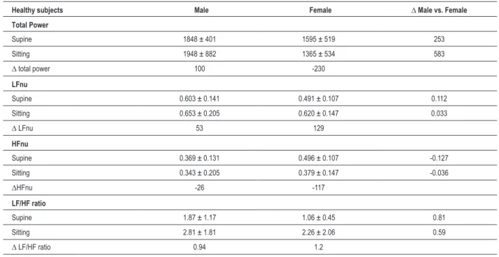
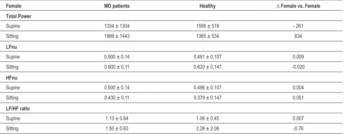
As per table 4, the control group showed no differences between gender and during changes in body position from supine to sitting. LFnu in the supine position was 22.8% higher for women in relation to men. Body position changes from supine to sitting lowered the HFnu by 30.8% among healthy females. The LF/HF ratio rose for men and women during changes in body position by 50% and 132%, respectively.
Inluence of gender and disease on HRV in healthy individuals and MD patients
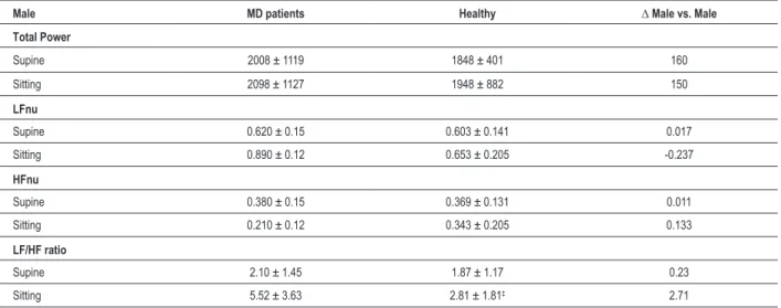
In the sitting position, male MD patients presented sympathovagal balances significantly high compared to healthy control individuals in 50.9% (Table 5). Influence of gender and disease on HRV in healthy individuals and MD patients is illustrated in Figure 1 and 2 and demonstrated in tables 5 and 6. Considering influences of disease and
gender on HRV in MD patients and healthy individuals, we observed significant gender-influence on the LF/HF ratio, HFnu in the sitting and supine position and LFnu in the supine position. Post hoc analyses show that gender significantly impacts MD patients and healthy individuals in different ways (p < 0.01). The low frequency domain in the sitting position, LFnu, was significantly influenced by the disease.
Discussion
This study proposes to assess inter-gender autonomic modulation of HR in patients with MD and healthy individuals in different body positions, as well as the influence of gender and disease and their interaction in both groups. HRV results in MD patients comparing genders suggest a difference in LFnu, HFnu and LF/HF ratio, with a decrease in parasympathetic modulation and increase in sympathetic modulation for males assessed in the sitting position. Sympathetic and parasympathetic modulations of the heart evaluated by LFnu, sympathovagal balance and LF/HF ratio increase for male MD patients with a change in body position. Sympathovagal balances are significantly high in male MD patients compared to healthy controls. HRV was affected by both gender and disease. Gender influenced LFnu in the supine position while the LF/HF ratio and HFnu was affected in both positions. Disease significantly influenced low frequency domain, LFnu.
In contrast to other research14-18, this study assesses HRV
during short time periods in sitting and supine positions, in addition to analyzing the magnitude of gender differences. Methodological procedures were based on the fact that Table 2 - Clinical characteristics of 17 patients with MD
Male (n=10) Female (n=7) All (n=17)
Length of time since diagnosis, years 9.5 ± 9.5 4.9 ± 3.4 7.6 ± 7.8 Degree of muscular impairment (MIRS)
Grade I - no muscular impairment 0 2 (28.6%) 2 (11.8%) Grade II - minimal signs, without distal weakness 3 (30%) 3 (42.8%) 6 (35.3%) Grade III - distal weakness 2 (20%) 2 (28.6%) 4 (23.5%) Grade IV - slight to moderate proximal weakness 5 (50%) 0 5 (29.4%) Grade V - severe proximal weakness 0 0 0
Data expressed as mean ± standard deviation. MIRS - Muscular Impairment Rating Scale17. MD - Myotonic dystrophy Table 1 – Anthropometric characteristics of 17 MD patients and 17 healthy individuals included in the study
MD CG p MD CG p
Female (n = 7)
Female (n = 7)
Male (n = 10)
Male (n = 10)
Age (years) 42.3 ± 13.8 40.14 ± 8.33 p > 0.05 38.2 ± 7.9 41.2 ± 2.9 p > 0.05 BMI (kg/m2) 25.1 ± 5.6 24.40 ± 1.2 p > 0.05 22.7 ± 3.1 24.5± 3.3 p > 0.05
Table 3 - Comparison of HRV in MD patients between supine and sitting positions
MD patients Male Female ∆ Male vs. Female Total Power
Supine 2008 ± 1119 1334 ± 1304 674 Sitting 2098 ± 1127 1999 ± 1443 99
∆ total power 90 665
LFnu
Supine 0.620 ± 0.15 0.500 ± 0.14 0.120 Sitting 0.890 ± 0.12* 0.600 ± 0.11‡ 0.290
∆ LFnu 0.120 0.100
HFnu
Supine 0.380 ± 0.15 0.500 ± 0.14 -0.120 Sitting 0.210 ± 0.12 0.430 ± 0.11‡ -0.220
∆ HF,nu -170 -70
LF/HF ratio
Supine 2.10 ± 1.45 1.13 ± 0.64 0.970 Sitting 5.52 ± 3.63* 1.50 ± 0.63‡ 4.02
∆ LF/HF ratio 3.42 0.37
Data expressed as mean ± standard error. * p < 0.01 between males in different positions and ‡ p < 0.01 between genders in sitting position. MD - Myotonic dystrophy; LFun - low frequency in normalized units; HFun – High frequency in normalized units; LF/HF - low frequency/ High frequency ratio or sympathovagal
balance; ∆ - delta or variation.
Table 4 – Comparison of HRV in healthy gender between supine and sitting positions
Healthy subjects Male Female ∆ Male vs. Female
Total Power
Supine 1848 ± 401 1595 ± 519 253
Sitting 1948 ± 882 1365 ± 534 583
∆ total power 100 -230
LFnu
Supine 0.603 ± 0.141 0.491 ± 0.107 0.112 Sitting 0.653 ± 0.205 0.620 ± 0.147 0.033
∆ LFnu 53 129
HFnu
Supine 0.369 ± 0.131 0.496 ± 0.107 -0.127 Sitting 0.343 ± 0.205 0.379 ± 0.147 -0.036
∆HFnu -26 -117
LF/HF ratio
Supine 1.87 ± 1.17 1.06 ± 0.45 0.81
Sitting 2.81 ± 1.81 2.26 ± 2.06 0.59
∆ LF/HF ratio 0.94 1.2
Data expressed as mean ± standard error. LFun - low frequency in normalized units; HFun – High frequency in normalized units; LF/HF - low frequency/ High
Gender inluence Inluência do Gênero
LF/H
F rati
o i
n s
upi
ne
MD
Heal
thy MD
Heal thy
LF/H
F rati
o i
n s
iti
ng
Males
Females p = 0.025
15.0
12.5
10.0
7.5
5.0
2.5
0.0
15.0
12.5
10.0
7.5
5.0
2.5
0.0
p = 0.001
p < 0.01
Figure 1 - Gender inluence on LF/HF ratio in MD patients and healthy individuals in both positions.
HRV measurements obtained with 5-minute recordings demonstrated good reproducibility, as well as being fast and easy to analyze24-26. Additionally, HRV responses to
postural changes are considered a better predictor of heart events27. Nevertheless, HRV is relatively simple
to assess, although results can be difficult to interpret28.
Modulation of heart rate is strongly influenced by several factors, including body position, humor, mental stress and environmental conditions29. HRV assessment also depends
on local conditions such as noise, temperature, spontaneous and quiet breathing30,31. As previously stated32, due to
physiological differences between males and females, it is essential to compare inter-gender differences in HRV indices. Finally, HRV must be analyzed considering frequency domains in normalized units. This is the most recommended
form of analysis since it accurately represents variations of sympathetic and parasympathetic modulation23.
To our knowledge, five previous studies on autonomic modulation of Heart Rate in patients with MD have been published. Discrepant results obtained in the past may be due to different analytical methods, autoregressive or fast Fourier, which were not interchangeable33,34, as well
as diverse postural positions during HRV measurement in each study. Inoue et al14 were the first authors to investigate
HRV in MD type 1 patients. They analyzed HRV using the autoregressive method in 10 MD patients (4 male and 6 female) without cardiac conduction disturbance, in addition to 10 age and gender-matched healthy controls at rest in the supine position. The authors found thatLFms2 and HFms2
decreased by 642% and 452% respectively, and that the Table 5 - HRV in male MD patients and healthy subjects in different body positions
Male MD patients Healthy ∆ Male vs. Male
Total Power
Supine 2008 ± 1119 1848 ± 401 160
Sitting 2098 ± 1127 1948 ± 882 150
LFnu
Supine 0.620 ± 0.15 0.603 ± 0.141 0.017 Sitting 0.890 ± 0.12 0.653 ± 0.205 -0.237
HFnu
Supine 0.380 ± 0.15 0.369 ± 0.131 0.011 Sitting 0.210 ± 0.12 0.343 ± 0.205 0.133
LF/HF ratio
Supine 2.10 ± 1.45 1.87 ± 1.17 0.23
Sitting 5.52 ± 3.63 2.81 ± 1.81‡ 2.71
Data expressed as mean ± standard error. ‡ p < 0.01 for signiicant difference between healthy individuals and MD patients in the sitting position. MD - Myotonic
dystrophy; LFun - low frequency in normalized units; HFun – High frequency in normalized units; LF/HF - low frequency/ High frequency ratio or sympathovagal
LF/HF ratio increased 158% in MD patients in relation to healthy individuals. The study did not consider the time since diagnosis and muscle impairment. However, the authors state that at the moment of investigation all patients were capable of walking and performing daily activities without assistance, corresponding to MIRS classification 1 to 4. The significant difference between MD patients and healthy individuals has never been confirmed by other studies. Flachenecker et al15 investigated the response of cardiovascular autonomic
function, including HRV, analyzed by the fast Fourier method, in MD type 2 patients. However, they recorded a reduction of 53.7% for LFms2 in MD type 2 patients compared to healthy
volunteers. Recent research has identified significant variations in severity, type, and distribution of electrical myotony in MD type 1 and MD type 2, as well as a correlation between muscle weakness and myotony in the two disorders35. Hence,
comparison between this study and the results obtained by these authors is not appropriate. In a study similar to ours, Di Leo et al17, investigated HRV in MD type 1 patients using the
autoregressive method with different body positions. In 23 MD patients and a control group, they observed a significant decrease in LFms2, a marker of sympathetic activity, for MD
patients in the supine position. The results of this study can only be partially considered for comparison with ours. The authors did not describe baseline characteristics of the control group, HRV analysis was performed using the autoregressive method and the MD group included men and women with a substantial age variation between 15 years and 51 years. These factors may influence results, in that physiological differences
are established by HRV in men and women of different ages. Other two previous studies applied the fast Fourier method to analyze HRV in patients with MD. In a multicenter study, Hardin et al16 evaluated 289 MD patients in the supine
position during short HRV acquisition periods. They did not include a control group since the study aims were to evaluate ambulatory ECG in a large and diverse population with MD type 1, in which clinical factors were associated to HRV by analyzing low frequency domains LFms2, HFms2 and
LF/HF ratio. Furthermore, the investigation was conducted at several centers. HRV data showed wide variability, with standard deviation greater than the mean and higher than commonly found in HRV studies. These authors observed a decrease in total power and predominance of sympathetic
drive compared to normal HRV values. Finally, Rakocević-Stojanović et al18 analyzed HRV in twenty patients and fifteen
healthy controls. The study does not clearly describe patient and control group characteristics such as age, gender, time since diagnosis and presence of minor or major disability. Total power fell by 64.6% in MD patients in relation to healthy controls, while LFms2, HFms2 and LF/HF ratios were
not significantly lower in MD patients. The authors concluded that sympathetic dysfunction may occur in patients with MD type 1.
This study builds on this prior investigation in several important ways. Firstly, time since diagnosis is critically important for the development of comorbidities in MD patients and was not considered in any previous studies,
Gender inluence Gender inluence Disease inluence Gender inluence MD MD MD MD Heal thy Heal thy Heal thy Heal thy p = 0.023
p = 0.025
p = 0.021 p = 0.022
H
F (nu)
s
upi
ne
H
F (nu)
s itti ng LF (nu) s upi ne LF (nu) s itti ng Males Females Males Females 0.8 0.8 0.8 0.6 0.6 0.6 0.4 0.4 0.4 0.2 0.2 0.2 0.0 0.0 0.0 1.2 1.0 0.8 0.6 0.4 0.2 0.0 1.2 1.0
Table 6 - HRV in Female MD patients and healthy subjects in different body positions
Female MD patients Healthy ∆ Female vs. Female
Total Power
Supine 1334 ± 1304 1595 ± 519 - 261
Sitting 1999 ± 1443 1365 ± 534 634
LFnu
Supine 0.500 ± 0.14 0.491 ± 0.107 0.009 Sitting 0.600 ± 0.11 0.620 ± 0.147 -0.020
HFnu
Supine 0.500 ± 0.14 0.496 ± 0.107 0.004 Sitting 0.430 ± 0.11 0.379 ± 0.147 0.051
LF/HF ratio
Supine 1.13 ± 0.64 1.06 ± 0.45 0.007
Sitting 1.50 ± 0.63 2.26 ± 2.06 -0.76
MD - Myotonic dystrophy; LFun - low frequency in normalized units; HFun – High frequency in normalized units; LF/HF - low frequency/ High frequency ratio or
sympathovagal balance; ∆ - delta or variation.
whereas this study defined time since diagnosis for the patient group. Secondly, it is important to perform HRV analyses separately for men and women, adopting a control group. Thirdly, using normalized units to study frequency domains is highly recommended in order to analyze the influence of sympathetic and parasympathetic autonomic modulation on the heart. All procedures should be adopted considering that there are no established HRV reference values. Finally, since HRV study is a matter of great interest in clinical and physiological research, HRV measurement must be obtained under rigorous conditions to reduce factors that may influence results. According to data provided by our results, a starting point on HRV in myotony dystrophy type 1 can be established.
This study contains several strengths; however, the main limitation lies in the small sample of participants and extrapolation of the results should be done carefully. An important objective of this work, not previously investigated, was the study of cardiac autonomic modulation in MD, considering both male and female patients and influence of postural body change.
The present findings suggest that in middle-aged males, MD patients who are not severely impaired and present a moderate duration of disease, sympathetic drive seems to be greater than in healthy matched individuals.
Potential Conflict of Interest
No potential conflict of interest relevant to this article was reported.
Sources of Funding
This study was funded by CNPq and FAPERN.
Study Association
This article is part of the thesis of master submitted by Thaise Araújo, from Universidade Federal do Rio Grande do Norte.
References
1. Day JW, Roelofs R, Leroy B, Pech I, Benzow K, Ranum LP. Clinical and genetic characteristics of a five-generation family with a novel form of myotonic dystrophy (DM2). Neuromuscul Disord. 1999;9(1):19-27.
2. Groh WJ, Groh MR, Shen C, Monckton DG, Bodkin CL, Pascuzzi RM. Survival and CTG repeat expansion in adults with myotonic dystrophy type 1. Muscle Nerve. 2011;43(5):648-51.
3. Bouhour F, Bost M, Vial C. Maladie de steinert. Presse Med. 2007;36(6 Pt 2):965-71.
4. Romigi A, Izzi F, Pisani V, Placidi F, Pisani LR, Marciani MG, et al. Sleep disorders in adult-onset myotonic dystrophy type 1: a controlled polysomnographic study. Eur J Neurol. 2011;18(9):1139-45.
5. Palmer PM, Neel AT, Sprouls G, Morrison L. Swallow characteristics in patients with oculopharyngeal muscular dystrophy. J Speech Lang Hear Res. 2010;53(6):1567-78.
6. McNally E M, Sparano D. Mechanisms and management of the heart in myotonic dystrophy. Heart. 2011;97(13):1094-100.
7. Groh WJ, Groh MR, Saha C, Kincaid JC, Simmons Z, Ciofaloni E, et al. Electrocardiographic abnormalities and sudden death in myotonic dystrophy type 1. N Engl J Med. 2008;358(25):2688-97.
9. Tsuji H, Larson MG, Venditti FJ Jr, Manders ES, Evans JC, Feldman CL, et al. Impact of reduced heart rate variability on risk for cardiac events. The Framingham Heart Study. Circulation. 1996;94(11):2850-5.
10. Stein PK, Kleiger RE. Insights from the study of heart rate variability. Annu Rev Med. 1999;50:249-61.
11. Hoikkala H, Haapalahti P, Viitasalo M, Vaananen H, Sovijarvi AR, Ylikorkala O, et al. Association between vasomotor hot flashes and heart rate variability in recently postmenopausal women. Menopause. 2010;17(2):315-20.
12. Olofsson BO, Niklasson U, Forsberg H, Bjerle P, Anderson S, Henriksson A. Assessment of autonomic nerve function in myotonic dystrophy. J Auton Nerv Syst. 1990;29(3):187-92.
13. den Heijer JC, van Dijk JG, Bollen WL, Bos JE, Wintzen AR. Assessment of autonomic function in myotonic dystrophy. J Neurol Neurosurg Psychiatry. 1991;54(6):531-4.
14. Inoue K, Ogata H, Matsui M, Hayano J, Miyake S, Kumashiro M, et al. Assessment of autonomic function in myotonic dystrophy by spectral analysis of heart-rate variability. J Auton Nerv Syst. 1995;55(1-2):131-4.
15. Flachenecker P, Schneider C, Cursiefen S, Ricker K, Toyka KV, Reiners K. Assessment of cardiovascular autonomic function in myotonic dystrophy tipe 2 (DM2/PROMM). Neuromuscul Disord. 2003;13(4):289-93.
16. Hardin BA, Lowe MR, Bhakta D, Groh WJ. Heart rate variability declines with increasing age and CTG repeat length in patients with myotonic dystrophy type 1. Ann Noninvasive Electrocardiol. 2003;8(3):227-32.
17. Di Leo R, Rodolico C, De Gregorio C, Recupero A, Cogliatore S, Annesi G, et al. Cardiovascular autonomic control in myotonic dystrophy type 1: a correlative study with clinical and genetic data. Neuromuscul Disord. 2004;14(2):136-41.
18. Rakocević-Stojanović V, Milovanović B, Ivić N, Ille T, Marjanovic I, Stevic Z, et al. Cardiac autonomic nervous system in patients with myotonic dystrophy type 1. Acta Myol. 2007;26(2):112-4.
19. World Medical Association. Declaration of Helsinki: ethical principals for research involving human subjects. As amended in Tokyo, 2004. Ferney-Voltaire, France: The Association; 2004. [Accessed on 2011 june 26]. Available from: http://www.wma.net/e/ethicsunit/helsinki.htm
20. Mathieu J, Boivin H, Meunier D, Gaudreault M, Bégin P. Assessment of a disease-specific muscular impairment rating scale in myotonic dystrophy. Neurology. 2001;56(3):336-40.
21. Ruha A, Sallinen S, Nissila S. A real-time microprocessor QRS detector system with a 1-ms timing accuracy for the measurement of ambulatory HRV. IEEE Trans Biomed Eng. 1997;44(3):159-67.
22. Loimaala A, Sievanen H, Laukkanen R, Parkka J, Vuori I, Huikuri H. Accuracy of a novel real-time microprocessor QRS detector for heart rate variability assessment. Clin Physiol. 1999;19(1):84-8.
23. Heart rate variability: standards of measurement, physiological interpretation, and clinical use. Task Force of European Society of C a r d i o l o g y a n d t h e N o r t h A m e r i c a n S o c i e t y o f Pa c i n g a n d Electrophysiology. Circulation. 1996;93(5):1043-65.
24. Lord SW, Senior RR, Das M, Whittam AM, Murray A, McComb JM. Low-frequency heart rate variability: reproducibility in cardiac transplant recipients and normal subjects. Clin Sci. 2001;100(1):43-6.
25. Hojgaard MV, Holstein-Rathlou NH, Agner E, Kanters JK. Reproducibility of heart rate variability, blood pressure variability and baroreceptor sensitivity during rest and head-up tilt. Blood Press Monit. 2005;10(1):19-24.
26. Dantas EM, Gonçalves CP, Silva AB, Rodrigues SL, Ramos MS, Andreão RV, et al. Reproducibility of heart rate variability parameters measured in healthy subjects at rest and after a postural change maneuver. Braz J Med Biol Res. 2010;43(10):982-8.
27. Carnethon MR, Liao D, Evans GW, Cascio WE, Chambless LE, Rosamond WD, et al. Does the cardiac autonomic response to postural change predict incident coronary heart disease and mortality? The Atherosclerosis Risk in Communities Study. Am J Epidemiol. 2002;155(1):48-56.
28. Low PA. Clinical autonomic disorders. Philadelphia: Lippincott-Raven Publishers; 1997.
29. Ewing DJ, Neilson JM, Shapiro CM, Stewart JA, Reid W. Twenty four hour heart rate variability: effects of posture, sleep, and time of day in healthy controls and comparison with bedside tests of autonomic function in diabetic patients. Br Heart J. 1991;65(5):239-44.
30. Pinna GD, Maestri R, Torunski A, Danilowicz-Szymanowicz L, Szwoch M, La Rovere MT, et al. Heart rate variability measures: a fresh look at reliability. Clin Sci. 2007;113(3):131-40.
31. Sandercock GR, Bromley PD, Brodie DA. The reliability of short-term measurements of heart rate variability. Int J Cardiol. 2005;103(3):238-47.
32. Acharya UR, Kannathal N, Hua LM, Yi LM. Study of heart rate variability signals at sitting and supine positions. J Body Mov Ther. 2005;9(2):134-41.
33. Chemla D, Young J, Badilini F, Maison-Blanche P, Affres H, Lecarpentier Y, et al. Comparison of fast Fourier transform and autoregressive spectral analysis for the study of heart rate variability in diabetic patients. Int J Cardiol. 2005;104(3):307-13.
34. Pichon A, Roulaud M, Antoine-Jonville S, de Bisschop C, Denjean A. Spectral analysis of heart rate variability: interchangeability between autoregressive analysis and fast Fourier transform. J Electrocardiol. 2006;39(1):31-7.