SUMMARY
Objective: To describe the preoperative upper limb lymphoscintigraphic pattern in women with breast cancer. Methods: hirty-seven patients undergoing lymphoscintig-raphy within 30 days of surgery were investigated. Lymphoscintigraphic studies of 37 upper limbs ipsilateral to surgery and 32 contralateral upper limbs were performed. he examination protocol consisted in obtaining static images of the upper limb in semi-lex-ion ater 10 minutes, and 1 and 2 hours ater subcutaneous injectsemi-lex-ion of 1 mCi (37 MBq) of Tc-99m-dextran in the dorsum of the hand. he velocity of axillary lymph node vi-sualization (I, visible at 10 minutes; II, 1 hour; III, 2 hours; and IV, invisible) and degree (intensity) of nodal uptake (a, marked; b, moderate; c,mild; and d, absent) were analyzed.
Results: Optimal lymphatic functional pattern (Ia) was observed in four (11%) patients, in the ipsilateral upper limb, and six (19%), in the contralateral upper limb. Worse con-dition was observed in three (8%) patients (IVd) in the ipsilateral upper limb and two (6%) patients in the contralateral upper limb. he remaining patients showed intermedi-ate stintermedi-ates of velocity and uptake intensity. Conclusion: his study found relevant changes in preoperative lymphoscintigraphy, demonstrating preexisting functional diferences in the lymphatic system.
Keywords: Lymphatic system; lymphedema; breast neoplasm; radionuclide imaging;
lymph node excision; lymphangiogenesis.
Study conducted at Centro de Atenção Integral à Mulher (CAISM) and Universidade Estadual de Campinas (UNICAMP), Campinas, SP, Brazil
Submitted on: 03/09/2011
Approved on: 07/23/2011
Correspondence to:
Laura Ferreira de Rezende Avenida Alexandre Fleming, 101 Cidade Universitária
“Zeferino Vaz” Campinas, SP, Brazil CEP: 13083-881 [email protected]
Conlict of interest: None.
©2011 Elsevier Editora Ltda. All rights reserved.
Preoperative upper limb lymphatic function in breast cancer surgery
LAURA FERREIRADE REZENDE1, FELIPE VILELA PEDRAS2, CELSO DARIO RAMOS3, MARIA SALETE COSTA GURGEL4
1 Post-doctorate, Gynecology, Obstetrics, and Mastology Department, Universidade Estadual Paulista (UNESP); Professor of Physiotherapy, Centro Universitário das Faculdades Associadas de Ensino (UNIFAE), SP, Brazil
2 Residency in Nuclear Medicine; Assisting Physician, Clínica de Medicina Nuclear Vilela Pedras, Rio de Janeiro, RJ, Brazil
INTRODUCTION
he lymphatic system is a component of the human body closely related to the venous system and of which we have limited scientiic knowledge1. It has important functions
and, among them, are control of macromolecular homeo-stasis, absorption of lipids, immunologic function, and control of tissue luids2. Its main function is the removal
of lipids and proteins from interstitial spaces. Removal of those elements, on the other hand, is possible through the capillary lymphatic membrane, which is more permeable than the epithelial membrane of blood vessels. hus, when failure of the lymphatic system occurs, the development of lymphedema is observed3.
he etiology and risk factors for the development of lymphedema in postoperative breast cancer patients seem multifactorial and are not fully understood. he risk of developing lymphedema is associated with axillary dissec-tion, axillary radiotherapy, obesity, extension of the sur-gical technique, infection4, age, number of lymph nodes
dissected, number of afected lymph nodes, and level of lymph node removal5. However, these associations were
not maintained in other studies6-8.
Little is known on the preoperative anatomic and functional characteristics of the lymphatic system, and, therefore, we consider that all patients have normal upper limb lymphatic function. However, the similar prevalence of postoperative lymphedema ater breast cancer surgery among patients with unilateral and bilateral axillary dis-section does not corroborate this hypothesis9.
Lymphoscintigraphy is currently proposed as the main diagnostic test to investigate the peripheral lymphatic sys-tem, allowing visualization of lymphatic vessels and lymph nodes, as well as the quantiication of lymphatic transport, being used in clinical practice to indicate and quantify the function of the lymphatic system, on the morphologic and functional point of view, determine the number of sentinel lymph nodes, and identify patients at risk for the develop-ment of lymphedema ater lymph node dissection10.
Currently, it is diicult to establish the ideal classiica-tion for the funcclassiica-tional status of the upper limb lymphatic system, since those scales are created empirically11. Some
authors have proposed an assessment of lymphatic trans-port through dynamic lymphoscintigraphy by analyzing the time of appearance of the radiopharmaceutical on the lymph node12, according to the quality of the image13, and
by static lymphoscintigraphy to visualize axillary lymph nodes11,14. he majority of studies established 10 minutes
as the normal time for transportation of the radiopharma-ceutical, considering 1 hour delayed transport, and two hours as seriously compromised transport15-17.
It is believed that prior assessment of the upper limb ipsilateral to the surgery allows the detection of anatomi-cal and functional abnormalities of lymphatic distribu-tion, enabling more accurate analysis of postoperative
changes in the ipsilateral upper limb18. Early evidence of
those changes would allow the development of preventive actions, stricter follow-up, and, possibly, perform early diagnosis and treatment. hus, the objective of the pres-ent study was to describe the preoperative lymphoscinti-lographic pattern of the upper limb in females with breast cancer before mastectomy with axillary dissection.
METHODS
his is a descriptive study that evaluated 37 women in the preoperative period of unilateral mastectomy with lymph node dissection in the three Berg levels for invasive breast carcinoma. hirty-seven lymphoscintigraphies of the up-per ipsilateral limb and 32 of the contralateral limb were performed. Patients with prior breast and axillary surgery, infection of the upper limb, lymphedema – diagnosed by a 2-cm diference in circumference of the upper limbs, were excluded. Other exclusion criteria were: bilateral surgery, indication of sentinel lymph node biopsy prior to axillary dissection, and preoperative radiotherapy.
he idea and realization of this study were based on the Helsinki Declaration19. his study was approved by the
Eth-ics on Research Committee of Faculdade de Ciências Médi-cas of Universidade Estadual de Campinas (UNICAMP) and all patients signed and informed consent, but two of them refuse to participate in the study.
LYMPHOSCINTIGRAPHIC TECHNIQUE
he lymphoscintigraphic protocol is not standardized and difers according to the diagnostic center performing it. Diferences include the choice of the radiopharmaceutical, type, and place of injection, use of static or dynamic evalu-ations, and intervals to obtain the images20.
Data pertinent to the lymphatic system were evalu-ated by lymphoscintigraphy on the Department of Nuclear Medicine of Hospital das Clínicas of UNICAMP, per-formed preoperatively, no more than 30 days before the surgery. he protocol of the test consisted of static images of the upper limb in semi-lexion obtained with a scinti-graphic camera ELSCINT SP4 or ELSCINT SP6, with the patient in dorsal decubitus, 10 minutes, 1 and 2 hours af-ter subcutaneous injection of 1 mCi (37 MBq) of Tc-99m dextran on the dorsum of the hand20. he upper limb was
maintained at rest in the interval between images. he in-jection was performed by nurses’ aides properly trained.
LYMPHOSCINTIGRAPHIC INTERPRETATION
Interpretation of the test is better done based on recogni-tion of abnormal distriburecogni-tion of the radiopharmaceutical and knowledge of the relative time that takes the radio-pharmaceutical to reach regional lymph nodes21. hus,
In the present study, the time it took the radiopharma-ceutical to reach axillary lymph nodes22 was called velocity,
and the accumulation (intensity) of the radiopharmaceuti-cal in axillary lymph nodes10, degree. Gloviczki et al.16 also
used these combined parameters to establish a classiica-tion criteria, since the literature does not have a standard criteria established for quantitative evaluation of lympho-scintigraphy.
According to this proposal, the ideal pattern of nor-malcy considered was Ia, and IVd as total disruption of lymphatic function. he remaining patterns relect inter-mediate stages of disruption. hus, images were evaluated according to Box 1.
Lymphoscintigraphic studies were analyzed by two nuclear experts on diferent moments and, in case of dis-agreement, the exam was analyzed by a third nuclear ex-pert. Concordance among evaluators, regarding degree and velocity, was excellent, according to the Kappa coeicient23.
RESULTS
hirty-seven patients with invasive breast carcinoma scheduled to undergo radical mastectomy were submit-ted to ipsilateral upper limb lymphoscintigraphic studies, while 32 underwent contralateral studies. Regarding tumor staging, three patients were EI, fourteen, EII, sixteen, EIII, three, EIV, and in one case, staging was not possible. Twen-ty-six of them (70%) had clinical axillary involvement. Other clinical characteristics are presented on Table 1.
Among the results of the ipsilateral upper limb, 18 women (49%) had velocity I, 14 (38%), velocity II, 2 (5%), velocity III, and 3 (8%), velocity IV. As for the degree, 8 women (22%) had degree a, 13 (35%), b, 13 (35%), c, and 3 (8%), d.
Among the results of the contralateral upper limb, 14 women (44%) had velocity I, and 14 (44%), II. Two women (6%) had velocity III, and 2 (6%), IV. Regarding the degree, 8 women (25%) had degree a; 13 (41%), b; 9 (28%), c; and 2 (6%), d.
Considering parameters of velocity and degree for a single classiication, it was observed that only 4 patients (11%) had classiication Ia, considered the normal pat-tern, on the ipsilateral upper limb versus 6 (19%) patients in the contralateral upper limb. hree (8%) and two (6%) patients were classiied IVd, respectively, considered complete disruption. he other patients were classi-ied as follows: 8 (22%) vs. 4 (12.5%), Ib, 6 (16%) vs. 4
(12.5%), Ic, 4 (11%) vs. 2 (6%), IIa, 3 (8%) vs. 7 (22%),
IIb, 7 (19%) vs. 5 (16%), IIc, 1 (3%) vs. 0, IIIa, and 1 (3%)
vs. 2 (6%), IIIb.
DISCUSSION
he objective of the present study was to describe the pre-operative functional pattern of the lymphatic system of the upper limb, through lymphoscintigraphy, in women with breast cancer to analyze intrinsic patient conditions as possible predictive factors to be considered in the de-velopment of lymphedema.
he initial diiculty of this study was to establish a re-producible classiication for the functional status of the upper limb lymphatic system, since those scales are cre-ated empirically11. We decided to group the analysis by
degree and velocity enabling static evaluation of the de-gree of capture of the radiopharmaceutical in the axillary lymph nodes and the transportation velocity of this drug through lymphatic vessels on the three moments ana-lyzed, as well as allowing its reproducibility.
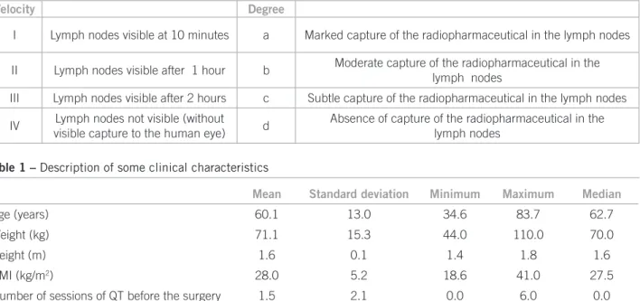
Box 1 – Classiication of velocity and degree of capture by lymph nodes in lymphoscintigraphy
Velocity Degree
I Lymph nodes visible at 10 minutes a Marked capture of the radiopharmaceutical in the lymph nodes
II Lymph nodes visible after 1 hour b Moderate capture of the radiopharmaceutical in the
lymph nodes
III Lymph nodes visible after 2 hours c Subtle capture of the radiopharmaceutical in the lymph nodes
IV Lymph nodes not visible (without
visible capture to the human eye) d
Absence of capture of the radiopharmaceutical in the lymph nodes
Mean Standard deviation Minimum Maximum Median
Age (years) 60.1 13.0 34.6 83.7 62.7
Weight (kg) 71.1 15.3 44.0 110.0 70.0
Height (m) 1.6 0.1 1.4 1.8 1.6
BMI (kg/m2) 28.0 5.2 18.6 41.0 27.5
Number of sessions of QT before the surgery 1.5 2.1 0.0 6.0 0.0
Cambria et al.12 proposed the evaluation of the
lym-phatic transportation through dynamic lymphoscintigra-phy by analyzing the time of emergence of the radiophar-maceutical in the lymph node. his scale also considered the transportation of the lymphatic movement, distribution of lymphatic pathways, and visualization of lymph nodes and lymphatic vessels. On the other hand, O’Mahony et al.13 proposed a classiication according to the quality of
the image, respecting the deinition of lymphatic vessels as very poor, poor, adequate, good, and excellent. Szuba et al.11 developed an empirical scale of static
lymphoscintig-raphy to visualize the axillary lymph nodes, considering normal pattern visible and symmetrical lymph nodes and, worse pattern as non-visible lymph nodes.
Lymphoscintigraphy is considered normal if discrete lymphatic vessels drain the extremity of the limb and if regional lymph nodes are visualized in up 1 hour14. Lane
et al.17 performed lymphoscintigraphic studies every 10
minutes for 1 hour. Weissleder and Weissleder15
consid-ered normal the appearance of the radiopharmaceutical in the lymph nodes in 10 minutes. Gloviczki et al.16 also
established 10 minutes as the normal time of transporta-tion of the radiopharmaceutical, considering 1 hour de-layed transportation and 2 hours seriously compromised transportation.
he intrinsic characteristic of lymphatic drainage is not considered when patients undergo surgery for breast can-cer, only postoperative factors are investigated as risk or not. his study demonstrated that the pattern considered ideal (Ia) was observed in only four patients (11%), in the ipsilateral limb, and in 6 patients (19%), in the contralat-eral limb.
hree patients (8%) had classiication IVd, on the ip-silateral limb, and two (6%), on the contralateral limb, considered the worse functional pattern because the ra-diopharmaceutical is not visible in any of the moments evaluated, demonstrating signiicant impairment of lym-phatic function. Of those, one patient had classiication IVd in both upper limbs. hree patients had BMI above 25 kg/m2, with staging indicating advanced disease; three
patients were older than 60 years, and the only young pa-tient had distant metastasis. hose clinical conditions ob-served agreed with Bourgeois et al.22 who observed that
age above 60 years and more than three lymph nodes in-volved represent variables that afect independently non-visualization of axillary lymph nodes. Two of those pa-tients underwent neoadjuvant chemotherapy.
In the present study, patients did not undergo radio-therapy, a known risk factor for lymphedema8,24, in which
signiicant clinical changes can be observed on preopera-tive lymphoscintigraphy of the ipsilateral limb, indicating it might have been caused by anatomical and functional patient-related or disease-related components as a conse-quence of advanced locoregional disease.
To evaluate the inluence of axillary disease on the changes observed, lymphoscintigraphic studies were per-formed on the contralateral limb, and relevant functional changes were also observed. hus, one can suggest that the variability on lymphatic functional status is also present on the upper limb considered normal.
Little is known about preoperative lymphoscinti-graphic study. It is believed that anatomical and functional changes present before the surgery would be important in the development of post-axillary dissection lymphedema. A single study reported an incidence of 7.5% of preopera-tive abnormalities in the lymphatic system, and 85% of those patients developed lymphedema approximately 34 months ater surgery22.
Baulieu et al.25 evaluated 32 patients with
post-opera-tive edema ater correcpost-opera-tive surgery of tibial fracture with lymphoscintigraphy performed between two and ten days ater the surgery, comparing with the healthy limb. Only patients with compromised lymphatic system developed lymphedema over a 3-month period. herefore, the au-thors inferred that preexistent functional changes in the lymphatic system justify the early development of lymph-edema.
he objective of this study was to evaluate preopera-tive lymphatic function in the upper limb, and the results observed, with 33 out of 37 patients of the ipsilateral limb with function outside the pattern considered ideal (Ia), and 26 out of 32 in the contralateral limb, suggest that the anantomo-functional characteristics of patients should be evaluated.
Considering the lymphoscintigraphic studies per-formed, 86% were classiied I or II, for velocity in the ip-silateral limb, and 87%, in the contralateral limb. As for the degree, 92% were degree a, b, or c, in the ipsilateral limb, and 97%, in the contralateral limb. hese classiica-tions could be considered normal in clinical practice, since they represent small changes in the lymphatic system. But women who were classiied III or IV, for velocity, and d, for the degree, should be referred to preventive programs in the immediate postoperative period, or even preopera-tively.
Correlating lymphoscintigraphic indings with com-mon complaints of changes in the upper limbs, which might indicate the onset of lymphedema, is important. hose complaints could reveal, subjectively, changes in the lymphatic system, indicating the need of evaluation for early diagnosis of lymphedema and follow-up conduct. Symptoms, such as feeling a heavy weight or hand being squeeze, increased volume, and changes in sensitivity, have been reported by patients with lymphedema and, rarely, by healthy women and, therefore, are useful early indicators of the development of lymphedema26, before the
Only the long-term follow-up of those patients can reveal whether the postoperative changes observed really indicate an increased risk for the development of lymph-edema. hus, patients with changes in preoperative lym-phoscintigraphy could be referred to lymphedema preven-tion programs earlier and with individualized attenpreven-tion; they could undergo weekly lymphatic drainage, receive orientation for auto-massage at home and preventive ori-entations.
hese results suggest the need lymphoscintigraphy studies in the immediate postoperative period to observe how preoperative indings behave ater axillary dissection and to the attempt of lymphangiogenesis.
CONCLUSION
his study observed relevant changes in preoperative lym-phoscintigraphy, demonstrating preexisting functional diferences in the lymphatic system.
REFERENCES
1. Szuba A, Pyszel A, Jedrzejuk D, Janczack D, Andrzejak R. Presence of functional axillary lymph nodes and lymph drainage within arms in women with ou without breast cancer-related lymphedema. Lym-phology 2007;40(2): 81-6.
2. Gashev AA, Zawieja DC. Physiology of human lymphatic contractil-ity: a historical perspective. Lymphology 2001;34:124-34.
3. Guyton AC. Tratado de Fisiologia Medica. 8ed. Rio de Janeiro: Ed. Guanabara Koogan, 1998.
4. Warren AG, Brorson H, Borud LJ, Slavin SA. Lymphedema: a com-prehensive review. Ann Plast Surg 2007;59:464-72.
5. Kiel KD, Rademacker AW. Early stage breast cancer: arm edema ater wide excision and breast irradiation. Radiology 1996;198:279-83. 6. Ozaslan C, Kuru B. Lymphoedema ater treatment of breast cancer.
Am J Surg 2004;18:69-72.
7. Soran A, D´Angelo G, Begovic M, Ardic F, Harlak A, Wieand S et al. Breast cancer-related lymphedema - what are the signiicant predictors and how they afect the severity of lymphdema? Breast J 2006;12(6):536-43.
8. Lee TS, Kilbreath SL, Refshauge KM, Herbert RD, Beith JM. Prog-nostic of the upper limb following surgery and radiation for breast cancer. Breast Cancer Res Treat 2008;110(1):19-37.
9. Stanton AW, Modi S, Mellor RH, Peters AM, Svensson WE, Levick JR et al. A quantitative lymphoscintigraphic evaluation of lymphatic function in the swollen hands of women with lymphoedema follow-ing breast cancer treatment. Clin Sci (Lond) 2006;110(5):553-61. 10. Szuba A, Shin WS, Strauss W, Rockson S. he third circulation:
ra-dionuclide lymphoscintigraphy in the evaluation of lymphedema. J Nucl Med 2003;44:43-57.
11. Szuba A, Strauss W, Sirsikar SP, Rockson SG. Quantitative radio-nuclide lymphoscintigraphy predicts outcome of manual lymphatic therapy in breast cancer-related lymphedema of the upper extremity. Nucl Med Commun 2002;23(12):1171-5.
12. Cambria RA, Gloviczki P, Naessens JM, Wahner HW. Noninvasive evaluation of the lymphatic system with lymphoscintigraphy: a pro-spective, semiquantitative analysis in 386 extremities. J Vasc Surg 1993;18(5):773-82.
13. O´Mahony S, Rose SL, Chivers AJ, Ballinger JR, Solanki CK, Barber RW et al. Finding an optimal method for imaging lymphatic vessels of the upper limb. Eur J Nucl Med Mol Imaging 2004;31:555-63. 14. Ter SE, Alavi A, Kim CK, Merli G. Lymphoscintigraphy. A reliable
test for the diagnosis of lymphedema. Clin Nucl Med 1993;18(8):646-54.
15. Weissleder H, Weissleder R. Lymphedema: Evaluation of qualita-tive and quantitaqualita-tive lymphoscintigraphy in 238 patients. Radiology 1988;167:729-35.
16. Gloviczki P, Calcagno D, Schirger A, Pairolero PC, Cherry KJ, Hal-let JW et al. Noninvasive evaluation of the swollen extremity:
ex-periences with 190 lymphoscintigraphy examinations. J Vasc Surg 1989;9(5):683-9.
17. Lane KN, Dolan LB, Worsley D, Mckenzie DC. Upper extremity lymphatic function at rest and during exercise in breast cancer survi-vors with and without lymphedema compared with healthy controls. J Appl Physiol 2007;103(3):917-25.
18. Scarsbrook AF, Ganeshan A, Bradley KM. Pearls and pitfalls of ra-dionuclide imaging of the lymphatic system. Part 2: evaluation of extremity lymphoedema. Brit J Radiol 2007;80:219-26.
19. Declaração de Helsinque III: ehtical principles for research involving human subject. Edinburgh; 2000. [cited 2000 oct 7]. Available from: http:www.ibemol.com.br/declarações/helsinque.
20. Yuan Z, Chen L, Luo Q, Zhu J, Lu H, Zhu R. he role of radionu-clide lymphoscintigraphy in extremity lymphedema. Ann Nucl Med 2006;20(5):341-4.
21. Howarth D. Increased Lymphoscintigraphy low pattern in the lower extremity under evaluation for lymphedema. Mayo Clin Proc 1997;72(5):423-9.
22. Bourgeois P, Leduc O, Leduc A. Imaging techniques in the manage-ment and prevention of posttherapeutic upper limb edemas. Cancer 1998;83(12 Suppl):2805-13.
23. Altman DG. Pratical statistics for medical research. London: Chap-man & Hall/CRC; 1991. p. 611.
24. Purushotham AD, Britton TMB, Klevesath MB, Chou P, Agbaje OF, Dufy SW. Lymph node status and breast cancer-related lymphede-ma. Ann Surg 2007;246:42-5.
25. Baulieu F, Itti R, Taieb W, Richard G, Martinat H, Barsotti J. Lym-phoscintigraphy. A predictive test of post-traumatic lymphede-ma of the lower limbs. Rev Chir Orthop Reparatrice Appar Mot 1985;71(5):327-32.