Universidade de Trás-os-Montes e Alto Douro
Repetitive DNA Sequences in Rodentia
Genomes
Its involvement in chromosome architecture reshuffling and in
genome functionality
Tese de Doutoramento em Genética Molecular Comparativa e Tecnológica
Ana Isabel do Paço Teixeira
Orientadora: Professora Doutora Raquel Chaves
Co-Orientadora: Doutora Filomena Adega
Composição do Júri:
Luis Herculano Melo de Carvalho Isabel Marques Carreira
Raquel Maria Garcia dos Santos Chaves José Eduardo Lima Brito
Agostinho Antunes
V
Este trabalho foi expressamente elaborado com vista à obtenção de grau de Doutor em Genética Molecular Comparativa e Tecnológica.
VII
Tese de doutoramento financiada pela
Fundação para a Ciência e Tecnologia, “Programa Operacional Potencial Humano –
POPH”, co-financiado pelo Fundo Social
Europeu (POPH/FSE) e por fundos nacionais (POPH-QREN).
IX
“A ciência será sempre uma busca e jamais uma descoberta. É uma viagem, nunca uma
chegada”.
XI
XIII
A
GRADECIMENTOSAo terminar esta etapa do meu percurso académico, olho para trás e vejo o quanto aprendi. Não adquiri apenas conhecimento! As pessoas com quem convivi e estabeleci laços de amizade foram imprimindo em mim novas formas de pensar e de ver o mundo. A pessoa que hoje sou não é mais que a soma de tudo o que vivi e consegui aprender. Desta forma, não posso deixar de prestar o meu mais profundo agradecimento a todos aqueles que contribuíram para a minha formação como pessoa, e que surpreendentemente não têm real consciência de como foram agentes ativos neste meu processo de crescimento.
À UNIVERSIDADE DE TRÁS-OS-MONTES E ALTO DOURO, na pessoa do magnífico Reitor Professor Doutor António Augusto Fontainhas Fernandes, instituição que me acolheu sempre durante a minha formação Académica, que se iniciou nos finais de 1998 ao ingressar na Licenciatura de Engenharia Zootécnica, continuando em 2004, com a realização
de um curso em Genética Molecular Comparativa e Tecnológica
(Pós-Graduação/Mestrado/Doutoramento). Olhando para trás, cerca de 35% da minha vida está relacionada com esta Universidade e por este motivo é imperativo agradecer-lhe. Sinto realmente que estou em casa na UTAD! Irei para sempre lembrar-me de todos os espaços verdes, locais encantados e misteriosos, onde consegui sentir paz e podia sempre recorrer quando precisava de calma. Irei recordar o cantar dos pássaros, principal razão pela qual ia tantas vezes ao nascer do sol para a Universidade, do orvalho ou a geada nas folhas, dos esquilos a saltar de árvore em árvore como se tivessem ventosas nas patas, da cidade de toupeiras ao pé do lago e do coaxar das rãs pela tardinha nos dias quentes. Mas principalmente do Pedrinhas, o meu local preferido, onde ficava maravilhada com as flores dos abrunheiros dos jardins ou das magnólias. Adorava o mês de Maio no Pedrinhas, com os seus dias longos e quentes, existindo aqui o conforto na sombra das grandes árvores sempre que o sol queimava. Mas também a sua igreja à noite, nos dias curtos de Dezembro, envolta em nevoeiro, sobressaindo as luzes dos candeeiros que a iluminavam. Irei realmente sentir saudades……
À ESCOLA DE CIÊNCIAS DA VIDA E DO AMBIENTE, na pessoa do Professor Doutor António Luis Herculano Melo de Carvalho, instituição que me acolheu durante o período que realizei trabalho de investigação referente ao meu Mestrado/Doutoramento.
XIV
À COMISSÃO PERMANENTE DO CONSELHO CIENTÍFICO DA ESCOLA DE CIÊNCIAS DA VIDA E DO AMBIENTE, na pessoa do seu atual Presidente, Professor Doutor Victor Machado Reis, pela aceitação da intenção de Doutoramento.
AO CENTRO DE GENÓMICA E BIOTECNOLOGIA DESTA UNIVERSIDADE, na pessoa da Professora Doutora Arlete Mendes Faia, expresso o meu profundo agradecimento pela disponibilização de meios para a realização desta tese.
ÀS COMISSÕES DE CURSO EM GENÉTICA MOLECULAR COMPARATIVA E TECNOLÓGICA, atualmente na pessoa da Professora Doutora Paula Lopes, pela disponibilidade e prontidão que sempre demonstraram na resolução de qualquer problema que surgisse.
À PROFESSORA DOUTORA RAQUEL CHAVES, minha orientadora, por me ter dado o possibilidade de participar na sua linha de investigação e com isso ter aprendido tudo o que sei hoje. Ter podido dar aulas e orientar estágios. Para além dos conhecimentos científicos que adquiri durante estes últimos sete anos, aprendi também consigo a ter os pés sempre assentes na terra. Agradeço-lhe sempre que me chamou à razão! Por vezes, estava tão envolta no trabalho que não me distanciava o suficiente para perceber com mais clareza as minhas falhas. E esteve sempre lá para me chamar a atenção. Irei sempre recordar a sua lucidez e inteligência. Foi a pessoa que mais contribuiu para o meu crescimento, pois os seus ensinamentos estão muito para além da minha vida académica.
À PROFESSORA DOUTORA FILOMENA ADEGA, minha co-orientadora e mestre, dedico um agradecimento muito especial. Em todos os momentos decisivos esteve sempre comigo! Sempre que não sabia como continuar era a ela que recorria. Agradeço-lhe pela alegria e pela sua preocupação com o meu bem-estar. Agradeço-lhe sobretudo também pela exigência, pois sem ela não teria aprendido tanto. Olhando para tudo que passamos juntas, não lhe posso só chamar co-orientadora, mas amiga, uma grande amiga! Pois sempre foi verdadeira comigo, disse-me sempre o que pensava, mesmo que eu não o quisesse ouvir! E isso mostra o quanto se preocupa comigo!
Ao Doutor MIROSLAV PLOHL e Doutora NEVENKA MEŠTROVIĆ, do Instituto Ruđer Bošković (Croácia, Zagreb) por me terem recebido no seu laboratório e me terem ensinado
XV
tanto. Obrigada também pela amizade e preocupação com o meu bem-estar. Agradeço igualmente aos restantes membros da sua equipa, Eva Šatović, Martina Pavlek e Brankica Mravinac com quem tive o prazer de trabalhar no período de tempo que passei lá.
AOS MEUS COLEGAS DE LABORATÓRIO, amigos para a vida e espero que para sempre. Refiro particularmente a Ana Borges, Ana Vieira da Silva, Cláudia Baptista, João Coutinho, Jorge Pereira, Karine Bouilly, Sandra Louzada, Sara Santos e Susana Meles (ordem alfabética). Tudo o que diga nunca chegará para expressar a estas pessoas o quanto as admiro e me são necessárias. E por não ter palavras suficientemente amplas que abranjam o agradecimento que lhes gostaria de prestar, dedico-lhes este poema de Vinícius de Morais.
Tenho amigos que não sabem o quanto são meus amigos. Não percebem o amor que lhes devoto e a absoluta
necessidade que tenho deles. A amizade é um sentimento mais nobre do que o amor,
eis que permite que o objeto dela se divida em outros afetos,
enquanto o amor tem intrínseco o ciúme, que não admite a rivalidade.
E eu poderia suportar, embora não sem dor,
que tivessem morrido todos os meus amores, mas enlouqueceria se morressem todos os meus amigos! Até mesmo aqueles que não percebem o quanto são meus amigos e o quanto a minha vida depende de suas existências ....
Vinícius de Morais (amigos)
Admiro particularmente a força da Ana Borges. Mesmo quando tudo lhe está a correr menos bem, consegue sempre incentivar e animar os outros. Defende arduamente quem ela considera amigo, e fico mesmo feliz por estar incluída nesse grupo.
Aprecio a sensibilidade da Ana Vieira da Silva. Até hoje, sem considerar a minha família, acho que foi a pessoa que me conheceu e compreendeu melhor. Foi com ela que descobri o gosto por viajar e conhecer novas culturas e modos de vida. Foi também com ela que vivi os momentos mais divertidos destes últimos oito anos. Esteve sempre do meu lado em tudo o que vivi de bom e mau. È uma pessoa particular, muito diferente de qualquer outra que conheço. Vê a bondade em toda a gente, o que por vezes a leva a desiludir-se.
XVI
Gosto da pessoa prática que é a Cláudia Baptista. Embora não conviva com ela todos os dias (o que lamento), segui seriamente todos os conselhos que ela me deu, principalmente no que diz respeito à escrita da tese e dos artigos. Penso que ela não tem consciência de quão seriamente ouvia o que ela me dizia.
Admiro a alegria e a autoconfiança do João Coutinho. Consegue sempre ter uma resposta para tudo! Sem se aperceber ajudou-me mil e uma vezes! Só o simples facto de me fazer rir tornava tudo muito mais fácil.
Para sempre vou lembrar-me do Jorge Pereira e da Sandra Louzada, meus grandes amigos. Adoro-os, são pessoas excecionais e confio neles mais do que em mim mesma. São realmente boas pessoas, em quem não consigo ver defeitos. Agradeço aos dois a ajuda que me deram em Inglaterra e particularmente ao Jorge a ajuda que também me deu na Croácia.
Agradeço à Karine Bouilly a amizade, a alegria e o apoio que sempre me deu quando precisava de tomar uma decisão. Irei para sempre recordar a sua companhia nas viagens que fizemos juntas. Foi mesmo divertido!
Admiro na Sara Santos a vontade altruísta que tem de ajudar os outros. Essa vontade é tão grande que por vezes se esquece dela própria. Tem sempre uma boa ideia para resolver qualquer problema, mas acho que ela não tem consciência do quanto me ajudou.
Agradeço à Susana Meles e Ana Cristina Silva, pela amizade e toda a disponibilidade que mostraram em ajudar-me. Com a Susana já vivi muitas experiências. Estivemos juntas desde o princípio e por isso lhe agradeço. À Ana Cristina, gostava de agradecer a alegria e o carinho que me transmitiu nos meus últimos dias no laboratório.
Ao João Castro, Raquel Matoso e Carlota Conceição, agradeço a simpatia e amizade. Estiveram comigo pouco tempo, mas tornaram a minha vida bem mais alegre.
Agradeço também à Daniela Ferreira e Ana Escudeiro. São a nova força do laboratório e por isso as reuni no mesmo grupo. Gostava de lhes agradecer por toda a ajuda que sempre me ofereceram.
AOS PROFESSORES DOUTORES JOSÉ EDUARDO LIMA-BRITO E ESTELA BASTOS, meus Professores na parte letiva deste curso, agradeço a simpatia e prontidão em ajudar sempre que necessário.
É impossível negar que na vida não passamos de simples folhas ao sabor do vento. Há um ano atrás, nunca suspeitaria que estaria agora a trabalhar na Universidade de Évora. Um novo
XVII
desafio nesta fase já por si só tão difícil do meu doutoramento. Por isso, não posso deixar de agradecer à professora Solange Oliveira pela confiança que depositou em mim e por me ter dado a oportunidade de trabalho que eu precisava. Agradeço também às doutoras Ana Alexandre e Marta Laranjo, por mostrarem confiança em mim e me incentivarem sempre. São realmente muito atenciosas comigo! Expresso também o meu agradecimento à dona Gertrudes Mariano, à professora Rosário Félix, ao professor José Manuel Martins e à doutora Carla Varandas, por serem sempre tão simpáticos para mim e terem ajudado na minha integração aquando da minha chegada a Évora. Mesmo muito obrigada!
Felizmente também, neste meu percurso por Évora fiz amigos a quem já devo muito! Pessoas que diminuem as minhas enormes saudades de casa, do Norte, de Vila Real!!! Que me fazem rir e dizer disparates, o que me faz tão bem! Refiro assim o Fabrício Macedo, Fernando Eliziário, Clarisse Brígido, Rodrigo Silva, Mónica Portella e Wellma Pedra (ordem alfabética). À Clarisse e ao Rodrigo, devo um agradecimento muito especial! São o meu apoio aqui, as pessoas que tem paciência de ouvir os meus problemas, que me tentam puxar para cima sempre que me sinto a bater no fundo! Fazem-me já muita falta e sem vocês tudo seria muito mais difícil. Muito, muito obrigada por tudo! Espero conseguir retribuir-lhes todo o esse carinho!
À MINHA FAMÍLIA, as pessoas que melhor me conhecem e que sempre me apoiaram durante toda a minha vida, não só nesta etapa, mas em todas as outras que fui superando. Agradeço o esforço que os meus pais, pessoas simples, filhos de lavradores, fizeram para que eu e a minha irmã estudássemos. Agradeço o estímulo, a confiança e o carinho que me deram todos os dias. Lamento no entanto, não poder dar-lhes a alegria de ter conseguido ter uma vida estável e por isso ficarem mais descansados em relação ao meu futuro. À minha irmã, e minha melhor amiga, agradeço o carinho, os conselhos, os estímulos, o incentivo, a confiança e a amizade. Agradeço também a ela e ao meu cunhado (Nuno) o privilégio de ser madrinha da Beatriz, a minha sobrinha. É uma menina feliz e espero sinceramente ser um bom exemplo para ela.
XIX
R
ESUMOUma das evidências mais claras revelada pelos projetos de sequenciação de genomas eucariotas, foi o seu elevado nível em sequências repetitivas de DNA, sendo a extraordinária variação no tamanho do genoma encontrada entre taxa atribuída à amplificação e eliminação diferencial destas famílias de sequências. No entanto, apesar da sua abundância, a(s) função(ões) que as sequências repetitivas desempenham nos genomas sempre esteve envolta em grande mistério, pois ao contrário dos genes, nunca foi atribuída a estas sequências a capacidade de codificar proteínas (pelo menos proteínas que não estejam envolvidas na sua própria replicação e integração no genoma, como é o caso dos Elementos Transponíveis). Por esta razão, estas sequências foram inicialmente designadas como “DNA lixo”, às quais nenhuma função era atribuída. Atualmente, as sequências repetitivas ganharam a merecida atenção e são agora consideradas como uma fração essencial dos genomas eucariotas, sendo reconhecidas como elementos reguladores importantes e também sendo implicadas na ocorrência de rearranjos cromossómicos, tendo assim uma função importante na evolução de genomas. Este foi precisamente o objetivo principal deste trabalho, contribuir para a compreensão da importância que as sequências repetitivas têm na evolução de genomas eucariotas. Com este propósito, aqui foi analisada a fração repetitiva de cinco espécies de roedores Cricetidae/Muridae, que apresentam cariótipos muito distintos, sendo nomeadamente estudadas sequências em tandem e dispersas: sequências DNA satélite (SatDNAs), Sequências Teloméricas Intersticiais (ITS) e Retrotransposões LINE-1. Uma análise detalhada sobre a distribuição e a natureza molecular da Heterocromatina Constitutiva (HC) foi também realizada para estes genomas de roedores.
A integração de todos os dados obtidos permitiu entender como os cinco genomas estudados evoluíram, bem como reconstruir os rearranjos cromossómicas ocorridos, nos quais as sequências repetitivas estão inquestionavelmente implicadas. De facto, todos os resultados obtidos aqui convergem para esta mesma conclusão. Nomeadamente, foi observada uma forte associação entre a distribuição e heterogeneidade da HC com o percurso evolutivo seguido por estes cariótipos. Uma análise detalhada feita neste trabalho, para duas das espécies estudadas, incidindo sobre a localização de regiões de “breakpoint” evolutivo e HC, revelou uma elevada coincidência entre estas regiões. Outros trabalhos, também focados na evolução cromossómica das restantes três espécies em análise aqui, relataram uma relação semelhante entre regiões de “breakpoint” e HC. Portanto, as sequências repetitivas localizadas na
XX
heterocromatina parecem estar intimamente envolvidas na ocorrência de rearranjos, quer promovendo diretamente reorganizações cromossómicas e/ou porque correspondem a regiões frágeis propensas à quebra. A análise de sequências repetitivas localizadas em regiões HC, especificamente satDNAs (sequências exclusivamente heterocromáticas), ITS (sequências localizadas principalmente nas regiões de HC) e LINE-1 (sequências frequentemente localizadas na HC), sugere realmente as sequências repetitivas como uma força motriz para a ocorrência de rearranjos cromossómicos. A natureza repetitiva das diferentes classes de sequências repetidas estudadas, por si só, favorece eventos de recombinação entre sequências homólogas em regiões não-homólogas, que podem culminar em rearranjos cromossómicos. No entanto, o papel na origem de reorganizações cromossómicas é particularmente sugerido para satDNAs, devido ao seu modo de evolução (evolução concertada), geralmente caracterizado por rápidas alterações na sua sequência de nucleótidos, variações no número de cópias e/ou movimentos intragenómicos, que resultam da ocorrência de diferentes eventos de recombinação (como “unequal crossing-over” ou “rolling circle replication/reinsertion”), podendo induzir quebras cromossómicas.
Adicionalmente, para além de sua importante função na reestruturação dos genomas, os resultados obtidos neste trabalho atribuem também outras funções às sequências repetitivas. Uma análise mais relacionada com a atividade transcricional de alguns satDNAs estudados, suporta o papel destas sequências em muitas outras funções, como no controlo da expressão génica, na remodelação da cromatina, na resposta celular ao stress e na função centromérica. As sequências LINE-1 têm também importantes funções no controlo da expressão génica, estando envolvidas no “imprinting” de genes e na inativação do cromossoma X. Assim, apesar de inicialmente consideradas sequências inertes, à luz de todos estes dados, é impossível negar que sequências repetitivas são cruciais para o bom funcionamento e evolução dos genomas eucariotas, destronando aos nossos olhos a importância dada no passado apenas a sequências codificantes. É agora realmente difícil de entender como estas sequências, tão abundantes nos genomas eucariotas, podem ter sido consideradas desnecessárias, só porque não lhes foi atribuída uma capacidade em codificar proteínas. Afinal, as sequências que codificam proteínas representam apenas uma pequena parte dos genomas (~ 1,5 % do genoma humano).
A presente tese resultou na elaboração de sete artigos que estão publicados, submetidos ou em preparação para submissão em revistas científicas internacionais indexadas.
PALAVRAS-CHAVE: Sequências Repetitivas de DNA, Evolução Cromossómica, Repetições em
XXI
A
BSTRACTOne of the clearest evidences that emerged from the eukaryotic genome sequencing projects was the high content in repetitive DNA sequences that these genomes harbour, being the extraordinary genome size variation found between taxa attributed to the differential amplification and deletion of these sequence families. However, despite its abundance, the role(s) that these sequences play in the genomes has always been shrouded in great mystery, as unlike genes, it was never assigned to them the ability to code proteins (at least proteins that are not involved in their own replication and genomic integration, as is the case of
Transposable Elements).For this reason, these sequences were initially designated as “junk”
DNA, with no function assigned. Presently, these sequences have won the deserved respect and are now regarded as a crucial fraction of eukaryotic genomes, recognized as important regulatory elements and also as being implicated in the occurrence of chromosomal rearrangements, with an important role in genome evolution. This was, precisely, the main goal of this work: to contribute to the understanding of the repetitive sequences significance in the evolution of eukaryotic genomes. For this purpose, it was analysed the repetitive genomic fraction of five Cricetidae/Muridae Rodentia species, with very distinct karyotypes, regarding tandem and dispersed repeats: Satellite DNAs (satDNAs), Interstitial Telomeric Sequences (ITSs) and LINE-1 Retrotransposons. A detailed analysis about the distribution and molecular nature of the Constitutive Heterochromatin (CH) of these rodent genomes was also performed.
The integration of all data allowed to understand how the five studied genomes evolved and to reconstruct the chromosomal evolutionary events elapsed, where the repetitive sequences were unquestionably involved. Indeed, all the results obtained here converge to this same conclusion. Namely, a strong association was observed between both the distribution and the level of CH heterogeneity with the evolutionary pathway that these karyotypes followed. In fact, for two of these species, a detailed analysis on the location of evolutionary breakpoint and CH regions revealed a very high coincidence between them. Other works focused on the evolution of the other three species, reported a similar relationship. Therefore, the repeats located in heterochromatin seem to be highly involved in the occurrence of chromosomal rearrangements, either by promoting directly chromosome reorganizations and/or because correspond to fragile regions prone to chromosome breakage. The analysis of the repeats located in CH regions performed here, namely satDNAs (exclusively
XXII
heterochromatic), ITSs (mainly located in CH regions) and LINE-1 (frequently located in CH), really suggest the repetitive sequences as a driving force in the occurrence of chromosomal rearrangements. The repetitive nature per se of the different classes of repeats studied favours recombinational events between homologous sequences in non-homologous regions, which may culminate in chromosomal restructurings. Nevertheless the role in the origin of chromosomal reorganizations is particularly proposed for satDNAs, due to its characteristic evolutionary mode (concerted evolution) generally marked by rapid sequence mutations, copy number variations and/or intragenomic movements, driven by different recombinational events (as unequal crossing-over or rolling circle replication/reinsertion), that may induce chromosome breakage.
Additionally, beyond its important function in genome restructuring, the data obtained in this work also suggest other roles to repetitive sequences. An analysis devoted to the transcriptional activity of some of the studied satDNAs supports the role of these sequences in many other functions, as in control of gene expression, chromatin remodelation, cellular response to stress and centromeric function. LINE-1 sequences as well have important functions in control of gene expression, acting in gene imprinting and in X-chromosome inactivation. Thereby, despite initially considered useless genomic elements, in the light of all this data, it is impossible to deny that repetitive sequences are crucial for proper functioning and evolution of eukaryotic genomes, dethroning to our view the importance given in the past just to the protein-coding sequences. It is really now difficult to understand how these sequences, so abundant in eukaryotic genomes, may have been considered unnecessary, just because a coding capacity was not reported. After all, the protein-coding sequences only account for a tiny part of genomes (~1,5% of the human genome).
The present thesis resulted in the elaboration of seven articles that are published, submitted or in preparation for submission to indexed international scientific journals.
K
EY WORDS:
Repetitive DNA Sequences, Chromosomal Evolution, Tandem Repeats, Long Interspersed Nuclear Elements-1, Rodentia.XXIII
G
ENERALI
NDEXAGRADECIMENTOS ... XIII RESUMO ... XIX ABSTRACT ... XXI
GENERAL INDEX ... XXIII INDEX OF FIGURES ... XXVII LIST OF PUBLICATIONS ... XXIX ABBREVIATIONS ... XXXII
ABBREVIATIONS OF SPECIES’ NAMES ... XXXIV
CHAPTER I.INTRODUCTION ... 1
I.1-Chromatin organization ... 4 I.1.1- Repetitive DNA sequences ... 5 I.1.1.1- Tandem repeats ... 7 I.1.1.1.1- Satellite DNA ... 7 I.1.1.1.2- Micro and minisatellites ... 12
I.1.1.1.2.1- Telomeric repeats... 12 I.1.1.1.3- Genomic impact and potential roles of tandem repeats ... 14 I.1.1.2- Dispersed repeats (transposable elements) ... 19 I.1.1.2.1- Mammalian LINE-1 retrotransposons ... 20
I.1.1.2.2-.Genomic impact and potential roles of mammalian LINE-1
retrotransposons ... 24 I.1.1.3- Tandem and dispersed repeats evolutionary relationship ... 26 I.2- Rodentia phylogeny: radiation and biogeography ... 28 I.2.1- Subamily Cricetinae: Cricetus and Phodopus genus ... 31 I.2.2- Subfamily Neotominae: Peromyscus genus ... 32 I.2.3- Subfamily Murinae: Praomys genus ... 32 I.3- Karyotype features of rodents ... 34 I.3.1- Muroidea karyotypes evolution ... 34
CHAPTER II.RESULTS AND DISCUSSION ... 37
XXIV
II.1.1- Hidden heterochromatin: characterization in the Rodentia species Cricetus cricetus, Peromyscus eremicus (Cricetidae) and Praomys tullbergi (Muridae) ... 43
II.1.2-.The involvement of repetitive sequences in the remodelling of
karyotypes: the Phodopus genomes (Rodentia, Cricetidae) ... 57
II.2- Satellite DNA ... 71
II.2.1-.Different evolutionary trails in the related genomes Cricetus cricetus
and Peromyscus eremicus (Rodentia, Cricetidae) uncovered by orthologous satellite DNA repositioning... 75
II.2.2-.Evolutionary story of a satellite DNA sequence from Phodopus
sungorus (Rodentia, Cricetidae) ... 85
II.2.3-.High-resolution organization of repetitive DNA sequences in
Phodopus roborovskii and Phodopus sungorus genomes (Cricetidae, Rodentia) ... 117 II.2.4-.Quest for the functional significance of a satellite DNA sequence
from Peromyscus eremicus (Cricetidae, Rodentia)… - in
preparation ... 151 II.3-LINE-1Retrotransposons ... 175
II.3.1- Line-1 Retrotransposons: from “parasite” sequences to Functional
Elements ... 179
CHAPTER III.GENERAL DISCUSSION AND FUTURE PERSPECTIVES ... 209
III.1- General discussion ... 211 III.1.1- Chromosomal location and molecular nature of the Constitutive
Heterochromatin ... 211 III.1.2- Characterization and evolution of Satellite DNA... 215 III.1.3- Telomeric repeats genomic distribution ... 219 III.1.4- LINE-1 retrotransposons genomic distribution ... 221 III.1.5- Concluding remarks ... 224
XXV
XXVII
I
NDEX OFF
IGURESFigure 1- Repetitive DNA sequences in eukaryotic genomes ... 6 Figure 2- Concerted evolution. ... 9 Figure 3- Satellite DNA evolution and diversification. ... 10 Figure 4- Library model ... 11 Figure 5- Possible mechanisms to explain the origin of large ITS blocks. ... 14
Figure.6-.Schematic representation of rat, mouse and human LINE-1
.retrotranposons ... 21 Figure 7- LINE-1 retrotransposition mechanism. ... 22 Figure 8- Rodentia Suborders. ... 29 Figure 9- Phylogenetic Tree of the superfamily Muroidea. ... 30 Figure 10- C-banding pattern in chromosomes of Cricetus cricetus, Peromyscus
eremicus, Phodopus roborovskii, Phodopus sungorus and Praomys tullbergi. ... 213
Figure.11-.Representative in situ hybridization of CCR4/10sat sequences on
Cricetus cricetus and Peromyscus eremicus chromosomes.…………. 216
Figure 12- Representative in situ hybridization of telomeric repeats (TTAGGG)n
on Phodopus roborovskii and P. sungorus chromosomes. ... 219
Figure.13-Table resuming all the results obtained in this work for the five studied
XXIX
L
IST OFP
UBLICATIONSThis thesis is based on the collection of the following articles throughout the PhD period:
Article 1:
Louzada S, Paço A, Kubickova S, Adega F, Guedes-Pinto H, Rubes J, Chaves R (2008) Different evolutionary trails in the related genomes Cricetus cricetus and Peromyscus eremicus (Rodentia, Cricetidae) uncovered by orthologous satellite DNA repositioning. Micron 39(8): 1149-1155.
Article 2:
Paço A, Adega F, Guedes-Pinto H and Chaves R (2009). The hidden heterochromatin: characterization in the Rodentia species Cricetus cricetus, Peromyscus eremicus (Cricetidae) and Praomys tullbergi (Muridae). Genetics and Molecular Biology 32 (1): 58-68.
Article 3:
Paço A, Chaves R, Vieira-da-Silva A and Adega F (2012). The involvement of
repetitive sequences in the remodelling of karyotypes: the Phodopus genomes (Rodentia, Cricetidae. Micron 46: 27-34.
Article 4:
Paço A, Adega F, Chaves R (2014). Line-1 Retrotransposons: from “parasite” sequences to Functional Elements. Submitted to Journal of Applied Genetics.
Article 5:
Paço A, Adega F, Meštrović N, Plohl M, Chaves R (2014). High-resolution
organization of repetitive DNA sequences in Phodopus roborovskii and P. sungorus (Cricetidae, Rodentia). In preparation.
Article 6:
Paço A, Adega F, MeštrovićN, Ploh M, Chaves R (2014). Evolutionary story of a
satellite DNA sequence from Phodopus sungorus (Rodentia, Cricetidae). In preparation.
Article 7:
PaçoA, AdegaF, ChavesR (2014). Quest for the functional significance of a satellite
XXX
From the work described resulted the following communications published in refereed proceedings of conferences:
1- Paço A, Adega F, Guedes Pinto H, Volobouev V and Chaves R (2007). Constitutive
heterochromatin characterization of the rodents Peromyscus eremicus, Cricetus cricetus and Praomys tullbergi. Chromosome Research 15(2): 41-42.
2- Meles S, Paço A, Filomena A, Guedes Pinto H and Chaves R (2008). Detailed
Constitutive Heterochromatin Map for Praomys tullbergi (Rodentia, Muridae) karyotype. Chromosome Research 16: 1035.
3- Louzada S, Vieira-da-Silva A, Paço A, Svatva K, Adega F, Guedes-Pinto H, Rubes J
and Chaves R (2008). Evolutionary chromosome repositioning of orthologous satellite DNA in the related genomes C. cricetus and P. eremicus (Rodentia, Cricetidae). Chromosome Research 16: 1046.
4- Paço A, Adega F, Guedes Pinto H, Volobouev V and Chaves R (2008). Molecular
analysis and Physical distribution of LINE-1 sequences in three rodent species, Cricetus cricetus, Peromyscus eremicus (Cricetidae family) and Praomys tullbergi (Muridae family). Chromosome Research 16: 1047.
5- Paço A, Adega F, Guedes-Pinto H and Chaves R (2009). Repetitive sequences: a
possible source of genome Remodelling. Chromosome Research 17 (Suppl 1): 166.
6- Paço A, Meštrovic N, Adega F, Plohl M and Chaves R (2010). Different organization
patterns of a satellite DNA sequence in closely related species, Phodopus sungorus and Peromyscus eremicus (Rodentia, Cricetidae). Chromosome Research 18: 727.
7- Paço A, Adega F, Meštrovic N, Silva A, Plohl M and Chaves R (2011). Construction
of repetitive DNA libraries from the genomes of two hamster species, Phodopus sungorus and Phodopus roborovskii (Cricetinae). Chromosome Research 19 (Suppl 1): S195.
From the obtained results are also deposited several DNA sequences in Genbank:
1- Cricetus cricetus LINE-1 partial sequence (Accession: HQ386006).
2- Peromyscus eremicus LINE-1 partial sequence (Accession: HQ386007).
3- Praomys tullbergi LINE-1 partial sequence (Accession: HQ386008).
4- Phodopus roborovskii Satellite DNA (PROsat) complete monomer variant clone 1
XXXI
5- Phodopus roborovskii Satellite DNA (PROsat) incomplete monomer clone 2
(Accession: KJ649145).
6- Phodopus Repetitive DNA (PsatDNA) clone 1 (Accession: KJ649146).
7- Phodopus sungorus Chromosome 1 Repetitive DNA (PSUchr1sat) clone 1
(Accession: KJ649147).
8- Phodopus sungorus Centromeric Repetitive DNA (PSUcentSat) clone 1 (Accession: KJ649148).
XXXII
A
BBREVIATIONSA, C, T, G Adenine, cytosine, thymine, guanine bp Base pair
CCP Comparative Chromosome Painting CENP-A Centromere protein A
CENP-B Centromere protein B CH Constitutive Heterochromatin cDNA Complementary DNA DNA Deoxyribonucleic acid
DIRs Dictyostelium intermediate Repeat sequences EN Endonuclease
FA-SAT Felis catus satellite DNA FISH Fluorescent in situ hybridization
het-ITSs Heterochromatic Interstitial Telomeric Sequences HIV-1 Human Immunodeficiency Virus
HOR Higher-order repeat HP1 Heterochromatin protein 1 HSF1 Heat Shock Factor 1
ITSs Interstitial Telomeric Sequences Kb Kilo bases
LINEs Long Interspersed Nuclear elements LINE-1 Long Interspersed Nuclear elements 1 My Million years
Mb Mega bases
mRNA Messenger RNA RE Restriction Endonuclease RT Reverse transcriptase ORFs Open Reading Frame RNA Ribonucleic acid rRNA Ribosomal RNA satDNA Satellite DNA
XXXIII s-ITSs Short Interstitial Telomeric Sequences RISC RNA-induced silencing complex
RIST RNA-induced transcriptional silencing complex RNP Ribonucleoprotein Particle
UTR’s Untranslated Regions TE’s Transposable Elements LTR Long Terminal Repeats
Non-LTR Non Long Terminal Repeats SIV Simian Immunodeficiency Virus TPRT Target Primed Reverse Transcription TSDs Target Site Duplications
XXXIV
A
BBREVIATIONS OFS
PECIES’
N
AMECCRCricetus cricetus
CGRCricetulus griseus
MAUMesocricetus auratus
MMUMus musculus
PERPeromyscus eremicus
PROPhodopus roborovskii
PSUPhodopus sungorus
PTU Praomys tullbergi RNO Rattus norvegicus
Chapter I
3 CHAPTER I.INTRODUCTION
In eukaryotic cells, genomic DNA is folded with histone and non-histone proteins forming the chromatin (Luger et al. 1997). During cell division, the condensed chromatin fibers are nominated as chromosomes, being this term firstly introduced in 1888 by Waldeyer (Zacharias 2001). The entire chromosome set, properly organized, of a species is known as karyotype. Organisms from different species present dissimilar karyotypes and this variation is due to numerical and structural chromosomal reorganizations (Sumner 2003). Some of the most spectacular karyotype differences occur among rodents, even among populations of the same species. The majority of the house mouse individuals have a karyotype consisting of 40 acrocentric chromosomes, however some populations, often in isolated places such as in Alpine valleys, presents a low chromosomal number as 2n = 22, by the formation of metacentric chromosomes from two acrocentric (Nachman and Searle 1995).
An important feature of the eukaryotic genomes is its high content in repetitive DNA sequences, whose functions are not yet completely understood. Nevertheless, several are the roles proposed to this genomic fraction, as its involvement in chromosomal rearrangements and consequently, in genome evolution (e.g. Moran and Gilbert 2002, Adega et al. 2009). This was, precisely, the main goal of this work; contribute to the understanding of the repetitive sequences significance in the evolution of genomes. For this purpose, several families of repeats were analysed in detail in various Rodentia species with very distinct karyotypes, namely Satellite DNAs, Interstitial Telomeric Sequences and LINE-1 Retrotransposons. A detailed analysis about the distribution and molecular nature of the Constitutive Heterochromatin for these rodent genomes was also performed.
To introduce the theme, a literature review embracing the current knowledge about the repetitive fraction in the eukaryotic genome, regarding its molecular characteristics, evolution mode and possible functions was undertaken and is presented in this chapter. The Results and Discussion chapter will be presented as individual papers being some of them already published, one submitted and others in preparation for submission. A general discussion is presented at the end, integrating and correlating all the data achieved.
4
I.1-Chromatin organization
In the eukaryotic genomes chromatin can be categorized as euchromatin or heterochromatin. Heitz (1928) was the first proposing this classification, based in the observation that throughout the cell cycle a chromatin fraction alters their degree of condensation (euchromatin), while the other fraction remains highly condensed (heterochromatin). Later, other features distinguishing these genomic compartments were identified. The euchromatin is enriched in unique coding sequences, while the heterochromatic fraction is referred to as gene poor, being mainly composed by repetitive DNA sequences such as clusters of satellite DNA and transposable elements (described in the next sections of this chapter) (Grewal and Jia 2007, Pezer and Ugarković 2008a). Besides, euchromatin and heterochromatin present characteristic histone-modification marks. The first is characterized by histone H4 acetylation and methylation of histone H3 at lysine 4, while the second is distinguished by the hypoacetylation and methylation of histone H3 at lysine 9 (Nakayama et al. 2001). The heterochromatin can also be considered as constitutive or facultative. The Constitutive Heterochromatin (CH) can occur as large blocks or discrete bands in different regions of the chromosome (detected by C-banding), however it is mainly found in large blocks near the centromere (Corradini et al. 2007, Probst and Almouzni 2008). Facultative heterochromatin is usually found at developmentally regulated loci where the chromatin state is changed in response to cellular signals and gene activity (Grewal and Jia 2007, Enukashvily and Ponomartsev 2013).
Several are the works reporting the ability of heterochromatin to propagate under epigenetic control to nearby DNA sequences, where a repression of these sequences occur in a process known as silencing (reviewed in Grewal and Jia 2007). This epigenetic control requires the methylation of histone H3 at lysine 9 and the subsequent association of chromodomain proteins, such as heterochromatin protein HP1, allowing histone modifications and chromatin assembly (Eymery et al. 2009). This process reduces the accessibility of the involved DNA sequences for being transcribed or for recombination (reviewed in Grewal and Jia 2007). Moreover, it was also reported that the heterochromatic surrounding of centromeres is necessary for their function, ensuring sister chromatid cohesion and kinetochore formation (e.g. Bernard et al. 2001, Nonaka et al. 2002, Obuse et al. 2004, Hall et al. 2012). The association between histone H3 methylated at lysine 9 and chromodomain proteins (e.g. HP1), allows the recruitment of cohesin complex that promote sister chromatid cohesion at
5
pericentromeric regions. HP1 facilitates as well the recruitment of kinetochore proteins such as Mis12 (reviewed in Grewal and Jia 2007). Heterochromatic rich regions are also considered as “hotspots” for the occurrence of structural chromosome rearrangements (John 1988, Chaves et al. 2004, Adega et al. 2009), which is mainly justified by the high molecular dynamics of the repetitive DNA sequences located in these regions (Adega et al. 2009). Therefore, the analysis of the heterochromatic fraction in a genome, regarding specifically its chromosome location and molecular heterogeneity (evaluation of repetitive sequences diversity located in heterochromatin), can be extremely important to understand the impact of repetitive sequences in genome evolution (e.g. Chaves et al. 2004, Adega et al. 2009).
I.1.1- Repetitive DNA sequences
Repetitive DNA sequences correspond to DNA elements that are present in multiple copies in a genome (e.g. Jurka et al. 2007). The accumulation of data from the 214 eukaryotic nuclear genomes completely sequenced up to now (data reviewed in January 2014, in http://www.ncbi.nlm.nih.gov/genome/browse/), shows that the repetitive sequences represent a significant portion of these genomes. Moreover, the differential amplification and deletion of their various families contribute significantly to the extraordinary genome size variation found between taxa (Petrov 2001, Boulesteix et al. 2006, Pritham 2009, Venner et al. 2009, Devos 2010, Sun et al. 2011), from 0.02 to 130 Gb within the Animal kingdown (Gregory 2011).
Based on the genomic organization of their copies, repetitive DNA sequences are classified as either tandem or dispersed repeats (reviewed in Slamovits and Rossi 2002, Jurka et al. 2007, Richard et al. 2008), as it is shown in figure 1. The first main type of repeats is organized in arrays of copies that can occur at few or in many different chromosomal locations. The second class, dispersed repeats, consist of sequences whose copies are highly scattered through the genome (Strachan and Read 2004). Each of these two classes of repetitive sequences can be itself divided into several subclasses (Figure 1). Here will be focused the largest of its subclasses, excluding the genic repetitive DNA sequences families. Within tandem repeats, three distinct subtypes exhibiting different properties, genomic distributions and length of copies and arrays are recognized: satellites, minisatellites and microsatellites (reviewed in Slamovits and Rossi 2002, Strachan and Read 2004). The dispersed repeats are represented by various types of transposable elements, presenting the
6
ability to transpose within the genome (Kazazian 2004, Jurka et al. 2007) as can be observed in figure 1.
Figure 1- Repetitive DNA sequences in eukaryotic genomes. This schematization collects the information
from several works (Slamovits and Rossi 2002, Feschotte and Pritham 2007, Jurka et al. 2007, Kapitonov and Jurka 2008, Richard et al. 2008, Kapitonov et al. 2009, Rebollo et al. 2012). Here there are only represented the largest subclasses of tandem and dispersed repeats, not including the tandem paralogue genes, ribosomal genes (tandem organization), retropseudogenes, transfer RNA genes or dispersed paralogue genes (dispersed organization).
7 I.1.1.1- Tandem repeats
Structurally, tandem repeats are formed by a sequential arrangement of copies, positioned one after the other (arrays). Two possible repeat orientations can be found in the genomes, head-to-tail repeats (direct repeats) and head-to-head repeats (inverted repeats) (Richard et al. 2008). Comparing the three subclasses of tandem repeats, the satellite DNA sequences have by far the largest arrays of copies, and this is the main feature that allows its differentiation from the micro and minisatellites (Plohl et al. 2008).
I.1.1.1.1- Satellite DNA
Satellite DNA sequences (satDNAs) correspond to highly tandemly repeated sequences that can be present in several millions of copies in a genome, organized into long arrays in the heterochromatic regions (Charlesworth et al. 1994). Indeed, satDNAs are the main constituent of Constitutive Heterochromatin (Ugarković and Plohl 2002, Chaves et al. 2004), being preferentially found in and around centromeres, but also assuming interstitial and terminal positions (reviewed in Adega et al. 2009). Historically, the first isolations of satDNAs were achieved by experiments with gradient centrifugation originating satellite bands, what justifies its name (Szybalski 1968). Currently, the more widespread strategy for the isolation of these repeats is the digestion of genomic DNA with restriction endonucleases, followed by sequence analysis of prominent cloned bands. This approach continued to be used even after the burst of large scale genome sequencing projects, since the high repetitive nature of satellites imposes serious limitations in assembling tandemly repeated motifs into large contigs, remaining these sequences underrepresented in outputs of genome sequencing projects (reviewed in Plohl 2010).
SatDNA repeat units (monomers) show a great variation in size, ranging from five nucleotides in human satellite III as well in some Drosophila satellites (Borstnik et al. 1994), up to several hundreds of base pairs (e.g. Modi 1993). However, it is considered that the preferential monomer length is 140-180 bp and 300-360 bp, since many satDNAs monomers in both plants and animals present these lengths (Schmidt and Heslop-Harrison 1998, Henikoff et al. 2001). In the same genome, several unrelated satDNA families presenting characteristic monomer extents and sequences can coexist, sharing only two common features, tandem arrangement of monomer repeats and heterochromatic localization (e.g.
8
Meštrović et al. 1998, Plohl 2010). The satellite DNA contribution to the total genomic content varies significantly among species, exceeding sometimes 50% of the total genomic DNA (Elder and Turner 1995, Schmidt and Heslop-Harrison 1998), and consequently these are involved in the enormous variation of genome size in eukaryotes (Doolittle and Sapienza 1980, Cavalier-Smith 1985, Gregory et al. 2007), as referred previously.
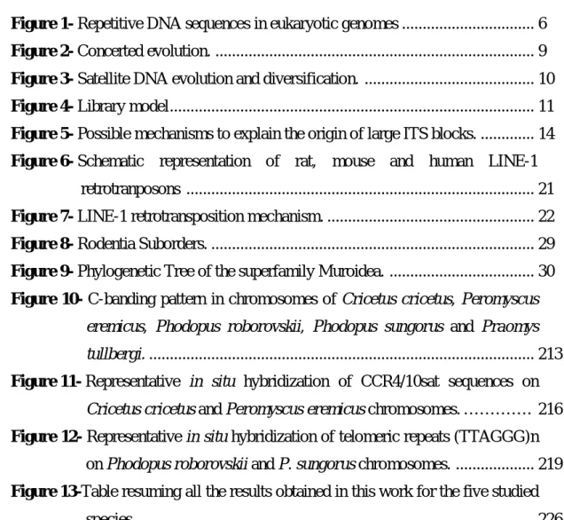
It is generally accepted that satDNAs follow the principles of concerted evolution (e.g. Palomeque and Lorite 2008, Plohl et al. 2008, Meštrović et al. 2013), indicating a non-independent evolution of satellite monomers within a genome, that results in an intraspecific homogenization of satDNAs (Elder and Turner 1995). This evolution mode is promoted by molecular drive, a complex process in which monomer mutations are spread or eliminated in the satellite arrays leading to repeats homogeneity (Figure 2), and concomitantly to its fixation in the individuals of a population (reviewed in Plohl 2010). This sequence homogenization occurs through mechanisms of non-reciprocal transfer within and between
chromosomes (as gene conversion, unequal crossing-over and rolling circle
replication/reinsertion that are dependent of intragenomic identity among satDNA monomers, and also transposon mediated exchange) (Walsh 1987, Thompson-Stewart et al. 1994, Elder and Turner 1995, Dover 2002). These homogenization mechanisms seem to act more efficiently within localized subsets of satellite monomers, decreasing their efficiency when occurring between different arrays on the same chromosome, homologous or heterologous chromosomes (Figure 2). This result on different rates of local and global sequence homogenization, showing adjacent monomers a higher degree of sequence similarity than those retrieved at random (reviewed in Plohl et al. 2008). In accordance, different works reported that when adjacent monomers are homogenized together may originate a new composite higher order repeat (HOR) unit, in which original monomers become subunits (e.g.
Willard and Waye 1987, Warburton and Willard 1990, Acosta et al.22010). These large
complex repeats generally show a high level of sequence identity, accumulating the constituent subunits
9 201co
Figure 2- Concerted evolution. Homogenization of a “mutated” satellite monomer within an array. Double
head arrows are correlated with the homogenization efficiency that is higher between homologous than non-homologous chromosomes. Adapted from Plohl et al. (2008).
constituent subunits a substantial sequence divergence (Palomeque and Lorite 2008). An example was reported for human alpha-satellite, presenting HORs highly homogeneous (97-100% of similarity), and internal subunits (alpha-satellite monomers) ~70% identical (Willard and Waye 1987, Roizes 2006). Based also on the different rates of sequence homogenization, theoretical models predicted that satDNA monomers at the array ends are more divergent than those located centrally (Smith 1976, Stephan 1989). The low efficiency of homogenization mechanisms in bordering regions of the satellite arrays leads to mutation accumulations in peripheral monomers (Mashkova et al. 1998, Schueler et al. 2005). The subsequent amplification of these monomers can originate novel satDNAs, as schematized in figure 3.
10
Figure 3- Satellite DNA evolution and diversification. (a) SatDNA is formed by tandem amplification of a
DNA sequence. (b) After amplification, sequence alterations and its homogenization lead to satellite sequence divergence relatively to the original sequence presented in a. (c) Due to the low efficiency of homogenization mechanisms in bordering regions of the satellite arrays, the amplification of highly mutated peripheral monomers can originate a new satDNA, and the process might repeat itself again. (d) During this evolutionary process, copy number of monomers can change significantly, what may eventually lead to extinction of some satDNAs. Adapted from Plohl (2010).
The described evolution mode of satDNAs results in species-specific satellite profiles due to differences in nucleotide sequence, monomer size, copy number variation or chromosome location (Charlesworth et al. 1994, Slamovits and Rossi 2002, Ugarković and Plohl 2002). Generally satDNAs present a highly dynamic molecular behaviour, being often reported satellite sequences only conserved in species belonging to a restricted taxonomic group (e.g. Martinez-Lage et al. 2005), a species (e.g. Stitou et al. 1999) or even a chromosome (e.g. Fátyol et al. 1994). Interestingly, despite the fact that the majority of satDNAs studied so far correspond to rapidly evolving genome components, some of these repeats seem to remain conserved during long evolutionary periods, with almost unaltered nucleotide sequences in the different genomes (e.g. Mravinac et al. 2002, Robles et al. 2004, Plohl et al. 2010). These few cases of satDNAs nucleotide sequence conservation highlight the complex behaviour of this genome fraction.The most extreme examples described until now are the mollusc BIV160 satellite family and the rodent PMsat, with about respectively 500 (Plohl et al. 2010) and 635 million years (My) (Louzada et al. 2014 submitted for publication). The basis for extreme conservation of some satDNAs is poorly understood. One assumption is that conservation can be a consequence of selection constraints imposed on satellite sequences, and/or it can be the result of slowing down mutation rates (e.g. Robles et al. 2004, Meštrović et al. 2006, Plohl et
11
al. 2010). The conservation of the satDNA nucleotide sequences can be also predicted by the concerted evolution mode, if “non desirable” mutations are preferentially eliminated instead of being spread throughout the satellite monomers (Dover and Flavell 1984, Ohta and Dover 1984, Strachan et al. 1985).
SatDNAs can vary dramatically in their number of copies among related species, and this high variation can be explained by the occurrence of molecular mechanisms involved in the homogenization of satDNA repeats (concerted evolution), namely unequal crossing-over and rolling circle replication/reinsertion (Slamovits and Rossi 2002). The variation in copy number of a set of satDNAs shared by related species was originally explained through the library model. This model postulates that related species share a satellite collection inherited from the ancestral genome, and in each species each satellite may suffer reduction or amplification in copy number, resulting in species-specific satDNA profiles (Fry and Salser
1977), as can be observed in figure 4. can becoiffere
Figure 4- Library model. In a genome several satDNA families can coexist, however, one of these families
often exhibits a higher copy number being considered as the major satellite. Adapted from Plohl et al. (2008).
As follows, one or few satDNAs can become highly represented in a species, whereas others remain as low copy number repeats. An experiment supporting this model was carried out by Meštrović et al. (1998) when studying different satDNAs shared by insect species from the genus Palorus. In this last work it was also demonstrated that satDNA copy number variations does not implies nucleotide sequence alterations. Species-specific profiles of satDNAs could result only by the variation of repeats copy number in the different genomes, independently of sequence nucleotide conservation (Meštrović et al. 1998, Mravinac et al. 2002, Bruvo et al. 2003).
12
The concerted evolution mode of satDNAs can also justify a distinct chromosomal location presented by orthologous satDNA sequences in related genomes (Hamilton et al. 1990, Slamovits and Rossi 2002). The homogenization of repeats between different chromosomal fields (within and between chromosomes) by molecular events as unequal crossing-over, rolling circle replication/reinsertion and transposon mediated exchange, can explain the different distribution of a satDNA in the genomes (Hamilton et al. 1990).
I.1.1.1.2- Micro and minisatellites
Micro and minisatellites are tandem repeated sequences composed by short repeat units, being the classification in one of these two categories mostly based on the length of their copies (reviewed by Richard et al. 2008). The size of the repetition motifs in microsatellites varies from 1 to 6 base pairs (bp) and therefore can be classified as mono, di, tri, tetra, penta or hexanucleotide repeats (e.g. (A)13, (GT)8, (GAT)7, (CTAG)6, (CATTG)5, (GGATCC)4). The classical microsatellites present only a single type of repeat unit, exhibiting an array size with up to 100 copies. However, these sequences can present more than a single type of repetitions and thus be named compound microsatellites (Schlötterer and Harr 2001).
Minisatellites present repeat units with up to tens of nucleotides and array sizes that may vary from 10 to 100 copies (reviewed in Slamovits and Rossi 2002). Both microsatellites and minisatellites are distributed throughout the genome, nevertheless minisatellites are also characterized by its (sub)telomeric location (Li 1997, Strachan and Read 2004), representing the telomeric repeats the major family of minisatellites (Strachan and Read 2004).
I.1.1.1.2.1- Telomeric repeats
Telomeres correspond to specialized nucleoprotein complexes (made up of DNA and proteins) that constitute the natural ends of eukaryotic linear chromosomes, displaying important roles, such as protection of the chromosome termini from degradation during DNA replication, prevention of deleterious end-to-end chromosome fusions, and participation in intranuclear chromosome positioning and segregation during cell division (Blackburn 2001). In the majority of eukaryotes, telomeric DNA is based on tandem arrays of simple short motifs with about 5-10 bp in length. The telomeric DNA of vertebrates and of some Bilateria
13
species, such as Mollusca, Annelida and Echinodermata consists of TTAGGG tandem repetitions (Ruiz-Herrera et al. 2008).
In addition to its characteristic terminal position, blocks of telomeric DNA repeats were already found at internal sites on the chromosomes of several vertebrate species, known as interstitial telomeric sequences (ITSs) (e.g. Meyne et al. 1990, Liu and Fredga 1999). The first cytogenetic evidence for the presence of ITSs in the karyotypes of vertebrate species was achieved by Meyne and collaborators (1990), using Fluorescent in situ Hybridization (FISH), that identified large ITS blocks preferentially located at the (peri)centromeric regions of the chromosome’s. According to their sequence organization and genomic location, two different types of ITSs can be identified in mammalian genomes, large blocks of heterochromatic ITSs (het-ITSs) and short ITSs (s-ITSs). The first type of ITSs presents several hundred kb of telomeric-like DNA, mainly located at heterochromatic (peri)centromeric regions. The s-ITSs length ranges from a few to a few hundred bp, and these repeats can be found in (peri)centromeric and interstitial regions of chromosomes (Ruiz-Herrera et al. 2008). Het-ITSs have been described in several mammalian species, such as primates of the genus Eulemur (Go et al. 2000), marsupials (Metcalfe et al. 2007), carnivores (Wurster-Hill et al. 1988), cetartiodactyls (Vermeesch et al. 1996), perissodactyls (Santani et al. 2002), chiropterans (Finato et al. 2000) and rodents (Bertoni et al. 1996, Ventura et al. 2006, Rovatsos et al. 2011). Outside the mammalian, this type of ITSs were also reported in amphibians (Wiley et al. 1992), reptiles (Pellegrino et al. 1999), fishes (Abuín et al. 1996) and birds (Nanda et al. 2002). Regarding s-ITSs, it is believed that they are probably present in all mammalian genomes (Ruiz-Herrera et al. 2008).
The origin of ITSs (het-ITSs or s-ITSs) is attributed to the occurrence of chromosomal rearrangements during karyotypes evolution (Figure 5), namely fusions (e.g. Slijepcevic 1998, Li et al. 2000) or pericentric inversions (e.g. Rovatsos et al. 2011). Specifically, the origin of (peri)centromeric ITSs are commonly explained by the occurrence of robertsonian-like fusions between acrocentric chromosomes, without loss of telomeric sequences. Besides, it is also suggested that s-ITSs can be generated through the insertion of telomeric DNA during the repair of double strand breaks (Nergadze et al. 2007, Ruiz-Herrera et al. 2008). The likely amplification of telomeric repeats through molecular mechanisms, such as unequal crossing-over, replication slippage or gene conversion, have been suggested to explain the large size presented by the het-ITS blocks (Ruiz-Herrera et al. 2008), as schematized in figure 5.
14
Figure 5- Possible mechanisms to explain the origin of large ITS blocks. Green blocks represent telomeric
sequences. The interrupted blue lines correspond to the breaks required for the occurrence of chromosomal reorganizations. (a) Origin of ITS blocks after the occurrence of fusion events. This process requires the inactivation of one centromere and the telomeres in the fusion point, for the correct segregation of the rearranged chromosome. (b) Origin of ITS blocks after the occurrence of a pericentric inversion. Chr fusion – Chromosome fusion, Cent and tel inact – Centromere and telomere inactivation, Per inv – pericentric inversion, ITS ampl – Interstitial telomeric sequences amplification. ITS amplification possibly occurs through molecular mechanisms as unequal crossing-over. This schematization collects informations from several works (Meyne et al. 1990, Slijepcevic 1998, Liu and Fredga 1999, Ruiz-Herrera et al. 2008, Rovatsos et al. 2011).
I.1.1.1.3- Genomic impact and potential roles of tandem repeats
Presently, repetitive sequences are gaining the respect deserved and are regarded as a crucial fraction of eukaryotic genomes, to which important functions have been assigned, like its recognition as important regulatory elements and also its involvement in the reorganization of genomes (e.g. Richard et al. 2008, Plohl 2010, Zhu and Pao et al. 2011, Hall et al. 2012, Enukashvily and Ponomartsev 2013). In fact, the extent of their regulatory importance is currently being explored in detail by The ENCODE Project Consortium, which have started with the human and mouse genomes (Mouse Encode Consortium 2012, The Encode Project Consortium 2012).
15
One of earliest functions suggested to satDNAs, if not the first, was its involvement in the centromeric activity, which was mainly based in their preferential (peri)centromeric location. In this regard, satDNAs localized within and around centromeres attract a considerable attention, having been proposed a role for these repeats in kinetochore assembling, spindle microtubule attachment and sister chromatid cohesion (e.g. Csink and Henikoff 1998, Henikoff et al. 2001, Sullivan et al. 2001). Additionally to its preferential location, the association of centromeric satDNAs with centromeric proteins also supports the involvement of these repeats in the centromeric functions (e.g. Plohl et al. 2008). The alpha-satellite in the human genome presents a sequence motif of 17 bp long, named CENP-B box, which is able to bind with the CENP-B protein, probably facilitating the kinetochore formation (e.g. Okada et al. 2007, Meštrović et al. 2013). Sequence motifs similar to the CENP-B box were also found in satDNAs from various organisms (e.g. Canapa et al. 2000, Lorite et al. 2004, Mravinac et al. 2005, Meštrović et al. 2013). Moreover, the molecular nature of satellite repeats led also to the suggestion that these sequences are the preferential form of DNA in functional centromeres and their flanking regions, because these sequences gather two features at the same time, sequence homogeneity over long DNA segments but also the potential to change very rapidly in time (e.g. Plohl et al. 2008, Plohl 2010). This dualism is considered particularly important for the interactions centromeric DNA sequences/proteins, since these interactions must remain stable; but to retain this stability over time the DNA sequence must also have the potential to coevolve with the rapidly changing protein component (Dawe and Henikoff 2006). Thus, the homogenization of monomer mutations within a (peri)centromeric satDNA array, which bring more efficiency for binding centromeric proteins, seems a crucial event in centromeric function (reviewed in Plohl et al. 2008). In accordance, it was assumed that both satDNA and protein evolution drive each other in the centromere, providing a stable but flexible system essential for centromeric activity (Dawe and Henikoff 2006). Nevertheless, the confirmation and completely recognition of the satDNA contribution to centromeric activity was achieved by the finding of transcription of these repeats, that shown that satDNA transcripts are structural elements of the functional centromere/kinetochore complex (e.g. Hall et al. 2012, Enukashvily and Ponomartsev 2013). Moreover, the satDNA centromeric transcripts are actually implicated in CENP-A deposition, a histone H3 variant found only at active centromeres (Bergmann et al. 2012). These discoveries add a new dimension to our current view of how (peri)centromeric sequences
16
participate in the formation, maintenance and function of specific structures such as centromeres.
Studies in the last decades shed light on a previous dark area where transcription was not accepted as a trait of satDNAs, mainly because these repeats are embedded in tightly packed heterochromatin. Presently, we all are aware of the fact that these repeats are transcribed, resulting in non-coding RNAs (e.g. Wong et al. 2007, Vourc’h and Biamonti 2011, Hall et al. 2012, Pezer and Ugarković 2012, Enukashvily and Ponomartsev 2013). In fact, the transcription of satDNAs seems to be a general phenomenon, having been reported satellite transcripts in several organisms including vertebrates, invertebrates and plants (Pezer and Ugarković 2012). So far, however, little is known about basic mechanisms of satDNA expression, its regulation and the function of all the generated transcripts (Pezer and
Ugarković 2008b, Vourc’h and Biamonti 2011, Enukashvily and Ponomartsev 2013).
Concerning the current knowledge, satDNA transcripts are usually heterogeneous in size and the transcription can proceed in both DNA strands or be strand-specific (Rudert et al. 1995, Rouleux-Bonnin et al. 1996). Some transcripts are present as polyadenylated RNA in the cytoplasm while others are found exclusively in the nucleus (Trapitz et al. 1988, Bonaccorsi et al. 1990). Besides, the transcription of some satDNAs is associated with the differentiation and development, and can be gender, age or tissue-specific, which provide clues indicating that these transcripts exhibit a regulatory role (Pezer and Ugarković 2012, Enukashvily and Ponomartsev 2013).
In addition to the centromeric functions, satDNA transcripts seem to have other and diverse roles. It has been shown that long single-stranded polyadenylated transcripts of human satellite III are directly involved in the cellular response to stress (Valgardsdottir et al. 2005). Some long satDNA transcripts can also function as ribozymes with self-cleavage activity (reviewed in Ugarković 2005). Moreover, diverse works reported as well that some satDNA transcripts can act as precursors of small interfering RNAs (siRNAs), which are recognized as having an important role in chromatin remodelation, leading to the heterochromatin formation and maintaining, and in the control of gene expression (reviewed in Vourc’h and Biamonti 2011, Enukashvily and Ponomartsev 2013). The mechanisms of chromatin modifications by siRNAs derived from satDNAs have been extensively studied in Schizosaccharomyces pombe, but it is believed that these mechanisms are conserved in other organisms as Drosophila, plants and, although with some doubts, in mammals (Pezer and Ugarković 2012, Enukashvily and Ponomartsev 2013). Double stranded RNAs of the S. pombe pericentromeric