Helena Maria Pinto Veiga
Dissertation presented to obtain the Ph.D degree in Biology
Instituto de Tecnologia Química e Biológica | Universidade Nova de Lisboa
Oeiras
Helena Maria Pinto Veiga
Dissertation presented to obtain the Ph.D degree in Biology
Instituto de Tecnologia Química e Biológica | Universidade Nova de Lisboa
Oeiras, December 2012
-
International Journals
Veiga H., Pinho M. G. (2009) Inactivation of the SauI type I restriction-modification system is not sufficient to generate Staphylococcus aureus strains capable of efficiently accepting foreign DNA.Applied and Environmental Microbiology.75 (10): 3034-8.
Pereira P.M.*,Veiga H.*, Jorge A.M., Pinho M.G (2010) Fluorescent reporters for studies of cellular localization of proteins in Staphylococcus aureus. Applied and Environmental Microbiology.76 (13): 4346-53.
*P.M.P. and H.V. contributed equally to this work.
Veiga H., Jorge A.M., Pinho M.G, (2011) Absence of nucleoid occlusion effector Noc impairs formation of orthogonal FtsZ rings during Staphylococcus aureus cell division. Molecular Microbiology.Jun; 80 (5): 1366-80.
-
Portuguese Journals
Veiga H., Pinho M.G (2012) Bacterial cell division: what it takes to divide a prokaryotic cell.Canal BQ. Nº9: 18-26.
Additional Publications
Atilano M.L.*, Pereira P.M.*, Yates J., Reed P., Veiga H., Pinho M.G.*, Filipe S.R* (2010) Teichoic acids are temporal and spatial regulators of peptidoglycan cross-linking in
Staphylococcus aureus.Proceedings of the National Academy of Sciences.107(44):18991-6 *A.M.L. and P.M.P and also M.G.P. and F.S.R. contributed equally to this work.
Contents
13
Abbreviations and Acronyms
15
Abstract
19
Resumo
25
Chapter 1
General Introduction
27 Bacterial cell division
28 Chromosome replication and segregation
28 The morphology of the bacterial chromosome
31 Spatial organization of the bacterial chromosome 32 Chromosome replication
34 Chromosome segregation
39 The FtsK/SpoIIIE family of DNA translocases
45 Assembling of the division septum
45 The Z-ring
47 Assembling of the Z-ring – Recruitment of the first group of divisome proteins
49 Maturation of the Z-ring
50 Constriction and closure of the division septum
52 Regulation of Z-ring assembling
52 Spatial and temporal regulation of the Z-ring
58 Regulation of Z-ring assembly according to cell cycle status
60 Staphylococcus aureus
60 Staphylococcus aureuscell division
62 Staphylococcus aureuspathogenicity and resistance to antibiotics
83
Chapter 2
Inactivation of the SauI Type I restriction-modification system is not sufficient to generateS. aureusstrains capable of efficiently accepting foreign DNA
85 Abstract
86 Introduction
88 Experimental procedures
88 Bacterial strains and growth conditions
88 Construction of hsdR null mutants
91 Electroporation
92 Transduction
93 Bacteriophage susceptibility assays
93 Results and discussion
93 Effect ofhsdRgene deletion on the transformation efficiency ofS. aureusstrains
97 Effect of deletion of the hsdR gene on S. aureus susceptibility to infection by
bacteriophages
98 Heat treatment of competent cells increases the transformation efficiency of hsdR
mutants.
99 Conclusion
100 References
103
Chapter 3
Fluorescent reporters for studies of cellular localization of proteins in Staphylococcus aureus
105 Abstract
106 Introduction
107 Experimental procedures
107 Bacterial strains and growth conditions
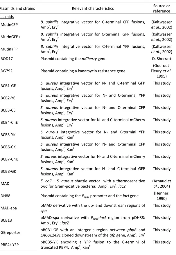
111 Construction of pBCB1-8 plasmids
112 Construction of pBCB13 plasmid
112 Construction of RNpGEreporter
113 Construction of RNpPBP4t-YFP
113 Construction of RNpPBP4-mCh
114 Construction of RNpEzrA-CFP
114 Construction of RNpPBP4t-YFP pEzrA-CFP
114 Construction of NCTCΔspaTetRmCh
115 Fluorescence microscopy
116 Results and discussion
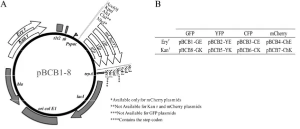
116 Construction of integration vectors for chromosomal expression of fluorescent derivatives
of staphylococcal proteins
119 Expression of fluorescent proteins by use of the pBCB series of plasmids
121 Covisualization of different fluorescent proteins expressed inS. aureuscells
123 Final remarks
124 References
127
Chapter 4
Absence of nucleoid occlusion effector Noc impairs formation of orthogonal FtsZ
rings duringStaphylococcus aureuscell division
129 Abstract
130 Introduction
133 Experimental procedures
133 Bacterial strains and growth conditions
133 Construction of the Noc null mutant
134 Complementation of Noc mutation
137 Construction of fluorescent derivatives ofS. aureusproteins
141 Fluorescence microscopy
142 Results
142 Chromosome segregation initiates prior to septum assembly in actively dividing
S. aureuscells
143 S. aureusNoc colocalizes with the origin proximal region of the chromosome
144 Noc mutants fail to avoid bisection of the chromosome, which results in DNA breaks
147 FtsZ polymerizes in multiple ring/arc structures in the absence of Noc
150 Perturbation of DNA replication/condensation results in the assembly of
non-orthogonal Z-rings
153 Discussion
158 References
163
Chapter 5
SpoIIIE and Slp mediate DNA translocation inStaphylococcus aureus
165 Abstract
166 Introduction
169 Experimental procedures
169 Bacterial strains and growth conditions
169 Construction of the SpoIIIE mutants
170 Complementation of the SpoIIIE mutation
171 Construction ofS. aureusstrains expressing a SpoIIIE fluorescent derivative
175 Construction of a Slp knockout mutant
176 Generation of anS. aureusSlp inducible mutant
176 Construction of Slp mutants in the DNA translocase domain
178 Construction ofS. aureusmutants lacking the DNA translocase activities from SpoIIIE and
Slp proteins
178 Construction of Slp fluorescent derivatives
180 Construction of aS. aureusstrain co-expressing fluorescent derivatives of SpoIIIE and Slp
181 Analysis of growth ofS. aureusstrains
183 Determination of the minimal inhibitory concentration (MIC)
183 Analysis of the expression of fluorescent proteins inS. aureus
184 Fluorescence microscopy
185 Results
185 Staphylococcus aureusexpresses two putative DNA translocases of the FtsK/SpoIIIE family
185 Lack ofS. aureusSpoIIIE causes chromosome condensation/segregation defects
187 S. aureusSpoIIIE can be found throughout the membrane or assembled in foci
190 Deletion ofS. aureusSlp causes a severe cell division phenotype
193 Slp is a multifunctional protein
195 Slp localizes early to the division septum
197 Slp localization depends on its N-terminal and linker domains
198 SpoIIIE and Slp have different localization patterns
200 The localization and expression of SpoIIIE and Slp is not interdependent
203 Deletion of both DNA translocases exacerbates the single mutants’ phenotypes
204 Discussion
208 References
213
Chapter 6
Concluding remarks and future perspectives
215 Concluding remarks and future perspectives
216 Development of genetic tools to better manipulateS. aureus
217 The direction of chromosome segregation definesS. aureusplanes of division
220 S. aureus SpoIIIE and Slp DNA translocases act in synergy to promote chromosome
segregation
223 References
Figures and tables Index
Chapter 1
31 Figure 1.1
The bacterial chromosome is spatially organized inside the cell 33 Figure 1.2
Precatenanes and dimers formed as a consequence of bacterial chromosome replication 43 Figure 1.3
B. subtilisSpoIIIE forms DNA conducting channels 57 Figure 1.4
Temporal and spatial regulation of Z-ring assembly by Min system and Nucleoid occlusion 61 Figure 1.5
Staphylococcus aureusmode of division
Chapter2
89 Figure 2.1
Strategy to deletehsdRusing pMAD vector 95 Figure 2.2
Genetic characterization ofhsdRmutants
90 Table 2.1
Primers 96 Table 2.2
Electroporation efficiency ofS. aureuswild-type andhsdRmutant strains transformed with plasmid pGC2 DNA extracted fromE. coliDH5α or fromS. aureusRN4220
98 Table 2.3
Efficiency of transduction and infection ofS. aureuswild type andhsdRmutant strains using phages 80α and ɸ75
Chapter 3
117 Figure 3.1
Map and nomenclature of pBCB series of plasmids 118 Figure 3.2
Map and mode of use of pBCB13 120 Figure 3.3
Expression of fluorescent proteins from the pBCB series of plasmids 122 Figure 3.4
108 Table 3.1
Plasmids and bacterial strains 110 Table 3.2
Primers
Chapter 4
144 Figure 4.1
(A) Chromosome segregation initiates prior to FtsZ assembly (B) Noc–YFP colocalizes with the nucleoid
146 Figure 4.2
Effects of Noc deletion inS. aureuscells and rescue bynocectopic expression 147 Figure 4.3
Noc deletion results in DNA breaks 149 Figure 4.4
In the absence of Noc, FtsZ forms multiple ring/arc structures on top of the nucleoid 151 Figure 4.5
Incubation ofS. aureuscells with chloramphenicol results in DNA condensation 152 Figure 4.6
FtsZ forms abnormal, non-orthogonal structures when DNA replication/condensation is impaired
157 Figure 4.7
Proposed model for cell division in three orthogonal planes
135 Table 4.1
Plasmids 136 Table 4.2
Bacterial strains 140 Table 4.3
Primers
Chapter 5
186 Figure 5.1
The absence of SpoIIIE causes nucleoid condensation and bisection by the septum 188 Figure 5.2
SpoIIIE molecules are usually distributed throughout the cell membrane and, in specific cases, assemble in foci
190 Figure 5.3
192 Figure 5.4
The absence of Slp protein causes severe cell division and chromosome segregation phenotypes
194 Figure 5.5
Slp C-terminal domain is not required to maintain normal cell and chromosome morphology 196 Figure 5.6
Slp localizes early to the division septa 198 Figure 5.7
Slp N-terminal and linker domains are both necessary for Slp localization 199 Figure 5.8
SpoIIIE and Slp do not colocalize in dividingS. aureuscells 202 Figure 5.9
SpoIIIE and the DNA translocase domain of Slp do not depend on each other for expression and localization
172 Table 5.1
Plasmids 174 Table 5.2
Bacterial strains 182 Table 5.3
Primers 203 Table 5.4
Phenotypes of SpoIIIE and Slp C-terminal single and double mutants
Chapter 6
219 Figure 6.1
Abbreviations and Acronyms
CFP Cyan fluorescent protein
CFU Colony forming units
GFP Green fluorescent protein
GlcNAc N-acetylglucosamine
IPTG Isopropyl-β-D-thiogalactopyranoside
KOPS FtsK-orienting polarized sequence
LA Luria-Bertani agar
LB Luria-Bertani broth
MCS Multiple cloning site
MIC Minimal inhibitory concentration
MRSA Methicillin resistantS. aureus MTS Membrane targeting sequence
MurNAc N-acetylmuramic acid
NBS Noc-binding sequence
NO Nucleoid occlusion
PBP Penicillin binding protein
PBS Phosphate Buffer Saline
PCR Polymerase Chain Reaction
PFGE Pulsed-field gel electrophoresis
PFU Plaque forming units
RBS Ribosome binding site
R-M Restriction-modification
SBS SlmA-binding sequence
SDS-PAGE Sodium dodecyl sulfate polyacrylamide gel electrophoresis
sfGFP Super-fast folding green fluorescent protein
SRS SpoIIIE recognition sequence
TSA Tryptic soy agar
TSB Tryptic soy broth
TUNEL Terminal deoxynucleotidyl transferase mediated X-dUTP nick end labeling
Van Vancomycin
VISA Vancomycin-intermediateS. aureusstrains
VRSA Vancomycin-resistantS. aureusstrains
WT Wild type
X-Gal 5-bromo-4-chloro-3-indolyl-β-galactopyranoside
Abstract
Bacterial cell division by binary fission involves several essential steps. Initially,
the mother cell duplicates in size and replicates its DNA, in preparation for division. As the
new sister chromosomes are synthesized, they are progressively segregated to the future
daughter cells and once the division site is cleared of the majority of chromosomal DNA,
the division septum starts assembling. The complete septum then constricts and,
ultimately, the mother cell splits into two identical daughters. The generation of equal,
viable progeny is strictly dependent on the regulation and coordination of these
processes, both in space and in time. It is particularly important that the division septum
is properly positioned at midcell and that chromosome segregation and cell division are
synchronized, to prevent fragmentation of the genome by septum closure over the
nucleoid.
Staphylococcus aureus is a particularly interesting model in which to study cell
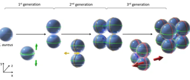
division as the cells are spherical and they divide in alternating perpendicular planes.
During three consecutive generations, the septa is sequentially placed in three orthogonal
planes, a process that is preceded by the segregation of the sister chromosomes along
three perpendicular axes. This mode of division is intrinsically different from that used by
the better studied rod-shaped organisms Escherichia coli and Bacillus subtilis, which
always segregate the chromosome in the same direction, parallel to the long axis of the
cell and place the septum at the same medial plane, to generate two identical daughter
cells.
This thesis focused on the study of the molecular factors and mechanisms
underlying cell division and chromosome segregation inS. aureusspherical cells, with the
aims of (i) investigating the role played by the nucleoid in the selection of S. aureus
division planes and (ii) assessing the role of DNA translocases in the coordination
between chromosome segregation and cell division. In order to perform these studies,
One of the limitations in genetically manipulatingS. aureuswas the fact that only
one strain, the highly mutagenized RN4220, could easily accept plasmid DNA isolated
fromE. coli, presumably as a result of a mutation in the hsdR gene of the Sau1 Type I
restriction-modification system. In order to obtain transformable variants of relevant S.
aureusstrains, thehsdRmutation was reproduced in different backgrounds. As described
in chapter two, deletion of hsdR gene in three different S. aureus strains was not
sufficient to make them readily transformable, indicating that transformability of S.
aureusRN4220 must also depend on other, unknown factor(s).
In order to increase the tool box of vectors that can be used for cell biology
studies inS. aureus, a series of plasmids that allow the expression of fluorescent fusions
to staphylococcal proteins were constructed, as described in chapter three. Additionally,
a new vector was constructed to allow the insertion of genes, under the control of the
IPTG-inducible Pspacpromoter, in the ectopicspalocus of theS. aureuschromosome. This
plasmid proved extremely useful for complementation studies that required a low gene
dosage, as well as for the construction of strains expressing fluorescent protein
derivatives at a distant place from their native locus. The latter is particularly important
for proteins encoded in essential operons, where it is critical to avoid polar effects on the
downstream genes. These tools were then used to study cell division and chromosome
segregation inS. aureus.
The first question that was addressed was the role of chromosome segregation in
the definition of the plane used for division. Before initiation of chromosome partioning,
most of the spherical staphylococcal cell is occupied by the nucleoid. Therefore, it is not
possible to define any putative division plane that divides the cell in equal halves without
bisecting the nucleoid. A nucleoid-free area, suitable for assembly of the division
apparatus, becomes available only upon segregation of the two newly formed
chromosomes. This implies that the axis of chromosome segregation restricts the number
of possible division planes to only one. Chapter four describes how the geometry of cell
division was found to be regulated by the nucleoid in a process mediated by theS. aureus
FtsZ, the first protein known to assemble at the division site, it was observed that in the
absence of Noc, FtsZ forms multiple ring structures that occupy different circular planes
of the spherical cells. These numerous FtsZ structures, which are not productive and do
not originate a complete septum, are formed on top of the chromosome, eventually
causing double-strand DNA breaks. These results show that, upon chromosome
segregation, the action of Noc is required to confine the localization of FtsZ and
consequently of the division septum to a single plane in the cell.
In addition to nucleoid occlusion, other regulatory mechanisms coordinate
segregation of chromosomes with cell division. The conserved proteins of the FtsK/SpoIIIE
family of DNA translocases for example, move DNA away from the division site before
cytokinesis. In chapter five of this thesis, it is shown that the twoS. aureus FtsK/SpoIIIE
proteins, named SpoIIIE and Slp (SpoIIIE-like protein), act through independent pathways,
most likely synergistically, to guarantee proper clearance of the chromosome from the
division site. SpoIIIE is a membrane protein that forms foci in the center of the septum
when its DNA translocase activity is required, while Slp is a multifunctional protein that
assembles early at the septum. Lack of SpoIIIE causes chromosome segregation defects
even during normal growth. In contrast, the C-terminal DNA translocase domain of Slp
seems to be required only in situations that impair normal chromosome replication/
segregation. The second function of Slp, performed by its N-terminal and linker domains,
is nevertheless critical for proper cell division, since lack of the entire Slp protein results in
severe cell division and chromosome morphological defects.
Overall, this study revealed the central role of the nucleoid in establishing the
division plane used for septum placement inS. aureus. It also highlighted the importance
of regulators that couple chromosome segregation with cytokinesis, like Noc or the DNA
Resumo
A divisão celular bacteriana por fissão binária envolve vários passos essenciais. Inicialmente, em preparação para a divisão, a célula mãe duplica de tamanho e replica o
seu DNA. À medida que os dois cromossomas são sintetizados, são progressivamente segregados para as futuras células filhas. Quando o local de divisão deixa de estar ocupado pela maior parte do cromossoma, o septo começa a formar-se, dividindo a célula mãe ao meio. Por fim ocorre a constrição do septo e a célula mãe separa-se em duas células filhas idênticas. A regulação e coordenação destes processos, tanto no
espaço como no tempo, é essencial para que seja gerada uma descendência viável e idêntica. É particularmente importante que o septo de divisão esteja perfeitamente posicionado no meio da célula e que os processos de segregação dos cromossomas e divisão celular estejam sincronizados, para evitar uma eventual fragmentação do genoma que aconteceria caso o septo se fechasse sobre o nucleoide.
Staphylococcus aureusé um modelo particularmente interessante para o estudo da divisão celular bacteriana porque tem uma morfologia esférica e divide-se alternadamente em três planos perpendiculares. Ao longo de três gerações consecutivas, o septo é sequencialmente colocado em três planos ortogonais, um processo precedido
pela segregação dos cromossomas ao longo de três eixos perpendiculares. Este modo de divisão é intrinsecamente diferente do utilizado por organismos mais estudados, como os bastonetes Escherichia colieBacillus subtilis,que segregam os cromossomas sempre na mesma direcção, paralela ao maior eixo da célula, e formam o septo sempre no mesmo plano, para gerar duas células filhas idênticas.
poder realizar estes estudos, foram desenvolvidas ferramentas de genética molecular para melhor manipularS. aureus.
Uma das limitações da manipulação genética de S. aureus é o facto de apenas uma estirpe, a altamente mutagenizada RN4220, poder facilmente aceitar DNA plasmídico isolado de E. coli, presumidamente em resultado de uma mutação no gene
hsdR do sistema de restrição-modificação Tipo I Sau1. De forma a obter variantes transformáveis de estirpes relevantes de S. aureus, a mutação no gene hsdR foi
reproduzida em “backgrounds” diferentes. Como descrito no capítulo dois, a deleção do gene hsdR em três estirpes diferentes de S. aureus não foi suficiente para as tornar transformáveis, o que indica que a transformabilidade da estirpe RN4220 deve depender de outro(s) factor(es) desconhecido(s).
Para aumentar o número de vectores disponíveis para os estudos de biologia
celular emS. aureus, foram construídos plasmídeos que permitem a expressão de fusões fluorescentes a proteínas de Staphylococcus, como descrito no capítulo três. Foi também construído um novo vector que permite a inserção de genes, sob o controlo do promotor indutivel Pspac, no locus ectópicospado cromossoma deS. aureus. Este plasmídeo provou
ser extremamente útil para estudos de complementação que requerem uma dose baixa do gene mutado, assim como para a expressão, emS. aureus, de derivados fluorescentes das proteínas de interesse, longe do seu locus nativo. Esta última aplicação é particularmente importante para proteínas codificadas em operões essenciais, onde é necessário evitar efeitos polares nos genes codificados a jusante do gene a alterar. Estas
ferramentas foram depois usadas para estudar a divisão celular e a segregação de cromossomas emS. aureus.
A primeira questão abordada neste trabalho prende-se com o papel desempenhado pela segregação dos cromossomas na definição dos planos usados para a divisão. Antes de se iniciar a segregação dos cromossomas, a maior parte do volume da
essencialmente livre de DNA, onde pode começar a ser formada a maquinaria de divisão. Isto implica que o eixo de segregação dos cromossomas restringe o número de planos de
divisão possíveis a apenas um. O capítulo quatro descreve a identificação do papel regulador do nucleoide na geometria da divisão celular, um processo que é mediado pela proteína Noc deS. aureus, homologa do factor mediador de “nucleoid occlusion” emB.
subtilis. Utilizando um derivado fluorescente do FtsZ, a primeira proteína a posicionar-se no local de divisão, foi observado que na ausência de Noc, o FtsZ forma numerosas
estruturas em forma de anel, que ocupam diferentes planos circulares da célula esférica. Estas estruturas de FtsZ, que não dão origem a uma maquinaria de divisão funcional e não originam septos completos, são formadas em cima do cromossoma e, eventualmente, resultam em quebras na cadeia dupla do DNA. Estes resultados mostram que, após a segregação dos cromossomas, a acção do Noc é necessária para confinar a
localização do FtsZ, e consequentemente do septo, a apenas um único plano na célula. Existem outros mecanismos de regulação, para além do processo de “nucleoid occlusion”, que coordenam a segregação de cromossomas com a divisão celular. Um exemplo são as proteínas conservadas da família de translocases do DNA FtsK/SpoIIIE,
que movem o DNA para longe do local de divisão antes da citocinese. No capítulo cinco desta tese, é mostrado que as duas proteínas FtsK/SpoIIIE deS. aureus, chamadas SpoIIIE e Slp (SpoIIIE-like protein), actuam através de vias independentes, e muito provavelmente de forma sinergística, para garantir que o local de divisão está livre de cromossomas. A proteína SpoIIIE é membranar e forma focos no centro do septo quando a sua actividade
como translocase do DNA é necessária. A proteína Slp, por seu lado, é multifuncional e localiza-se cedo no septo. A ausência de SpoIIIE causa defeitos na segregação de cromossomas mesmo durante o crescimento normal. Em contraste, o domínio C-terminal da proteína Slp, responsável pela actividade de translocação do DNA, parece só ser necessário em situações de replicação/segregação anormal dos cromossomas. A segunda
Este estudo revelou o papel central do nucleoide no estabelecimento do plano de divisão onde se forma o septo deS. aureus. Este estudo também realçou o importante
Bacterial cell division
Bacterial cells are no longer viewed as simple and disorganized reaction chambers
of homogenously distributed enzymes. It is now well established that these organisms
possess a complex subcellular organization, maintained by specialized temporal and
spatial regulatory systems. Moreover, the three classes of cytoskeletal elements (tubulin,
actin and intermediate filaments), previously considered exclusive to eukaryotic cells,
have also been shown to be present in bacteria. It is this dynamic and highly regulated
interior organization that assures the orderly and coordinated progression of the cell
cycle, essential for growth, chromosome segregation and cell division.
During the course of the cell cycle and in preparation for division, which usually
occurs by binary fission, bacteria double their mass, replicate their DNA and segregate the
two newly formed chromosomes. A division septum then assembles at a predetermined
site between the chromosomes, the cell constricts and, ultimately, the mother cell splits
into two identical daughters. This apparently straightforward mode of division has been
studied since the beginning of the twentieth century. Early investigations, using light and
electron microscopy, allowed the observation that, prior to bacterial division, an annular
disc (now known as the septum) was formed in the middle of the cell. This structure
closed, like an iris diaphragm, by invagination of the cytoplasmic membrane and ingrowth
of the cell wall, until partitioning of the cell is complete (Knaysi, 1941, Chapman & Hillier,
1953). However, it was only at the end of the 1960s, with the application of genetic tools,
that the essential proteins and molecular mechanisms behind bacterial division started to
be characterized. In pioneering work, Hirota and co-workers isolated Escherichia coli
thermosensitive mutants that failed to form the division septum when grown at
nonpermissive temperatures (Hirotaet al., 1968). This group of mutant strains included
filamentation temperature-sensitive (fts) mutants, which are unable to divide and
therefore filament/elongate at the restrictive temperature, as well as partition (par)
mutants that, in addition to the filamentation phenotype, had abnormally positioned
were later shown to localize at the septum. Critical among them is FtsZ (Bi & Lutkenhaus,
1991). This bacterial tubulin homologue is the earliest protein known to assemble at the
division septum. It polymerizes into a cytoskeletal contractile structure, the Z-ring, which
constitutes the initial position marker of the division site and is responsible for the
recruitment of the other components of the division apparatus (Bi & Lutkenhaus, 1991,
Goehring & Beckwith, 2005). Only a small group of bacteria divide in the absence of FtsZ
(Reviewed in (Erickson & Osawa, 2010)).The Z-ring orchestrates cytokinesis in most
prokaryotic cells, similarly to the actin-myosin ring in eukaryotes (Laporteet al., 2010).
FtsZ polymerization has to be tightly regulated in space and time to ensure
generation of equal progeny and faithful transmission of hereditary information. The
spatial control of FtsZ polymerization usually involves the identification of the midpoint of
the cell, although in some developmental situations, like sporulation, division occurs
asymmetrically (Barak & Wilkinson, 2007). An efficient temporal coordination between
septum placement and chromosome replication/segregation assures chromosome
integrity.
In the following sections, the current knowledge on the different steps of the
bacterial cell division cycle are summarized: (i) DNA replication and segregation; (ii) FtsZ
ring assembly; (iii) Z-ring maturation after orderly recruitment of the divisome
components; (iv) septal invagination with constriction of the envelope layers and (v)
septum closure and splitting of the daughter cells.
Chromosome replication and segregation
The morphology of the bacterial chromosome
The majority of bacteria possess a single circular chromosome that is not
enclosed in a specific membrane-surrounded compartment, but occupies a distinct region
in the cytoplasm that, due to its functional resemblance to the eukaryotic nucleus, is
chromosome is approximately 1.6mm, which largely exceeds the size of the cell (2µm
length and 1µm diameter). For that reason, the DNA is kept in a highly compacted
architecture (Odijk, 1998).
The first level of compaction is ensured by topoisomerases that catalyze the
coiling of the DNA duplex around itself (Wang, 2002). As a result of topoisomerases
activity the circular bacterial chromosome is organized in numerous supercoiled domains
that emanate from the central core and have an average length of 10Kb inE. coli(Postow
et al., 2004). The boundaries of these loops are variable and randomly distributed over
the chromosome. Moreover, each domain is independent from the others, which means
that, a break in a single loop does not affect the superhelicity of the others (Postowet al.,
2004). These supercoiled domains are disrupted during replication and transcription and
rapidly reestablished afterwards, with the introduction of negative supercoils. The
torcional tension accumulated in the loops is the source of valuable free energy used in
the processes that requires melting of the DNA strands (Denget al., 2005).
Supercoiling by itself is very effective in the reduction of the cellular volume
occupied by the chromosome. Nonetheless, other factors contribute to the balance of
forces that establish the degree of compaction. Important roles are attributed to the
forces exerted by a crowded cytoplasm (Zimmerman & Murphy, 1996) and to the
transertion process (insertion of proteins into the membrane as their genes are
transcribed and translated) that counteracts compaction by pulling DNA loops towards
the cell membrane (Woldringhet al., 1995). Additional very important players include a
group of nucleoid-associated ‘histone-like’ proteins (NAPs) and the SMC/MukB
complexes.
NAPs are a large and heterogeneous family of low molecular weight proteins that
bend or twist the DNA, which includes: HU (heat unstable protein), H-NS (Histone-like
nucleoid structuring protein), IHF (integration host factor) and Fis (factor for inversion
different NAPs, some acting as compacting agents, and others as antagonists of
compaction (reviewed in (Rimsky & Travers, 2011, Browninget al., 2010)).
The SMC (structural maintenance of chromosome) family, conserved in the
different branches of life, includes the well-studiedB. subtilisSMC protein (Brittonet al.,
1998) and itsE. colihomologue MukB (Nikiet al., 1991), both of which are involved in the
condensation of the chromosome. SMCs function in complex with two non-SMC subunits.
MukB interacts with MukE and MukF (Yamazoeet al., 1999), while SMC complexes with
ScpA and ScpB (Mascarenhas et al., 2002, Soppa et al., 2002). All prokaryotic SMC
proteins have the same structure. They are composed of globular N- and C-terminal
domains separated by two long coiled-coil regions and a central flexible hinge. The two
coiled-coil motifs fold back onto each other so that the terminal domains form a
functional ABC-type ATPase. Then the two SMC molecules dimerize through their hinges
domains to form a V-shapped structure with a large opening angle (Melbyet al., 1998).
Inactivation of SMC or MukB causes temperature-sensitive growth, nucleoid
decondensation and the formation of anucleated cells (Brittonet al., 1998, Moriyaet al.,
1998, Nikiet al., 1991). In addition,B. subtilis smcmutant cells are hypersensitive to DNA
gyrase inhibition (Lindow et al., 2002). If excessive supercoiling is induced in these
mutants, by elimination of topoisomerase I (Sawitzke & Austin, 2000) or overexpression
of topoisomerase IV (Tadesseet al., 2005), the phenotypes are suppressed. This shows
that, as expected, the bacterial SMC/MukB complexes are involved in compaction of the
chromosome, most likely by affecting DNA supercoilling. As discussed later, this
SMC-mediated chromosome compaction also affects proper chromosome segregation
(Danilovaet al., 2007, Sullivanet al., 2009, Gruber & Errington, 2009).
The final V-shaped structure of SMC/MukB is the key for SMC-mediated DNA
condensation. The two arms of the SMC dimer can embrace and join DNA segments upon
ATP-induced closing of the V-shaped structure. The N/C-terminal domains of one dimer
can also interact with other dimers originating large nucleoprotein complexes that
Spatial organization of the bacterial chromosome
In the 1990s, with the advent of site-specific DNA labeling methods, it became
possible to investigate the three-dimensional arrangement of the bacterial chromosome
inside the cell. These studies showed that, inE. coli andB. subtilis, the origin (oriC) and
terminus (ter) of DNA replication present a bipolar and very reproducible pattern of
localization during the cell cycle (Webbet al., 1997, Nikiet al., 2000). Likewise, in several
bacteria, individual loci of the circular chromosome were consistently found at defined
cellular locations in between the regions occupied by the origin and terminus (Telemanet
al., 1998, Viollier et al., 2004, Niki et al., 2000). This shows that there is a correlation
between the position of a locus on the chromosomal map and its spatial localization
within the cell, i.e., that the bacterial chromosome is organized and orientated in the cell.
This chromosomal organization is species dependent. Slow growing E. coli cells with a
single non-replicative chromosome, haveoriCandterpositioned close to the midcell with
the left and right chromosome arms in opposite cell halves (Figure 1.1) (Wang et al.,
2005, Wang et al., 2006, Nielsen et al., 2006b). In contrast, Caulobacter crescentus
chromosome is positioned along the long axis of the cell, with the origin and terminus
near opposite cell poles (Figure 1.1) (Viollieret al., 2004).
As discussed later, the linear chromosomal arrangement of loci is maintained in each cell cycle as a consequence of an orderly movement of the DNA regions during chromosome segregation.
Chromosome replication
Bacterial chromosome replication initiates at a single defined site, the origin of
replication (oriC). Conserved motifs within this region, the DnaA boxes, are recognized by
the AAA+ ATPase bacterial initiation protein, DnaA, which induces the local separation of
the double-stranded DNA and is therefore required for initiation of DNA replication
(Kaguni, 2006). This event is followed by DnaC-mediated assembling of two DnaB
helicases that separate further the DNA duplex to open two symmetric replication forks.
Replication starts after complete loading of the additional components of the bacterial
replication complex, the replisome (reviewed in (Mott & Berger, 2007, Reyes-Lamotheet
al., 2012)).
It is essential that the chromosome is replicated to completion only once during
each division cycle and that replication occurs before cell division. This is controlled at the
level of DnaA-oriC interaction by the action of several restrictive mechanisms that
modulate the DnaA ligation to oriC according to the physiological state of the cell
(reviewed in (Boyeet al., 2000, Donachie & Blakely, 2003)).
The assembling of the complete replisome marks the beginning of the elongation
phase of chromosome replication. The replication forks progress bi-directionally through
each arm of the circular chromosome, until they meet at the terminus region (ter). With
the movement of these forks the unreplicated DNA undergoes an increase in twist that
can be accommodated as positive supercoiling ahead of the fork or, occasionally, due to
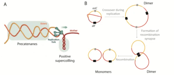
the rotation of the replication fork, diffuse behind to form precatenanes (the two new
sister chromosomes interwrapped) (Figure 1.2A). DNA gyrase acts to remove the positive
supercoils accumulated ahead, whereas topoisomerase IV assumes two functions,
removal of positive supercoils and decatenation (Postowet al., 2001, Espeli & Marians,
2004, Schvartzman & Stasiak, 2004). As the replication forks approach each other near
inaccessible to DNA gyrase. Therefore, the positive supercoiling accumulated ahead of
the forks, diffuses behind, forming precatenanes that, with the end of replication, are
converted in catenanes (interlinked sister chromosomes). These final links have to be
removed by topoisomerase IV (Sherratt, 2003).
Figure 1.2. Precatenanes and dimers formed as a consequence of bacterial chromosome replication. (A) Progression of the replication fork (green arrow), with concomitant unwinding of the DNA double helix, results in the accumulation of positive supercoils ahead of the fork. When this positive tension diffuses behind the fork, the newly replicated strands of DNA rotate and interwrap, originating precatenanes. DNA gyrase and topoisomerase IV remove the positive supercoils accumulated ahead, while decatenation is only performed by topoisomerase IV. Adapted from (Reyes-Lamothe et al., 2012). (B) Crossover events between two growing circular chromosomes produce chromosome dimers. Site-specific recombination at thedifsite (black box), located in ter region, resolves dimers to monomers. This reaction is catalyzed by two tyrosine recombinases (XerC and XerD inE. coli) and requires FtsK inE. coli. Dark red: mother chromosome; light red and orange: newly replicated daughter chromosomes.
Replication of a circular chromosome can also produce chromosome dimers, as a
consequence of crossing over during homologous recombination of the newly replicated
sister chromosomes (Figure 1.2B) (Lesterlin et al., 2004). This has been estimated to
happen, in E. coli, once every six generations (10-15% of a population) (Steiner &
Kuempel, 1998, Peralset al., 2000). Before segregation, chromosome dimers have to be
converted to monomers by the combined action of two tyrosine recombinases, XerC and
region (Lesterlin et al., 2004). In E. coli, the action of the XerCD and Topoisomerase IV
chromosome-resolving enzymes depends on the cell division protein FtsK that, as
discussed in more detail later, establishes an important link between the end of
chromosome replication/segregation and the beginning of cell division.
In rapid growth conditions, some bacteria, likeE. coliandB. subtilis, can undergo
multi-fork DNA replication to ensure that the time it takes to synthesize and segregate
new chromosomes does not exceed the mass doubling time. During multi-fork
replication, the mother cell initiates more than one round of replication before division.
This gives a “head-start” to its progeny that, in this situation, are able to complete
chromosome replication/segregation in fast dividing cells (Cooper & Helmstetter, 1968).
Chromosome segregation
In marked contrast with eukaryotic cells, bacteria segregate their chromosome
progressively in parallel with replication. Immediately after oriC replication, the newly
formed origin regions are separated and move to conserved positions in the incipient
daughter cells. Subsequently, as each new locus is replicated, the two new copies are
condensed and incorporated into the nucleoid in formation. In this way, the DNA loci are
organized accordingly to the order of replication, which assures the maintenance of the
original orderly arrangement of the chromosome (Figure 1.1) (Nielsen et al., 2006a,
Viollier et al., 2004). Following decatenation and, eventually, chromosome dimer
resolution, segregation is concluded, with the movement of the terminus regions away
from the cell-division site, to allow cytokinesis.
Despite decades of study, the mechanisms that mediate bacterial chromosome
segregation remain elusive. Bacteria do not possess an obvious mitotic-like apparatus.
However, the observation of rapid and directed movement of the new oriC regions into
opposite directions, led to the proposal of an active mechanism as the driving force for
segregation, for which different models were proposed. It was hypothesized that
energy of DNA polymerization (Lemon & Grossman, 2001). However, this model required
the two replisomes to be immobile in the cell and was therefore ruled out after the
finding that replisomes in fact track along a stationary chromosome (Bates, 2008,
Reyes-Lamothe et al., 2008). Alternatively, the transcription process was implicated in DNA
segregation. RNA polymerase is an abundant and powerful motor protein with a rather
stationary behavior. Since the majority of genes are oriented away from the oriC, the
combined effect of many transcription reactions could result into the movement of the
newly synthesized DNA (Dworkin & Losick, 2002). However it is now known that inhibition
of transcription with rifampicin, does not prevent proper chromosome segregation (Wang
& Sherratt, 2010). For a short period, the actin-like cytoskeletal protein MreB, involved in
the determination of the bacterial cell shape, was also implicated in the partioning
process (Kruseet al., 2006, Soufo & Graumann, 2003, Kruseet al., 2003). However, it was
shown that segregation defects observed upon MreB depletion result from the
MreB-induced morphological cell alterations rather than from the direct involvement of this
protein in DNA segregation (Karczmareket al., 2007, Wang & Sherratt, 2010).
Other studies provided evidence for the role of the chromosomal encoded
ParABS system in chromosome segregation. TheparABSlocus, constituted by twotrans
-acting proteins, ParA and ParB, and a cis-acting site parS, was first identified in several
low-copy number plasmids as the essential component of their partioning system. ParB
protein binds to the cis-acting centromere analog parS and spreads into the flanking
regions forming a nucleoprotein complex. This complex then recruits the ATPase ParA
protein, which is thought to assemble into dynamic polymeric structures to mediate
segregation (reviewed in (Schumacher, 2008, Gerdeset al., 2010)). The presence of ParA
and ParB homologues in the chromosome of several bacteria (but notE. coli) (Livnyet al.,
2007, Gerdes et al., 2000) raised the hypothesis that chromosomally encoded Par
proteins might function in the process of chromosome segregation, in a similar way to the
plasmid segregation system. This idea was reinforced by the observation that insertion of
various chromosomalparABShomologues in an otherwise unstable plasmid increases its
Grossman, 1998). However, there are only few cases in which this system was shown to
be truly essential for chromosome segregation. The most compelling evidence was
obtained in Vibrio cholerae and Caulobacter crescentus, which segregate their new
chromosomes asymmetrically. In both of these bacteria, theoriCis located at one pole,
(the “old” pole) and, after replication, one copy remains at that site while the other
transverses the entire length of the cell, to be positioned at the opposite end (new pole).
(Fogel & Waldor, 2005, Mohl & Gober, 1997).V. cholerae,one of the few bacteria with
two circular chromosomes, each with their own ParABS system, was shown to require all
Par components to properly segregate the oriC region of its larger chromosome
(Chromosome I). ParBIbinds to threeparSsites in the vicinity of theoriC,and this region
is moved across the cell by ParAI. Time-lapse analysis shows that a ParAI structure
stretches throughout the cell from the new pole to the old one and, then, progressively
retracts, pulling the ParBI-parScomplex (Fogel & Waldor, 2006). InC. crescentus, ParA is
also required for the segregation of a chromosomal region containing the oriC and the
ParB-parS complex (Toroet al., 2008). Notably, in both species, the ParABS systems are
primarily involved in the segregation of the oriCregions. Most likely, segregation of the
rest of the chromosome relies on other, still unknown, mechanisms.
Contrary toV. choleraeandC. crescentus, inactivation of Par system in B. subtilis
has only mild effects in chromosome segregation. The B. subtilis genome encodes ten
parSsites, eight of which have high affinity for Spo0J (ParB homologue) and are located in
the origin-proximal region of the chromosome (Lin & Grossman, 1998, Breier &
Grossman, 2007). Spo0J binds to these sites and spreads over a distance of
approximately 800 Kb, to form a nucleoprotein complex that appears as a single
fluorescent focus, when visualized by fluorescence microscopy using a fluorescent fusion
to Spo0J (Linet al., 1997). A Spo0J mutant produces 1 to 2% of anucleated cells (Iretonet
al., 1994) while in 15% of new born cells oriCregions are positioned closer together than
in the wild-type (Leeet al., 2003, Lee & Grossman, 2006). Deletion of Spo0J disrupts the
organization of a large (~1Mb) region surrounding the replication origin. On the contrary
(Iretonet al., 1994) and its absence only affects the position of a discrete region next to
the oriC (Sullivan et al., 2009). All these results argue against the idea that B. subtilis
parABS system is responsible for exerting the motor force that drives chromosome
segregation. In fact, it was recently shown that the role exerted by Spo0J in chromosome
partitioning is rather indirect. The parS-Spo0J nucleoprotein complex targets the SMC
complex to the origin of replication, which ensures proper condensation of the newly
synthesized DNA, necessary for proper chromosome segregation (Sullivan et al., 2009,
Gruber & Errington, 2009). In an independent and complementary pathway, Spo0J also
regulates Soj role in chromosome replication. Soj, due to its ability to alternate between a
monomeric form and an ATP-bound dimeric form, functions as a dynamic molecular
switch that regulates the activity of the replication initiator protein DnaA. Initiation of
replication is stimulated by the Soj dimer and inhibited in the presence of the monomeric
form. Generally, due to the presence of Spo0J, that stimulates Soj ATPase activity and
therefore the conversion of the Soj dimer to monomer, the equilibrium favors the
presence of the monomeric conformation (Murray & Errington, 2008, Scholefieldet al.,
2011). However, at some point in the cell cycle, Soj “escapes” Spo0J control by an
unknown mechanism, so replication can occur.
The effect of Spo0J/Soj in sporulation is also dependent on Soj-regulation of the
initiation of replication. For a long time it has been known that in the absence of Spo0J,
Soj represses the expression of genes required for the early stages of sporulation, which
blocks the switch from vegetative growth to spore differentiation (Ireton et al., 1994).
This happens because, without Spo0J control, Soj dimer stimulates DnaA which, in turn,
activates a sporulation checkpoint protein, Sda, that blocks sporulation (Murray &
Errington, 2008). Spo0J and Soj not only regulate the entrance into sporulation but, as
soon as this pathway is activated, also ensure that the future spore receives its copy of
the chromosome. Soj drives the movement ofparS-Spo0J nucleoprotein complex towards
the pole (in the future spore) where it is tethered by the sporulation protein RacA to the
Until now, all efforts devoted to finding a conserved machine-like apparatus that
depends on specific proteins to move bacterial DNA during segregation have not been
successful. All of the identified systems seem to act more at the level of organization and
proper positioning of the origin of replication than on the movement of the bulk of the
chromosome. Therefore, alternative models, presenting chromosome segregation as a
passive process that depends on physical aspects of DNA organization, have been
receiving more attention. It has been suggested that chromosome segregation is a
self-organizing process, driven by separation of transertion domains (Woldringh, 2002). As
explained earlier, numerous chromosomal regions are linked to the cell envelope due to
the coupling of co-transcriptional translation and translocation of membrane proteins
(transertion). According to this model, the transertion complexes of each daughter
chromosome create two independent proteolipid domains that are located in opposite
cell halves. Continuing synthesis of DNA, and thus addition of new transertion complexes
between the domains was thought to push the two nucleoids away from each other
(Woldringh, 2002). However, this implies that the movement of each DNA region is
gradual, which is not what is observedin vivo(Webbet al., 1998, Viollieret al., 2004).
The most plausible and attractive model presented to date, suggests that the
driving force for chromosome segregation in bacteria is entropy (Jun & Mulder, 2006, Jun
& Wright, 2010). Mathematical modeling analysis indicates that, when long polymeric
chains (the chromosome molecules) are confined to a compartment with little free space
(the bacterial cell), the polymers strongly self-avoid and therefore separate
spontaneously, in a physiologically relevant timescale, without the need for any other
force except entropy. That is, entropy alone can induce segregation of replicated
chromosomes. The same is not applicable to plasmids. Due to their small size, plasmids
do not suffer the same constriction forces as chromosomes inside the cell. Therefore,
plasmids cannot solely rely on the entropic forces to segregate, which may explain why
they possess such easily identifiable partitioning systems. This model of entropic
exclusion of sister chromosomes also implies that the primary role SMC/MukB proteins is
entropic forces. In agreement, aberrant chromosome organization, due to the absence of
functional SMC/MukB, leads to altered chromosome segregation (Danilovaet al., 2007,
Sullivanet al., 2009, Gruber & Errington, 2009).
The FtsK/SpoIIIE family of DNA translocases
Bacteria possess regulatory mechanisms to ensure that septum formation only
occurs after proper clearance of the chromosomes from the division site (discussed in
more detail below). Nevertheless, in some situations, particularly when chromosome
dimers are formed, the septum starts assembling over non-segregated DNA. To guarantee
that all genetic material is properly cleared from the division site before septum closure,
bacteria rely on the action of DNA translocases (Kaimer & Graumann, 2011). These
double strand DNA transporters, which belong to the FtsK/SpoIIIE family, localize at the
leading edge of the constricting septum, to remove all DNA standing in the way of the
septum (Yuet al., 1998a, Biller & Burkholder, 2009, Kaimeret al., 2009) or form channels
in already assembled septa, to rescue trapped DNA (Wu & Errington, 1994, Bathet al.,
2000). In addition, some FtsK/SpoIIIE proteins also help to resolve the physical
chromosomal links formed during replication (Steineret al., 1999).
FtsK/SpoIIIE DNA translocases share a common organization in three domains
(Bigotet al., 2007). A membrane localized N-terminal domain, a soluble carboxy-terminal
region and a central linker of variable length that connects the N- and C-terminal
domains. The poorly conserved N-terminal domain often mediates functions unrelated
with DNA translocation, while the C-terminal domain forms an ATP-dependent DNA
pumping motor (Ausselet al., 2002, Kaimeret al., 2009, Masseyet al., 2006, Bathet al.,
2000).
The E. coli DNA translocase, FtsK, is a multifunctional protein that links three
important cell division processes: resolution of chromosome links, segregation of sister
chromosomes and formation of the division septum. Its amino-terminal membrane
to localize the protein to the septum and is part of the divisome, the multiprotein
complex that orchestrates bacterial cell division (see sections ahead). Cells expressing
only FtsKNcan survive, however, in the absence of this domain, the division machinery
cannot properly assemble at the septum and thus, the cells present a filamentation
phenotype, typical ofE. colicells that cannot divide (Wang & Lutkenhaus, 1998, Yuet al.,
1998a, Draperet al., 1998). The FtsK linker domain (FtsKL), with approximately 600 amino
acids, extends from the septum into the cytoplasm, separating FtsKNfrom the soluble
C-terminal motor domain (FtsKC). This linker was initially considered to be just a spacer,
however, it is now known to be required for the establishment of essential interactions
between FtsK and several other components of the divisome (Bigotet al., 2004, Dubarry
& Barre, 2010). Contrary to the other two domains, the soluble C-terminal FtsK region
(FtsKC) is dispensable for septum synthesis. Instead, it bears the important roles of
promoting chromosome dimer resolution and removing DNA from the site of constriction
(Yuet al., 1998b, Liuet al., 1998, Steineret al., 1999). FtsKCcan be subdivided into three
regions (Yateset al., 2003). Theand domains, that constitute the DNA translocation
motor, and the regulatory domain that interacts with DNA and with the XerD
recombinase. The/subdomains of different FtsK molecules assemble into a hexameric
ring with a large central channel capable of accommodating a double-stranded DNA
molecule (Masseyet al., 2006). This complex uses the energy of ATP hydrolysis to move
chromosomal DNA with a velocity of approximately 5 Kb/s (Masseyet al., 2006, Peaseet
al., 2005, Saleh et al., 2004). The correct direction of translocation, crucial for the
accurate sorting of chromosomal DNA, is determined by FtsK ability to “read” sequences
on the DNA. The chromosome carries preferential FtsKC loading sites, known as KOPS
(FtsK-orienting polarized sequences), which are polarized towards the terminus region
(Bigot et al., 2005, Levy et al., 2005). The subdomain, which is connected to the /
motor by a small flexible linker (Sivanathan et al., 2006), recognizes these KOPS motifs,
promoting the loading of FtsK/ in a precise orientation. This interaction directs DNA
translocation by FtsK towards the dif recombination site, a specific region within the
2005, Bigotet al., 2006, Loweet al., 2008, Sivanathanet al., 2009, Grahamet al., 2010).
FtsK also promotes chromosome dimer resolution in combination with the site-specific
tyrosine recombinases XerC and XerD. The FtsKC-mediated DNA translocation, helps to
align the sister dif sites, bound by XerC and XerD, to form a productive recombination
synapse (Corre & Louarn, 2002, Steiner et al., 1999, Capiauxet al., 2002, Massey et al.,
2004). Additionally, FtsK interacts with and activates XerD to perform a first pair of
strand exchanges that result in an intermediate Holliday junction. This Holliday junction is
then resolved into a crossover product, independently of FtsK, by XerC-mediated strand
exchanges (Yates et al., 2003, Barreet al., 2000, Aussel et al., 2002, Yates et al., 2006,
Graingeet al., 2011). FtsK, like other DNA translocases, strips off proteins from the DNA
as translocation occurs (Crozat et al., 2010). Interestingly however it stops DNA
movement when it encounters the XerCD-difcomplex, thus preventing the removal of the
tyrosine recombinases (Grahamet al., 2010). FtsK is also thought to assist in the removal
of the catenation links that persist between sister chromosomes after replication. FtsKC
was shownin vitro,to interact with the ParC subunits of topoisomerase IV and stimulate
the decatenation activity of this enzyme (Espeli et al., 2003, Bigot & Marians, 2010).
Moreover, the FtsK-XerCD-dif recombination machinery seems to be able to unlink
catenated DNA monomers when topoisomerase IV function is compromised (Ip et al.,
2003, Graingeet al., 2007).
The processes of chromosome unlinking, chromosome segregation and septum
formation have to be tightly coordinated to ensure faithful transmission of the genetic
material. Therefore, it seems very advantageous that regulation of these processes is
centered on one protein. In the last few years, the role of FtsK as a cell division
checkpoint has been the subject of discussion. Is FtsK able to delay septation until
chromosomes are completely and correctly segregated? It seems plausible that it can do
so, since otherwise it would have a short period to clear DNA from the division site. FtsK
does not transport DNA before membrane fusion (Dubarry & Barre, 2010) and the
XerCD-dif complex is only activated after septum invagination has already started (Kennedyet
FtsKCis enrolled in the process of DNA translocation, the linker becomes stretched and
cannot establish the necessary contacts with the other cell division proteins, which delays
septum constriction. Only once the activity of the C-terminal motor is no longer required,
are the interactions reestablished, allowing division to proceed (Grainge, 2010, Dubarryet
al., 2010).
In B. subtilis, two DNA translocases, SpoIIIE and SftA, act at different stages to
coordinate chromosome segregation and cell division. SftA, like FtsK, clears DNA before
septum constriction and SpoIIIE moves trapped chromosomes after septum closure.
SpoIIIE is best known for its essential role in spore development (Wu & Errington,
1994). During sporulation, an asymmetric septum is formed at the cell pole, dividing the
cell into a large mother cell and a small forespore (future spore). When this septum
assembles, only the origin-proximal region of the chromosome is positioned in the
forespore space. Approximately two-thirds of the chromosome are still located in the
mother cell. SpoIIIE uses the energy of ATP hydrolysis to actively transport, through the
closed septum, the remaining two-thirds of the DNA destined to the forespore (Wu &
Errington, 1997, Wuet al., 1995, Bathet al., 2000, Burtonet al., 2007). This DNA crosses
two membranes and a peptidoglycan layer (Becker & Pogliano, 2007) and, like with FtsK,
the direction of translocation, is determined by SpoIIIE-mediated recognition of specific
DNA sequences. These sequences, known as SRS (SpoIIIE recognition sequences), are
polarized towards the terminus region (Ptacin et al., 2008, Becker & Pogliano, 2007).
SpoIIIE has a transmembrane N-terminal domain and is usually distributed throughout
the cytoplasmic membrane. Only in the presence of septum-entrapped chromosomes, do
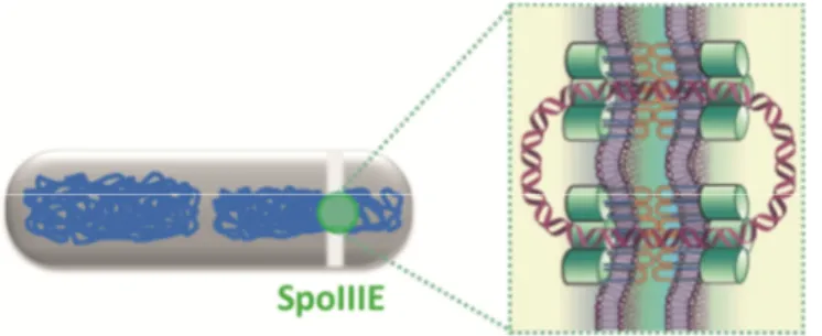
the SpoIIIE molecules assemble around the DNA, forming multimeric translocation
channels (Figure 1.3) (Wu & Errington, 1997, Ben-Yehudaet al., 2003a). These are seen by
fluorescence microscopy, as a bright foci at the middle of the septum (Wu & Errington,
1997, Ben-Yehudaet al., 2003a). The two arms of the entrapped circular chromosome are
transported, at the same time, by two independent SpoIIIE channels. Each channel is,
membrane) joined through their extracellular loops located in the peptidoglycan layer
(Masseyet al., 2006, Burtonet al., 2007, Becker & Pogliano, 2007) (Figure 1.3). It is not
yet clear, however, how the final loop of the circular DNA molecule is translocated across
the septum. Since the DNA does not seem to break to allow translocation, most likely the
channels open/merge for the chromosome to pass. Recent data suggests that the SpoIIIE
molecules can only form stable channels when their C-terminal motor interacts with the
DNA (Fleming et al., 2010). Possibly, once the terminus region reaches the septum and
the DNA motor dissociates from the DNA, the channels destabilize and a large pore is
opened for final DNA translocation (Fleminget al., 2010).
Figure 1.3.B. subtilisSpoIIIE forms DNA conducting channels.During sporulation, to translocate the last two thirds of the chromosome from the mother cell to the forespore, across two membranes and a peptidoglycan layer, SpoIIIE forms DNA conducting channels. Most likely, each SpoIIIE channel, is composed of two communicating SpoIIIE hexamers (one within each membrane) joined through their extracellular loops located in the peptidoglycan layer. Each channel can only transport one double-stranded chromosome arm. Green: SpoIIIE; Grey: SporulatingB. subtiliscell; Blue and Violet: DNA. Adapted from (Wu & Errington, 2008).
Importantly, SpoIIIE has a second function during sporulation. Its N-terminal
domain mediates the two sporulation processes that require membrane fission:
asymmetric division and spore engulfment (Sharp & Pogliano, 1999, Sharp & Pogliano,

