C
fo
Chromos
or thera
T
some in
peutic d
Ana
Tese de D
nstabilit
drugs to
a Filipa B
Doutoram
ty in Fa
o preve
diseas
Brinco de
mento em
2012
nconi A
ent the p
se
Oliveira
Ciências
Anemia:
progres
Ponte
s Bioméd
: search
ssion of
icas
hing
f the
Ana Filipa Brinco de Oliveira Ponte
Chromosome instability in Fanconi Anemia: searching for
therapeutic drugs to prevent the progression of the disease
Tese de Candidatura ao grau de Doutor em
Ciências Biomédicas submetida ao Instituto
de Ciências Biomédicas Abel Salazar da
Universidade do Porto.
Orientadora – Professora Doutora Beatriz
Porto
Categoria – Professora Auxiliar
Afiliação – Instituto de Ciências Biomédicas
Abel Salazar da Universidade do Porto
Co-orientador – Professor Doutor Félix
Carvalho
Categoria – Professor Catedrático
Afiliação – Faculdade de Farmácia da
Universidade do Porto
Os REQ Facu Micro Estes Tecn 3990 estudos a QUIMTE/Lab uldade de F oscopia, do s estudos nologia (FC 03 / 2007). presentado boratório d Farmácia d o Instituto de contaram CT), através s nesta te e Toxicolo do Porto e e Ciências B com o ap s da atribuiç ese foram gia do De no Labora Biomédicas poio finance ção de uma realizados epartamento atório de C s Abel Salaz eiro da Fu a Bolsa de s no Labo o de Ciênc itogenética, zar. undação pa Doutorame oratório As cias Biológ , Departam ara a Ciên ento (SFRH ssociado gicas da mento de ncia e a H / BD /
Author’s declaration
The author states to have afforded a major contribution to the conceptual design, technical execution of the work, interpretation of the results and manuscript preparation of the published or under publication articles included in this thesis.
Publications
According to nº 2, alínea a, do artigo 31º do Decreto-Lei nº 230/2009 the following published or under publication articles were prepared in the scope of this thesis:
Articles in international peer-review journals
Ponte F, Carvalho F and Porto B (2011) Protective effect of acetyl-L-carnitine and a-lipoic acid against the acute toxicity of diepoxybutane to human lymphocytes. Toxicology, 289 (1): 52-52.
Ponte F, Sousa R, Fernandes AP, Gonçalves C, Barbot J, Carvalho F and Porto B (2012) Improvement of genetic stability in lymphocytes from Fanconi Anemia patients through the combined effect of α-lipoic acid and N-acetylcysteine. Orphanet Journal of Rare Diseases (in press).
Abstracts in international peer-review journals
Ponte F, Sousa R, Carvalho F, Porto B (2011) α-Lipoic acid decreases chromosome instability in lymphocytes from Fanconi Anemia patients. Toxicology Letters, 205 Suppl, S108.
Ponte F, Sousa R, Carvalho F, Porto B (2011) The combined used of a-lipoic acid plus N-acetyl-cysteine as an effective cocktail for decreasing chromosome instability in Fanconi Anemia patients. Chromosome Research, 19 (1) S25.
Sousa R, Ponte F, Porto B (2011) Accuracy in the cytogenetic diagnosis of Fanconi Anemia and other chromosome instability syndromes. Chromosome Research, 19 (1) S36.
Ponte F, Carvalho F, Porto B (2010) Acute toxicity of diepoxybutane to mononuclear leukocytes. Multifactorial mechanisms? Toxicology Letters, 196 Suppl, S86.
Ponte F, Carvalho F, Porto B (2009) Acute toxicity of diepoxybutane to human mononuclear lymphocytes. Toxicology Letters 189, Suppl, S121.
Sinto-te como um rio
na exata medida
que desvendas caminhos
misteriosos.
Da nascente até ao mar
cruzas-te com obstáculos
na tentativa vã
de pararem a torrente
mas teu rio corre inteiro
cálido, rebelde, impetuoso
sem se perder na paisagem
que desperta outras correntes.
Quando o objetivo é seguir
o caminho é sempre em frente.
Sentes a limpidez das águas
que correm nas tuas veias
e teu caudal majestoso
quando em ti confluem rios
que com o mesmo fulgor
têm pressa na chegada.
Ficas contente e sorris
mas segues o teu traçado
e quando estás já tão perto
quase a desaguar
olhas para ti surpresa
dizes com voz sublime:
- afinal sou uma gota
majestoso
é o mar!
Sinto-te navegar...
Agradecimentos
Agradeço à Rosa tudo o que passámos juntas no nosso pequeno e ao mesmo tempo, grande Laboratório de Citogenética. Todas as alegrias partilhadas (que foram muitas) no decorrer destes 4 anos, as tristezas de não conseguirmos o que queríamos, as brigas e discussões científicas, que por vezes ainda mais confusão geravam nas nossas cabeças, e de onde mais tarde saía sempre uma luz, a amizade que criámos e sei que perdurará para além da ciência. As viagens hilariantes mundo fora, sempre com a anemia de Fanconi ao lado, nunca serão esquecidas. Obrigada por me fazeres ainda gostar mais de ciência e de anemia de Fanconi. Sei que sem ti não seria igual. Obrigada por partilhares comigo os teus conhecimentos. Por me ouvires quando estava desanimada e me sentia incapaz de continuar. As tuas palavras sempre me ajudaram e passaram além do trabalho. Foi um prazer encontrar-te e espero que os nossos caminhos se cruzem sempre e o nosso objetivo final seja cumprido. Tenho a certeza que sem ti nada disto seria possível, pelo menos desta forma. És uma pessoa incrível.
Agradeço à Sara Teixeira e à Débora Franco, as minhas colegas mais novas do laboratório de Citogenética, todo o incentivo que me deram e a alegria que proporcionaram no laboratório, o convívio sempre salutar de onde sempre saía ciência! Foi bom ajudar e receber o vosso apoio. Um agradecimento muito especial à nossa “informática” sempre de serviço, a Lara Andrade! A tua ajuda foi preciosa! Sempre pronta a resolver os problemas do meu PC, que foram muitos, e a ajudar a utilizar softwares como o EndNote e na formatação da tese! À Maria, apesar do pouco tempo de convívio, foi bom ter-te aí também e teres-nos adotado como o teu laboratório! À Joana, apesar de por lá passar pouco tempo, o pouco tempo em
que lá esteve mais que uma hora, foi sempre surreal! Ao nosso primeiro membro masculino, o João, obrigada por conseguires tantos doentes para nós.
Ao Nuno Santos um muito obrigado por toda a ajuda informática prestada! Foste incansável, amigo, compreensivo e nunca te “revoltaste” pelos imensos pedidos de ajuda… ultimamente diários, muitos deles de simples resolução, o que demonstra as minhas imensas capacidades na área da informática! Bem, isto sem ti também tinha sido bastante mais complicado! E, no fim de tantas horas passadas em frente ao PC, até ficámos amigos!
Um agradecimento especial à Alice Porto pela dica para a capa da tese! Gostei!
À Ana Paula agradeço a dedicação em cuidar do nosso pequeno recanto, sem ela não teria sido tão fácil trabalhar.
Obrigada a todos os clínicos que trabalharam comigo nestes anos, dos quais saliento o Dr. Barbot, a Dra. Ana Paula Fernandes e a Dra. Cristina Gonçalves que tanto fizeram por conseguir amostras de sangue dos nossos meninos doentes. Obrigada ao serviço de dadores do HGST e às simpáticas enfermeiras que sempre me ajudaram. Obrigado aos dadores, por contribuírem para este estudo. Obrigada à Sandra Maia do Laboratório de Imunologia do ICBAS por nos ajudar nas recolhas de amostras de sangue.
À Maria João Valente, à Helena Faria e à Diana Dias da Silva agradeço ter-vos conhecido no meio da confusão do Laboratório de Toxicologia da FFUP! Como foi possível no meio de tanta gente encontrar-vos como amigas? Obrigada por tudo, pelas partidas que por vezes me deixaram louca, e pela ajuda e convívio nos nossos maravilhosos Eurotox, dos quais saliento o de Barcelona e o de Paris!
Agradeço às colegas mais experientes, Vera, Renata e Helena Pontes, a ajuda no início, quando comecei os trabalhos no Laboratório de Toxicologia. As dicas, a paciência e os vossos ensinamentos foram preciosos. Ao Ricardo Oliveira um grande obrigada pela ajuda prestada, ao ensinar-me a fazer a determinação das caspases, que não foi fácil. Ao Professor Fernando Remião e à Professora Helena Carmo obrigado por serem sempre prestáveis e me ajudarem, quando nunca foi vossa obrigação. À Engenheira Elisa obrigada pelos caramelos, que tanto adoçaram os nossos dias, e obrigada pelos seus elogios sempre muito simpáticos! À doutora Maria de Lourdes Bastos obrigada por me ter deixado integrar a fantástica equipa do Laboratório de Toxicologia. À restante equipa do laboratório obrigada pelo bom ambiente criado e o espirito de interajuda.
À Dona Conceição, à Dona Júlia e à Ana Maria um obrigado por permitirem que que o laboratório de Toxicologia fosse utilizável! Sem a vossa cooperação seria impossível fazer ciência!
Agradeço às minhas amigas, Lília Pereira, Sara Assunção, Ana Lúcia Pedrosa, Raquel ou Tinka, Diana Ledo, Marta Ledo, Filipa Moreira, Filipa Cortes, Isabel Gonçalves, Adriana Moura, Carla Rocha, Nídia Rocha e amigos, Jorge Martinho, Rui Amorim, Carlos Martins, André Ferreira, Joel Pereira, Gonçalo Barreto, João Sousa, Thiago Silva, Diogo Oliveira e Silva, Hugo Oliveira, Pedro Borges e Samuel Reis as palavras de apoio e carinho que me foram dando nestes anos. Obrigada por compreenderem que nem sempre foi possível estar perto e por acreditarem em mim e no meu trabalho. Hoje todos vocês sabem o que é anemia de Fanconi, o que me deixa extremamente satisfeita. Agradeço, principalmente às meninas, nunca me deixarem desistir e serem sempre boas ouvintes. Muitos acompanharam-me desde o início e
sabem o quanto passei para estar aqui hoje, obrigada por serem meus amigos de verdade.
Ao Dr. Miguel Ricou agradeço nunca me ter deixado desistir da vida académica. De sempre me ter incentivado e mostrado que tinha capacidades para fazer um doutoramento brilhante. De me ter mostrado que a vida é mais que um trabalho e que do trabalho teria de tirar o melhor partido, para assim ser mais feliz. Penso que consegui. Obrigada por toda a ajuda, que sabe que foi essencial para hoje ser uma pessoa bem sucedida.
Ao meu irmão Ricardo, o Brinco, obrigada por me pores sempre o astral para cima e me achares a mana genial, mais uma das vedetas da casa! As tuas brincadeiras, mesmo nos meus momentos de tristeza, sempre me fizeram sorrir. Obrigada por existires na minha vida. Espero para ti ser um exemplo de luta e persistência, mostrando-te que com garra vencemos tudo. Para mim já sabes que és o maior exemplo de felicidade. E isso, mesmo que não tenhas dado conta, foi-me contagiando. Óbvio que continuo à espera da minha casa feita por ti, meu projecto de Arquitecto!
Obrigado Pai por, mesmo por vezes ausente fisicamente, estares sempre tão perto. És o meu guru. Em ti sempre procurei palavras sábias e os caminhos certos a percorrer. Nunca me deixaste desistir com as tuas palavras, por vezes austeras, mas sempre racionais. E a ti também te consegui passar o “bichinho” das doenças raras. Também tu hoje sabes falar de anemia de Fanconi. Os teus sorrisos aquando dos meus pequenos momentos de vitória encheram-me o coração de uma felicidade inexplicável. Sem ti, não teria sido igual.
Mãe, a tua presença constante, foi preponderante nesta minha luta, nem sempre fácil. As palavras de apoio e coragem foram incessantes. Obrigado por acreditares que um dia isto seria mesmo verdade. Sempre me mostraste que eu ia conseguir, mesmo quando eu acreditava que era impossível. Desculpa o mau humor antes dos congressos, os “não vou conseguir”, seguidos de umas lágrimas e tu sempre lá estiveste, ao meu lado, com um sorriso de uma mãe que olha com orgulho para a sua filha.
Sem vocês os três este caminho seria outro, talvez não tão bem sucedido. Obrigada pelos valores e amor que sempre me transmitiram e ajudarem-me a construir a pessoa e cientista que hoje sou.
Às avós Maria e Georgete obrigada pela preocupação e por mesmo não sendo fácil para vocês entenderem aquilo que faço, me darem valor. À avó Georgete agradeço ainda a velinha que acendia nos meus momentos cruciais, para que tudo me corresse sempre bem! Ao avô Zé, obrigado por teres gostado tanto de mim e te orgulhares da neta que fui para ti. Estarás sempre no meu coração. Gostava tanto que aqui estivesses neste momento tão especial na minha vida… Sinto a tua ausência e vou sentir falta de ouvir os teus parabéns emocionados… Aos tios e tias obrigada pelos incentivos e pelos parabéns constantes aquando das minhas pequenas conquistas.
Ao Boris obrigado por seres um ouvinte sempre tão atento e fiel. És o meu leal companheiro e a ti agradeço toda a atenção e companhia que sempre me fazes, nos bons e maus momentos. Em ti procurei o amor que só os animais de estimação sabem oferecer…e o conforto e o calor quando nem as palavras dos humanos me faziam sentir mais calma.
Ao Professor Félix agradeço todo o apoio que sempre me deu. Lembro-me dos dias em que entrava no seu gabinete cabisbaixa e triste, porque os resultados nunca eram aquilo que eu esperava. As suas palavras de incentivo foram fulcrais para que nunca desistisse: “Mas estes resultados são fantásticos! Parabéns Filipa!”. Obrigada por toda a ajuda, compreensão e por todos os ensinamentos que me transmitiu e ainda me ter incentivado a ser melhor. Se hoje percebo alguma coisa de toxicologia a si o devo. Se hoje sou persistente aprendi consigo. Se tenho ambição e coragem também. Obrigada por acreditar que um dia iriamos chegar a um resultado positivo.
À professora Beatriz agradeço tudo. Não há palavras que descrevam a gratidão que por si tenho. Todas seriam poucas… Mesmo assim arrisco a dizer que se hoje sou feliz no meu trabalho a si o devo. O amor à ciência também. A dedicação à anemia de Fanconi. Obrigada por todos os sorrisos, que foram muitos, quando eu estava desanimada e não acreditava em mim. Por todas as conversas científicas e não só, que sempre me ajudaram a percorrer este caminho. Se hoje sinto amor à camisola e desejo continuar esta luta, a si o devo. É o meu exemplo.
Sei que tive sorte em ter o Professor Félix e a Professora Beatriz como orientadores. Além de me terem transmitido todo o saber, cada um da sua área, são pessoas excecionais. Sem eles tenho a certeza que os resultados que obtive não seriam os mesmos. Obrigada
Table of Contents
Preface 1 Abbreviations 5 Resumo 7 Abstract 9PART I – GENERAL INTRODUCTION 11
Introduction: Fanconi anemia, from the past towards present 13 1. Clinical features of Fanconi anemia 15
1.1. Fanconi anemia phenotype 15
1.1.1. Congenital malformations 15
1.1.2. Hematologic manifestations 17
1.1.3. Impairment in immunological function 18
1.1.4. Cancer susceptibility 21
1.2. Fanconi anemia genotype 23
1.2.1. The FA pathway 25
1.3. Diagnostic tests for Fanconi anemia 27
1.3.1. The chromosomal fragility test 27
1.3.2. Other diagnostic methods 28
1.4. Fanconi anemia treatments 30 2. Diepoxybutane as a golden standard test for Fanconi anemia diagnosis and as an oxidative stress inducer – Implications on Fanconi anemia research 31
2.1. DEB: why is it a golden standard test for Fanconi anemia diagnosis? 31
2.2. DEB cytotoxicity: biotransformation as a potential cause 32
2.3. DEB cytotoxicity: generation of reactive oxygen species and oxidative stress 34
2.4. DEB cytotoxicity: relation with mitochondrial dysfunction 35 3. Oxidative stress in Fanconi anemia: from cells and molecules towards prevention against chromosome instability 36
3.2. Redox related functions of FANC proteins 38 3.2.1. FANCA, FANCC and FANCG: link with xenobiotic processing pathway 38 3.2.2. FANCD2 and FANCJ: multiple identities and redox-related roles 40 3.3. Mitochondrial dysfunction in Fanconi anemia cells 41 3.4. In vitro and in vivo experimental therapies – Antioxidant agents as
protectors of chromosome instability
44
PART II – OBJECTIVES OF THE THESIS
47
PART III – ORIGINAL RESEARCH
51
Chapter I. Protective effect of acetyl-L-carnitine and α-lipoic acid against the acute toxicity of diepoxybutane to human lymphocytes
53 Chapter II. Improvement of genetic stability in lymphocytes from Fanconi Anemia patients through the combined effect of α-lipoic acid and N-acetylcysteine
63
PART IV – GLOBAL DISCUSSION
81
PART V – FINAL CONCLUSIONS AND FUTURE PERSPECTIVES
95
Preface
Four years ago it was straightforward to choose Fanconi anemia (FA) as a model for studying chromosome instability (CI) disorders and relate them with oxidative stress (OS). In the Laboratory of Cytogenetics of Institute of Biomedical Sciences (ICBAS) a special focus has long been given to FA. The blood samples that arrived there were frequently to confirm the diagnosis of FA. Thus, the interest for this disease slowly “grew” inside me and the will to learn more about it became natural. The relation with OS came later, after reading several papers about this disease, in which the main theory to explain the phenotype and the molecular aspects of the disease was impairment in DNA repair system. Notwithstanding this postulate, in my perspective, many aspects were not explained based on it and a conjunction of the two theories would make much more sense. The works of Pagano and co-workers were very important for establishing my perspective. So, the idea about antioxidant treatments simply arose. I even remember the first discussion about this with Professor Beatriz and since the beginning we believed that we could aim to solve some of the FA problems, finding the right antioxidant! And the work began! But I didn't know back then how difficult it was to study such a rare disease...
The works in the Laboratory of Toxicology of Faculty of Pharmacy of University of Porto (FFUP) were the beginning of a long and not always happy journey. It took almost one year to establish the right techniques and procedures. The next step was to test a variety of drugs in healthy donors, in the hope of finding a successful one to test in the blood of FA patients. One after another and the results were always negative…One year later, after testing 8 or 9 compounds, I got the first positive result and a big smile in my face! "Maybe I will achieve something", I thought. Working in FFUP, although it was very difficult, due to the scarcity of space and available material (we were 20 colleagues working at the same time in a class laboratory!!!), it was funny, even when everything went wrong and I wanted to do something and I wasn’t able to! Working in FFUP taught me to deal with stress and to work as a team, a very important lesson to a future scientist.
In the beginning of the third year of my PhD I came back to laboratory of Cytogenetics. The work with FA patients was finally about to start! First we tested one compound with good results! All the good results were celebrated with lots of smiles, because getting blood samples from FA patients was always so difficult… I remember all the phone calls to all the clinicians of North of Portugal to remember them that we needed blood samples of all their patients… The work was hard and the phone calls were almost daily! We had to move clinicians to help us. And the work went on… We waited many weeks for blood samples, we spent many hours on the microscope and in the end, I was tired but happy. Then we tried a combination of compounds and the work gained another shape. The results were simply unbelievable! We wanted to jump but at the same time we
were afraid. Was this real?? More blood please! More calls and the confirmation arrived! It was true, we found something. And the recognition started. First, with the European Cytogenetic Conference where I was selected to present our work and 3 months later the unthinkable happened. I sent an abstract for Fanconi Anemia Research Fund Scientific Symposium, with our results, but to be selected for an oral presentation seemed out of range…I didn’t know the real importance of our results. And one day, when I opened my e-mail I saw an answer. I read once, twice, three times until I believe in those words! I had been selected for an oral communication! My colleague Rosa and I screamed, laughed, jumped, cried…and we ran off the lab to go tell Professor Beatriz! After this day I started to believe in me… In October we went to Barcelona for the congress. It was one of the most satisfying experiences in my life. To speak for 300 FA researchers and to listen "congratulations!" from everyone got me with a huge smile on my face and made me feel like I was living a dream. And there, in Barcelona, I saw that my work was valid and more important, that I wanted to do more to save the life of these children, that born fated to an early death. The FA children and parents were there, and seeing them was very exciting and gave us another strength to go on. And of course, getting to know Professor Pagano was another unthinkable happening! Our guru, the one that always associated FA with OS, found me to personally say that he shares the same idea about the work I was developing and that he also has proposed this in his new paper! Could this be real?! And, with this meeting, arose the collaboration for an European Project…
Working with rare disease is not easy. We are a small country and the scarcity of patients is a big challenge. It needs dedication, patience and, above all, to know how to wait! We don´t work with cell lineages, we have to wait for the right patient! We don’t put solutions in a machine, click the button and make the results show up! We have to pass many hours and many days counting chromosomes on the microscope, like an ant collecting its food. We don´t have a true government support. We don’t have projects accepted for founding, with the justification that it is not a priority to study rare diseases. Once it was even stated that our work was esoteric! We do almost everything by our own! And, sometimes, I feel misunderstood. So, this journey was hard, but now I can smile and think that it is so much better than I thought it would be! Four years ago this was a dream, now I know it’s true, and this fills my heart. I feel very close, but I know I won't stop until my work can significantly contribute to save these children.
I devote this thesis to all FA patients and parents, especially to those that continue their hard life battle.
Abbreviations
1O 2 – Singlet oxygen 3-AT – 3-amino-1,2,4-triazole 8-OHdG – 8-hydroxy-2’-deoxyguanosine ALC – Acetyl-L-carnitineAML – Acute myeloid leukemia ANC – Absolute neutrophil count
ATM – Ataxia telangiectasia mutated protein AIDS – Acquired immune deficiency syndrome BMF – Bone marrow failure
BMT – Bone marrow transplant CDU – Cyclohexyl-3-dodecyl urea CHX – Cycloheximide
CI – Chromosome instability CYP – Cytochrome
DC – Dendritic cells
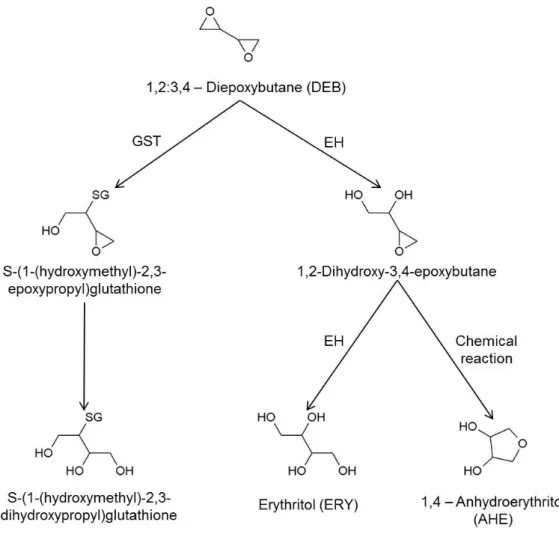
DEB – 1,2:3,4-diepoxybutane
DHPLC – Denaturing high performance liquid chromatography DHLA – Dihydrolipoic acid
EBV – Epstein-Barr virus EH – Epoxide hydrolase EPO – Erythropoietin FA – Fanconi anemia
FARF – Fanconi Anemia Research Fund FOXO 3a – Forkhead box 3a
G-CSF – Granulocyte colony-stimulating factor
GM-CFS – Granulocyte macrophage colony-stimulating factor GR – Glutathione reductase GSH – Reduced glutathione GPx – Glutathione peroxidase GST – Glutathione S-transferase GSTP1 – Glutathione S-transferase P1 GSTT1 – Glutathione S-transferase T1 H2O2 – Hydrogen peroxide
HClO – Hypochlorous acid Hb – Hemoglobin
HbF – Fetal hemoglobin HD – Healthy donor
HO-1 – Heme oxygenase 1
HSTC – Hematopoietic stem cell transplantation ICL – Interstrand cross-link
IFAR – International Fanconi Anemia Registry IFN-γ – Interferon gamma
IL – Interleukin
IR – Ionizing radiation LPS – Lipopolysaccharides MDS – Myelodysplasic syndrome mEH – Microsomal EH
MLPA – Multiplex ligation-dependent probe amplification MMC – Mitomycin C
mtDNA – Mitochondrial DNA NAC – N-acetylcysteine
NF-kB – Nuclear factor kappa-light-chain-enhancer of activated B cells NK – Natural killer
O2•- – Superoxide anion
•HO – Hydroxyl radical OS – Oxidative stress
PCR – Polymerase chain reaction PRDX3 – Peroxyredoxin 3
RBC – Red blood cells
ROS – Reactive oxygen species SCC – Squamous cell carcinoma sEH – Soluble EH
SH – Sulfhydryl
SOD – Superoxide dismutase TNF-α – Tumor necrosis factor alfa TNF-β – Tumor necrosis factor beta UVA – Ultraviolet A
Zn2+ – Zinc
α-LA – α-Lipoic acid
Resumo
A anemia de Fanconi (AF) é uma doença genética rara, caracterizada pela falha progressiva da medula óssea e elevada predisposição para o cancro, o que implica uma esperança média de vida reduzida. A instabilidade cromossómica (IC) é um dos principais defeitos celulares, e a hipersensibilidade das células AF a agentes “crosslinking”, tais como o diepoxibutano (DEB), constitui um marcador único para o diagnóstico. Geneticamente a AF é uma doença muito heterogénea, com 15 genes até agora caracterizados (FANC-A, B, C, D1, D2, E, F, G, I, J, L, M, N, O, P). A função destes genes está ligada principalmente à reparação de “DNA crosslinks”. O aumento da IC, e consequente aumento do risco para a malignidade, mostram a importância de compreender onde e como falham os mecanismos envolvidos na defesa celular. Além do conhecido envolvimento dos genes AF na reparação do DNA, vários estudos relacionam o fenótipo AF com desequilíbrio redox, demonstrando que o stress oxidativo (SO) é uma das principais causas para os defeitos celulares. Estudos recentes demonstram também defeitos ao nível das mitocondrias das células AF, o que está intimamente relacionado com o aumento da produção de espécies reativas de oxigénio e pela concomitante depleção das defesas antioxidantes, das quais a glutationa (GSH) é um conhecido biomarcador. Além disso, está bem documentada na literatura a relação entre a toxicidade do DEB e o SO, bem como a relação destes com disfunção mitocondrial. Daí que a caracterização das defesas antioxidantes contra a toxicidade induzida pelo DEB possa ser útil para uma melhor compreensão do que está a falhar nas células AF.
Na presente tese o grande objectivo foi conseguir uma abordagem profilática com recurso a antioxidantes, com o intuito de atrasar a progressão dos sintomas clínicos causados pela IC em doentes AF. O plano de trabalhos foi dividido em duas partes distintas mas com objetivos complementares. Na parte I o propósito global foi contribuir para uma melhor compreensão da toxicidade induzida pelo DEB em linfócitos humanos, através do estudo da contribuição de alguns parâmetros bioquímicos potencialmente envolvidos na reatividade deste composto. Este estudo foi realizado em suspensões de linfócitos humanos isolados de dadores saudáveis que foram expostos ao DEB e a uma variedade de compostos, como a ciclohexil-3-dodecil ureia (CDU), a elaidamida, o zinco (Zn2+), a
N-acetilcisteína (NAC), a acetil-L-carnitina (ALC), o ácido lipóico (α-LA), o ácido tânico, a 3-amino-1,2,4-traizole (3-AT) e a cicloheximida (CHX). Na parte II o propósito global foi selecionar os melhores compostos testados na parte I e que poderiam ser utilizados para diminuir a IC espontânea e induzida pelo DEB em linfócitos de doentes AF. Dos compostos inicialmente testados somente a ALC, o NAC, o α-LA e o Zn2+ foram
dadores saudáveis e, numa segunda fase, em culturas de linfócitos primários de doentes AF.
Os ensaios efetuados demonstraram alguns novos e importantes resultados. Relativamente à toxicidade induzida pelo DEB foi claramente evidenciado, pela primeira vez, que a exposição aguda de suspensões de linfócitos humanos a este agente resulta na depleção severa da GSH e na perda de ATP, seguida de morte celular. Além disso, foi demonstrado que a ALC contribui para um significativo efeito protetor da toxicidade induzida pelo DEB, efeito esse potenciado pelo α-LA. Coletivamente, estes resultados contribuíram para aumentar o nosso conhecimento sobre a toxicidade induzida pelo DEB e que foi bastante útil para aplicar nos estudos subsequentes. Os resultados obtidos considerando o objetivo final revelaram que um cocktail com α-LA e NAC melhora drasticamente a estabilidade genética em culturas de linfócitos de doentes AF in vitro, diminuindo a IC em 60% e 80% em culturas primárias de doentes AF e doentes AF com mosaicismo e/ou quimerismo, respetivamente. Os resultados finais apresentados nesta tese sugerem que o cocktail estudado deve ser usado como uma medida profilática para atrasar a progressão dos sintomas clínicos da doença causados pela IC, e portanto atrasar a falha progressiva da medula óssea e diminuir a suscetibilidade ao desenvolvimento de cancro. Desta forma, este estudo revela uma elevada importância clinica, pelo que é esperado que inspire os clínicos a começar ensaios clínicos do tipo III, especialmente porque tanto o α-LA como o NAC são compostos pouco dispendiosos e que apresentam um perfil farmacocinético bom e seguro, tendo sido já aprovados para uso humano.
Abstract
Fanconi anemia (FA) is a rare genetic disorder, characterized by progressive bone marrow failure and increased predisposition to cancer, which accounts for a reduced life expectancy. Chromosome instability (CI) is a major cellular defect and the unique hypersensitivity of FA cells to interstrand crosslinking agents, such as diepoxybutane (DEB), is an exclusive marker for the diagnosis. Genetically FA is a very heterogeneous disease, with 15 genes so far characterized (FANC-A, B, C, D1, D2, E, F, G, I, J, L, M, N, O, P). Functions of these genes are mostly attributed to DNA interstrand crosslinks repair. The increased CI with consequent increased risk of malignancy highlights the importance of understanding where and how the mechanisms involved in cellular defense fail. Apart from the known involvement of FA genes in DNA repair, several studies have related redox imbalances with FA phenotype, showing that oxidative stress (OS) is a major feature in FA’s cellular defect. Recent studies point to defective mitochondria in FA cells, which is closely related with increased production of reactive oxygen species and concomitant depletion of antioxidant defenses, of which glutathione (GSH) is a well-known biomarker. Besides, established literature also relates DEB toxicity to OS and mitochondrial dysfunction, and so characterization of oxidant defenses against this toxicity can also help to understand what is failing in FA cells.
In the present thesis the big aim was to get an antioxidant prophylactic approach to delay progressive clinical symptoms in FA patients caused by CI. The work plan was divided in two distinct parts, with complementary objectives. In part I the global purpose was to contribute for a better understanding of DEB-induced toxicity to human lymphocytes, by studying the putative contribution of biochemical pathways postulated to be involved in the reactivity of this compound. This study was done in freshly isolated human lymphocyte suspensions from healthy donors (HD) exposed to DEB and to a variety of compounds, such as cyclohexyl-3-dodecyl urea (CDU), elaidamide, zinc (Zn2+), N-acetylcysteine
(NAC), acetyl-L-carnitine (ALC), α-lipoic acid (α-LA), tannic acid, 3-amino-1,2,4-triazole (3-AT) and cycloheximide (CHX). In part II the global purpose was to select the best compounds found in part I that could turn out to be useful to prevent spontaneous and DEB-induced CI in lymphocytes from FA patients. From the compounds firstly tested only ALC, NAC, α-LA and Zn2+ were selected to test in primary lymphocyte cultures from HD
and FA patients, to study the spontaneous and DEB-induced CI.
The results showed some novel and important findings. Relatively to DEB induced toxicity it was clearly evidenced, for the first time, that acute exposure of freshly isolated human lymphocytes to DEB results in severe GSH depletion and loss of ATP, followed by cell death. Moreover, it was demonstrated that ALC elicits a significant protective effect on
DEB induced toxicity, which was potentiated by α-LA. Collectively, these findings contribute to increase our knowledge of DEB-induced toxicity and they were very useful to apply in the subsequent studies. The obtained results considering the ultimate goal revealed that a cocktail of α-LA and NAC can drastically improve the genetic stability in FA lymphocytes in vitro, decreasing CI by 60% and 80% in cultures from FA patients and FA mosaic/chimera patients, respectively. The final results presented in this thesis suggest that the studied cocktail can be used as a prophylactic approach to delay progressive clinical symptoms in FA patients caused by CI, which can culminate in the delay of the progressive bone marrow failure and decrease in the predisposition to cancer development. Therefore this study is of great clinical importance and it is expected that it will hopefully inspire clinicians to begin phase III clinical trials, especially because α-LA and NAC are inexpensive drugs that present a good and safety profile, being already approved for human use.
Introduction: Fanconi anemia, from the past towards present
Fanconi anemia (FA) was first described in 1927 by the Swiss pediatrician Guido Fanconi (Fanconi, 1927), who described a familial form of aplastic anemia in three brothers between the ages of 5 and 7 with short stature, hypogonadism, skin pigmentation and pancytopenia. Fanconi’s observations formed the basis for the diagnosis of FA for many years and included hyperpigmentation, pancytopenia, small stature, skeletal malformations, urogenital malformations and familial occurrence. In earlier times, FA children had the inevitable outcome of death, due to progressive aplastic anemia, with no supportive care available. In the first part of the twentieth century, the advent of modern blood banking allowed the clinician to stem the immediacy of anemia with transfusions. Another major problem became infection, even with the development of antibiotics. Neutropenic infections were generally not well tolerated and typically not cured with antibiotics alone, and many FA children succumbed to bacterial and fungal infections. Finally, even when a child could be supported through the huge problem of aplastic anemia, the looming problem of acute myelogenous leukemia (AML) inevitably and inexorably presented itself. It was the exceptional rare patients who survived to adulthood (Green and Kupfer, 2009).
During many years the diagnosis of FA was only based on clinical observed features and most cases remained unknown. In 1981 the diagnosis gained accuracy through the work of Auerbach and co-workers (Auerbach et al., 1981). It was well evidenced, for the first time, that FA lymphocytes were hypersensitive to the clastogenic effect of DNA crosslinking agents, such as mitomycin C (MMC) and diepoxybutane (DEB). Since that time, the chromosome fragility test with MMC, and especially with DEB, provides the unique marker for the diagnosis of FA. According to these findings, this cellular characteristic can be used to identify pre-anemic cases, as well as patients with aplastic anemia, who do not have characteristic physical signs. Besides, it is now known that FA patients may also present a phenotype similar to Seckel syndrome, Nijmegen breakage syndrome, Dubowitz syndrome, Holt-Oram syndrome, thrombocytopenia absent radius syndrome, Townes-Brocks syndrome, Saethre-Chotzen syndrome (TWIST1 mutation), velocardiofacial syndrome, Diamond-Blackfan anemia, and dyskeratosis congenita (Auerbach, 2009). Therefore, the clinician must recognize the considerable overlap of FA phenotype with these other syndromes and not be misled by preexisting ‘diagnostic labels’. The issues of misdiagnosis and therefore mismanagement have decreased with the applicability of chromosome fragility test, as a gold standard for the diagnosis.
FA is found in all populations and ethnic groups. It has been widely reported to have a carrier frequency of 1 in 300 in Europe and United States, the incidence being
approximately three per million births. The true gene frequency is likely to be considerably higher than this; a low estimate can result from an incomplete ascertainment of positive cases before the widespread application of chromosomal breakage tests for FA diagnosis. In 1982, in order to study a large number of patients with a so rare genetic disorder, and find the full spectrum of diverse features of the syndrome, including chromosome instability (CI), the International Fanconi Anemia Registry (IFAR) was established at The Rockefeller University. The registry serves as a centralized repository for clinical and genetic information on patients with FA, as well as biological samples from patients. Patients with one or more clinical features associated with FA are referred to the IFAR by their physicians. Patients in the IFAR have the diagnosis confirmed by chromosomal breakage studies, mostly using the DNA crosslinking agent DEB. In agreement with IFAR, in 1989 was founded the Fanconi Anemia Research Fund (FARF), Inc., to provide support to FA families and to raise money for scientific research.
With the support of IFAR and FARF and with the progressive advances of technology and science, particularly in the field of molecular biology, FA is now a well-known genetic disease. So far 15 genes have been characterized and cloned, which are responsible for the known FA complementation groups (A, B, C, D1, D2, E, F, G, I, J, L, M, N, O, P) (Kim et al., 2011). The discovery of such genes gave rise to new knowledge about the pathogenesis of the disease and also provided new methodologies for the accurate diagnosis. Approximately 85% of all IFAR patients with known complementation group are defective in one of the three most common disease-causing genes FANCA, FANCC, FANCG. The FA proteins have been extensively studied and divided according to their function. It is consensual that FA genes and proteins function as a FA pathway in the repair of DNA damage, most probably caused by excess of oxidative damage. Although this field has undergone a long way within the past decades, there is still much to learn. Elucidation of the complexities of the FA pathway and its relation with oxidative pathways will ultimately allow for more individualized and efficacious treatment of FA patients.
Recent years have revolutionized the medical care of FA patients. Blood counts can be improved with androgens administration and hematopoietic growth factors can be used in cases of neutropenic infections (Guinan et al., 1994; Rackoff et al., 1996). However, hematopoietic stem cell transplantation (HSCT) is at present, the only curative therapy for the hematologic manifestations. Although HSCT is being performed for almost 30 years, and it is, till now, the truly hope for FA patients, it was only in recent years that such approach has been done more safely and successfully. Even with greater survival of children into adulthood as a result of HSCT the potential development of solid tumors, such as squamous cell carcinomas (SCC) of the head, neck and genitourinary track, remains a serious problem.
We are now in the era of genomics, proteomics, gene therapy, embryonic stem cells and induced pluripotent cells, and all these new fields are being used for future therapies and for a better understanding of FA. Despite all improvements in therapy, FA patients continue, nowadays, to die very early due to progressive bone marrow failure (BMF) and cancer development. It is time to do something more, to avoid such inexorable fate and increase the hope. And the way must pass through prevention, although that hasn’t been properly addressed in FA. Sometimes, it is with simple thoughts, simple methods and simple ways that science makes great advances, as we hope to contribute with the present thesis.
1. Clinical features of Fanconi anemia
1.1. Fanconi anemia phenotype
FA is a rare genetic disorder with an estimated incidence of 1:360,000 births, based on an assumed carrier frequency of 1:300 and an autosomal recessive model (Swift, 1971). In some populations, like Ashkenazi Jewish, Spanish Gypsy and black South African, the carrier frequency of FA is estimated at around 1:100 (Callén et al., 2005; Kutler and Auerbach, 2004; Morgan et al., 2005). This disorder is characterized by several congenital malformations, progressive BMF and higher predisposition to cancer. However, the clinical phenotype is not always conclusive for the diagnosis, since 25% of FA patients are phenotypically normal (Shimamura and Alter, 2010). With a so complex clinical condition the average life expectancy of FA patients is around 20 years, and patients reaching the age of 50 are extremely rare (Kalb et al., 2006).
1.1.1. Congenital malformations
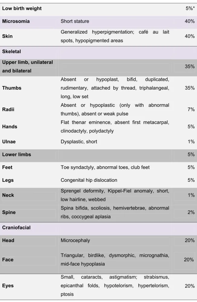
The likelihood of physical abnormalities is approximately 75%, though generally these are not a cause of mortality in individuals with FA (Alter and Kupfer, 2002 (update 2011)). The most commonly reported abnormalities and their frequencies are described in Table 1.
Table 1. Congenital malformations in patients with Fanconi anemia (adapted from Shimamura and Alter, 2010).
Low birth weight 5%*
Microsomia Short stature 40%
Skin Generalized hyperpigmentation; café au lait
spots, hypopigmented areas 40%
Skeletal
Upper limb, unilateral
and bilateral 35%
Thumbs
Absent or hypoplast, bifid, duplicated, rudimentary, attached by thread, triphalangeal, long, low set
35%
Radii Absent or hypoplastic (only with abnormal
thumbs), absent or weak pulse 7%
Hands Flat thenar eminence, absent first metacarpal,
clinodactyly, polydactyly 5%
Ulnae Dysplastic, short 1%
Lower limbs 5%
Feet Toe syndactyly, abnormal toes, club feet 5%
Legs Congenital hip dislocation 5%
Neck Sprengel deformity, Kippel-Fiel anomaly, short,
low hairline, webbed 1%
Spine Spina bifida, scoliosis, hemivertebrae, abnormal
ribs, coccygeal aplasia 2%
Craniofacial
Head Microcephaly 20%
Face Triangular, birdlike, dysmorphic, micrognathia, mid-face hypoplasia
Eyes
Small, cataracts, astigmatism; strabismus, epicanthal folds, hypotelorism, hypertelorism, ptosis
20%
Renal
Kidneys: horseshoe, ectopic or pelvic, abnormal, hypoplastic or dysplastic, absent; hydronephrosis or hydroureter
20%
Gonads
Males Hypospadias, micropenis; undescended testes,
absent testes 25%
Females Bicornuate uterus, malposition, small ovaries 2% Developmental delay Intellectual disability, developmental delay 10%
Ears Hearing loss, abnormal shape 10%
Cardiopulmonary Congenital heart defect 6%
Gastrointestinal
Esophageal, duodenal, jejunal atresia; imperforate anus; tracheoesophageal fistula; annular pancreas; malrotation of the gut
5%
Central nervous system
Small pituitary, pituitary stalk, interruption syndrome, absent corpus callossum, cerebellar hypoplasia, hydrocephalus, dilated ventriculus
3%
* Percentages are calculated from 2000 cases reported in the literature from 1927 to 2009. Frequencies are approximate, since many reports did not mention physical description.
The clinical manifestations can be highly variable. Some patients can have many congenital malformations at the same time, while in others none of such findings are observed at the time of diagnosis. Furthermore, other non-FA disorders can also have many of the clinical findings associated with FA. In conclusion, the presence or absence of physical malformations does not fully establish, neither rule out the diagnosis of FA.
1.1.2. Hematologic manifestations
Hematologic abnormalities in FA patients typically occur within the first decade of life, at a median age of 7 years, but are highly variable. Thrombocytopenia is often associated with elevated levels of fetal hemoglobin (HbF) and macrocytosis, and usually precedes onset of anemia or neutropenia. Pancytopenia generally worsens over time, and can be present as early as in the newborn period. Sweet syndrome (neutrophilic skin infiltrations) has been reported in a few individuals with FA and myelodysplasic syndrome (MDS).
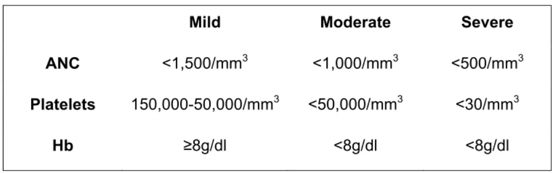
BMF is clinically manifested by blood counts that are below age-appropriate values due to decreased effective marrow hematopoiesis. While many patients progress to frank aplastic anemia, others may remain at mild abnormal levels indefinitely. Clinical surveillance and therapeutic management are guided by the severity of the cytopenias, the stability of the blood counts, the presence of morphologic and cytogenetic marrow abnormalities, and potentially high-risk genotypes such as FANCC, FANCD1/BRCA2 or FANCN mutations (Shimamura, 2008). BMF was classified into three broad categories, depending upon the degree of cytopenia(s) (Table 2). These definitions are more than semantic; they also define points at which different clinical management options should be considered.
Table 2. Severity of bone marrow failure (adapted from Shimamura, 2008).
Mild Moderate Severe
ANC <1,500/mm3 <1,000/mm3 <500/mm3 Platelets 150,000-50,000/mm3 <50,000/mm3 <30/mm3
Hb ≥8g/dl <8g/dl <8g/dl
ANC, absolute neutrophil count; Hb, hemoglobin.
Severe BMF may lead to death and requires HSCT. It has a peak hazard rate of about 5% per year at age of 10 years (Alter and Kupfer, 2002 (update 2011); Auerbach, 2009). Data from the IFAR suggest that BMF develops in up to 80% of patients (Seif, 2011).
1.1.3. Impairment in immunological function
The human immune system recognizes, eliminates, and protects the body from viral and bacterial infections, as well as from transformed cells (pre-cancer cell) (Masserot et al., 2008). A variety of cells are actively involved in this process of immune surveillance, including B and T-lymphocytes, natural killer (NK) cells, dendritic cells (DC), macrophages, and polymorphonuclear leucocytes. FA, as mentioned before, is characterized by pancytopenia, which means lower levels of leucocytes, platelets and hemoglobin (Hb), and is a cancer prone disease. One of the first studies that characterized impairment in the immune system of FA patients was reported in 1982.
Hersey and co-workers, (Hersey et al., 1982) found that the immune function of one FA patient revealed selective defects in NK cell activity, which is known to be important in surveillance against tumors. This case report suggested that the absence of NK activity was secondary to a defect in interferon release from lymphocytes on exposure to tumor antigens. They considered that these defects may be an important predisposing factor in the development of malignancy, not only in this patient but possibly other patients with FA. Lebbé and co-workers (Lebbé et al., 1993) reported the same results in another patient, where NK activity was undetectable even with a developing carcinoma.
Another study revealed that both lymphoblasts and fibroblasts from FA patients demonstrated a reduction in interleukin (IL) 6 production. IL-6 is an interleukin that acts as both a proinflammatory and anti-inflammatory cytokine. It is secreted by T cells and macrophages to stimulate immune response. This study showed that this lymphokine is not induced by tumor necrosis factors α and β (TNF-α and TNF-β) in FA cells, as is the case in normal cells. It was suggested that the observed deficiency in IL-6 production may account for one of the major characteristics of FA disease, the defect in differentiation of the hematopoietic system (Rosselli et al., 1992). The same group also showed that, in comparison to normal cells, TNF-α is overproduced by FA lymphoblasts from the four genetic complementation groups A, B, C and D. Indeed, up to an eight-fold increase in TNF-α is observed in the growth medium of FA cells. Moreover, addition of anti-TNF-α antibodies partially corrects the FA hypersensitivity to MMC. Treatment of FA cells with IL-6, which partially restored an almost normal sensitivity to MMC, also reduced the TNF-α overproduction in FA lymphoblasts (Rosselli et al., 1994). With this study, the authors concluded that abnormal TNF-α production seems to be associated with the FA genetic background, which was later confirmed by authors of the same group (Briot et al., 2008). One important activity of this cytokine is its cytotoxic/cytostatic effect on normal and cancer cells (Sugarman et al., 1985). Moreover, TNF-α enhances intracellular and extracellular superoxide anion (O2•-), production, and can induce DNA breakage and cell
death (Rubin et al., 1988; Yamauchi et al., 1989). This protein is of interest in the context of the FA phenotype, not only for its relation with IL-6 expression, but also for the large spectrum of its biologic activities (Schindler et al., 1990), which are completely deregulated in FA cells. Further investigations using FA mouse models revealed that Fancc(-/-) mice underwent excess inflammatory response, as a result of hematopoietic suppression, that was corrected by wild-type Fancc gene, suggesting a potential role of the FANCC protein in innate immunity (Sejas et al., 2007). Fancc(-/-) mice challenged in vivo with lipopolysaccharides (LPS) at doses that induce septic shock have increased peripheral blood levels of inflammatory mediators (Sejas et al., 2007), although it remains unknown what cell type(s) is responsible for this response.
As mentioned above, a number of clinical studies indicate that FA patients have altered levels of circulating cytokines. In addition, it has been suggested that FA patients may have an increased susceptibility to a variety of pathogens (Fagerlie and Bagby, 2006), although it is unclear whether this observation is a result of a subtle immunodeficiency or secondary to leukopenia from evolving BMF. A study of Liu and co-workers, (Liu et al., 2012) provides compelling evidence for a cell-autonomous defect in Fancc(-/-) macrophages. Specifically, functions requiring dynamic cytoskeletal changes are impaired, including adhesion, migration, and phagocytosis, as well as in vivo inflammatory monocyte mobilization and recruitment. Macrophages are a primary line of defense in the innate immune system (Rees, 2010). The biologic functions of macrophages are complex, including elimination of pathogens via phagocytosis and cytokine/chemokine production and repairing damaged tissues during inflammation (Gordon and Taylor, 2005; Rees, 2010). Most of these functions require macrophages to migrate to an inflammatory site, and these dysfunctions could explain the cytokine deregulation and increased susceptibility to pathogens.
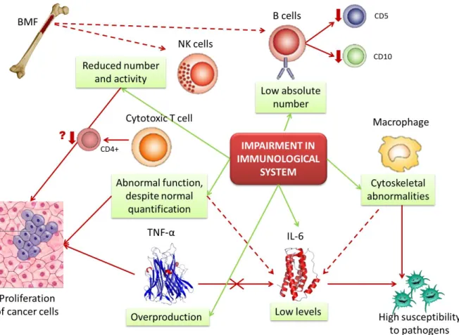
Recently it was performed a study that reported a cross-sectional immunological assessment in 10 children with FA, representing the first attempt at a comprehensive quantitative and functional evaluation of immune function (Myers et al., 2011). The study reported a significant, novel, and previously unappreciated abnormality in cytotoxic T cell function, despite normal quantitative evaluation, and significantly reduced NK cell number and function in the majority of these children, this last result being confirmed by previous studies. Additionally, Castello and co-workers (Castello et al., 1998) demonstrated a decrease in CD4+ T lymphocytes, which was not detected in Myers’ study. Moreover, Myers’s cohort of patients demonstrated a quantitative abnormality in the B cell compartment, with significantly lower absolute number of B cells relative to age-based normal. The phenotype of the remaining B-cells was nearly identical in these patients, showing reduced percentage of CD5 and CD10 expressing B cells. This particular B-cell phenotype would be expected in adults, especially at advanced age (Bleesing, 2004). As B and NK cells mature in the bone marrow, they may probably be most affected as BMF develops (Myers et al., 2011). Figure 1 resumes the information known till now about impairment in immunological system in FA patients and possible relation with FA phenotype.
Figure 1. Impairment in immunological function of Fanconi anemia patients. Possible interactions among immunological cells, cytokines and Fanconi anemia phenotype (original figure). Fanconi anemia patients show an impairment in immunological system, characterized by low absolute number of B lymphocytes, abnormal function of T lymphocytes, reduced number of NK cells, cytoskeletal abnormalities in macrophages, low levels of IL-6 and overproduction of TNF-α. In turn, abnormal function of T lymphocytes, reduced number of NK cells and overproduction of TNF-α increase the probability of cancer cells proliferation. The typical deregulation of IL-6 can be aggravated by the abnormal function of T lymphocyte and macrophages, the cells that produce this cytokine. Additionally, overproduction of TNF-α could be regulated by IL-6 activity, but this event apparently doesn’t occur. Therefore, with abnormal macrophages and IL-6 production, FA patients have high susceptibility to pathogens and, thus, to infections. In BMF conditions, B cells and NK cells are more affected, as they mature in bone marrow, so their number and activity keep impaired. BMF, bone marrow failure; IL-6, interleukin 6; NK, natural killer; TNF-α, tumor necrosis factor alpha.
1.1.4. Cancer susceptibility
In addition to the early onset of BMF, FA is a cancer prone disorder, being the one with higher risk of cancer development among BMF syndromes. Hematologic malignancies are the first to appear and approximately 10% of patients develop leukemia (Kutler et al., 2003). Over 90% of these hematologic malignancies and pre-malignancies are myeloid in origin, representing a 600-fold increase in risk of AML and more than a
5,000-fold risk increase for MDS compared to the general population (Shimamura and Alter, 2010). In a review of the literature reporting clinical cases of patients with FA, 9% developed AML and 7% developed MDS (Alter et al., 2003). The median age of leukemia diagnosis in FA patients is between 11 and 14 years, with almost all cases arising before 25 years old (Alter et al., 2003; Rosenberg et al., 2003).
Table 3. Malignancies in FA cases cited in the literature between 1927 and 2001 (adapted from Alter, 2003).
Characteristics All Leukemia MDS* Solid tumors Liver tumors
No. of cases 1301 116 89 68 37
% of total 100 8.9 6.8 5.3 2.8
Male:female 711:578 70:46 46:42 23:45† 22:15
Ratio 1.2 1.5 1.1 0.5† 1.5
FA diagnosis age (yrs)
Mean 8.3 10.1† 11.3† 12.7† 9.2
Median 7 8.6 9.3 9 7
Range 0-48 0.13-28 0.2-43 0-44 3-48
Complications age (yrs)
Mean _ 14.5 15.7 22.6† 15.7
Median _ 14 14 25.5 13
Range _ 0.13-29 1.8-43 0.2-45 6-48
No. of report deceased 488 84 44 41 30
% of report deceased 38 72 51 61 81
Estimate median
survival age (yrs) 20 16 21 31
† 16
Age on general
population (yrs) 68 68
_
47-68 68
* Includes 13 patients who subsequently developed leukemia; † P<0.01 compared with
Hematologic malignancies are not the only cause of early death in patients with FA. Nonhematologic malignancies are especially striking in these patients. They are unusually young when they develop cancer and the incidence of the malignancy probably would be considerably higher if patients had a longer life expectancy. Most of the nonhematologic tumors in FA patients are solid tumors, especially SCC of the head and neck and anogenital regions. The risk of head and neck SCC is 1000-fold greater in patients with FA than that of the general population, and also occurs at an earlier age (Myers et al., 2011). Epidemiologic analyses strongly suggest that solid tumors will become the predominant clinical problem of post-transplanted FA patients. HSCT is becoming available for a growing number of patients, because of an increased pool of alternative donor options and new transplant protocols. The result is an improved probability of survival to an age when the incidence of solid tumors begins to increase (Auerbach, 2009). There is also a probably of development of liver tumors, especially in patients receiving androgen treatment for BMF. This information is summarized in Table 3.
Interestingly, patients with no congenital anomalies have the highest risk of AML (cumulative incidence 23.7%) (Rosenberg et al., 2008). Significant dysmorphia is more closely associated with BMF and with a much lower total cancer cumulative risk (1.4%), although more seriously affected children may die of BMF before attaining their full cancer risk (Rosenberg et al., 2008). Importantly, in approximately 25% of patients, FA is identified only after the leukemia diagnosis as a result of associated complex cancer-related cytogenetic aberrations or excessive therapy-cancer-related toxicity (Alter, 2007; Gyger et al., 1989).
1.2. Fanconi anemia genotype
Based on somatic cell fusion studies, at least 15 complementation FA groups have been identified each one with a corresponding gene. Each of these genes, when biallelically mutated, causes FA. Gene denomination and chromosome location, as well as their proportion among FA patients are summarized in Figure 2 and Table 4 based on the work of Alter and Kupfer (Alter and Kupfer, 2002 (update 2011)) and Green and Kupfer (Green and Kupfer, 2009).
Tabl Gree Com e 4. The 15 en and Kupf mplementatio group FA-A FA-B FA-C FA-D1 FA-D2 FA-E FA-F FA-G 5 FA genes fer, 2009). on Respo ge FAN FAN FAN BR FAN FAN FAN XR s and their onsible ene C NCA NCB NCC RCA2 NCD2 NCE NCF CC9 location (a Chromosom location 16q24.3 Xp22.31 9q22.3 13q12.13 3p25.3 6p21-22 11p15 9q13 dapted from me % o attribu mutatio ge 60% ~2 ~1 ~3 ~3 ~3 ~2 ~1 Figure 2. frequency complem (genes). F and FANC the most c population frequency considerin ethnic gro m Alter and of FA table to on in this ene - 70% 1 2% (M 4% 3% 3% 3% 2% 0% Relative y of the FA mentation g FANCA, FA CG mutation common in n: however, y can be var ng the race oup. d Kupfer, 20 Referen (Apostolou 996; Lo Te al., 199 Meetei et a (Strathdee 1992 (Howlett e 2002 (Timmers 2001 (de Winter 2000 (de Winter 2000 (de Winter 2000 A roups ANCC ns are general the riable and 011 and nces u et al., n Foe et 96) l., 2004) e et al., ) et al., ) et al., ) r et al., ) r et al., ) r et al., )
FA-I KIAA1794 15q25-26 ~1% (Dorsman et al., 2007; Sims et al., 2007; Smogorzewska et al., 2007) FA-J BRIP1/BACH1 17q22-24 ~2% (Levitus et al., 2005; Levran et al., 2005; Litman et al., 2005)
FA-L PHF9 2p16.1 ~0.2% (Meetei et al., 2003)
FA-M FANCM 14q21.3 ~0.2% (Meetei et al., 2005)
FA-N PALB2 16p12 ~0.7% (Reid et al., 2007;
Xia et al., 2007)
FA-O RAD51C 17q22 ~0.2% (Vaz et al., 2010)
FA-P SLX4 16p13.3 ~0.2%
(Kim et al., 2011; Stoepker et al.,
2011)
These genes account for over 95% of all known FA patients. Some patients do not appear to have mutations in any of these 15 genes, so it can be anticipated that additional FA genes will be discovered in the future.
1.2.1. The FA pathway
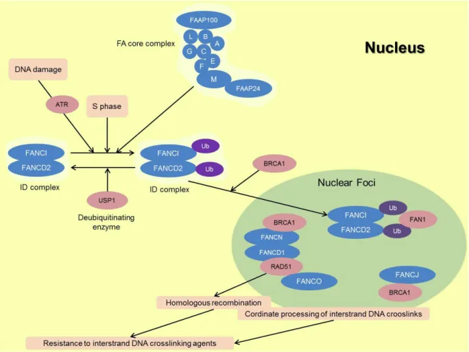
FA genes and proteins function together in a common pathway, involved in DNA repair and in the maintenance of genomic stability (Bogliolo et al., 2002b). The encoded proteins for each gene can be subdivided into three groups: (1) eight proteins that make up the core complex of the upstream FA proteins, FANCA, FANCB, FANCC, FANCE, FANCF, FANCG, FANCL and FANCM; (2) the FANCD2 and FANCI proteins, which compose the ID complex; and (3) the downstream effector proteins, FANCD1, FANCJ, FANCN, and presumably FANCO and FANCP (Kim et al., 2011; Moldovan and D'Andrea, 2009; Vaz et al., 2010).
Figure 3. Schematic representation of the FA pathway and associated proteins in response to DNA damage (original figure). A cascade of events starts with DNA damage, being activated the FA core complex, which in turn activates de monoubiquitination of ID complex. These events activate other FA proteins, culminating in the rearrangement of the damage through different repair processes.
In a simple manner and summarizing all this information (Figure 3) the eight upstream FA proteins together with FAAP100 and FAAP24 form a complex with ubiquitin ligase activity, termed the FA core complex. This complex is required for monoubiquitination of FANCD2 and FANCI in response to DNA interstrand crosslink (ICL) lesions during replication in S phase. FANCD2 and FANCI thereafter localize to DNA repair foci together with FA effector proteins and other proteins. BRCA1 is required for FANCD2 foci formation in response to DNA damage. FANCC, FANCE, and FANCG also form nuclear foci and co-localize with FANCD2. All these factors are required for cellular resistance to interstrand DNA crosslinking agents, through homologous recombination, trans-lesion synthesis and some other unknown mechanisms. A few other gene products were found to be associated with the FA protein complexes and are required for FA activation. Thus, it is presumed that their inactivation would lead to FA; however, patients
with such mutations have not yet been described (Hucl and Gallmeier, 2011; Moldovan and D'Andrea, 2009).
Interestingly, many of the FA proteins contain no recognizable motifs. Therefore, discovering their contributions to the FA pathway and the main function of the FA pathway will be an important challenging in the future (Green and Kupfer, 2009).
1.3. Diagnostic tests for Fanconi anemia
Considering the high genetic FA variability and the vast clinical phenotype, with a great overlap with other BMF syndromes, and the particularity that 25% of FA patients are phenotypically normal, a rapid and correct clinical diagnosis is difficult and may be delayed or even missed. Fortunately more accurate diagnostic methods were developed, the most important being the chromosomal fragility test.
1.3.1. The chromosomal fragility test
Schroeder and co-workers were the first to report that FA is a CI syndrome (Schroeder et al., 1964). They first suggested the use of spontaneous chromosomal breakage as a cellular marker for FA, but subsequent studies of CI in more FA patients showed these findings to be inconsistent. Years later, Auerbach and co-workers evidenced that FA cells have an unique hypersensitivity to the clastogenic (chromosome breaking) effect of cross-linking agents and so this characteristic became a reliable cellular marker for the diagnosis of FA (Auerbach et al., 1981). DEB and MMC are the most widely used agents for the diagnosis, but DEB demonstrated to be the one that proportionated more accuracy.
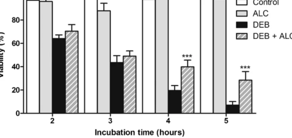
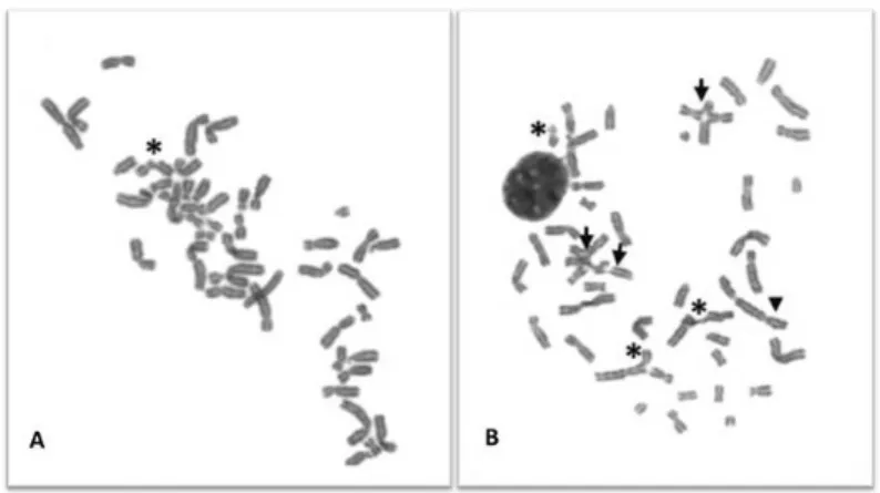
The chromosomal fragility test with DEB is the cytogenetic method for excellence of FA diagnosis (Auerbach et al., 1989). Peripheral blood lymphocytes are cultured in the presence and absence of DEB and a minimum of 50 cells arrested in metaphase are scored and analyzed for chromosomal breakage (the most used parameter is the number of breaks per cell) and formation of tri- and tetraradial figures. These figures are the hallmark of FA diagnosis, since this parameter is the only one that can differentiate FA from the other CI syndromes. Results are compared with those of normal control cells. Normal cells are able to correct most of the damage and are not severely affected by DEB, whereas FA cells show marked chromosome breakage, as can be seen in Figure 4. This test can also be performed prenatally on cells from chorionic villi or amniotic fluid (Auerbach et al., 1981).
F D p o ly c c t r b brea popu the c agen same be m been inher susp T-lym accu exam G2/M Figure 4. C DEB-induce patient. Ch observed w ymphocyte can be see culture expo the tri- and represents t break (aster The exis kage tests. ulations of s cases), whi nts and the e agents (A more useful n shown to rited mutati picious of m mphocytes. urately differ 1.3.2. Ot Cell cycl mines cell c M after cultu Chromosom ed lympho romosomes with an op culture exp en (asterisk osed to 0.0 d tetraradia the hallmar risk). stence of m . Somatic somatic cell
ich can pre other show Auerbach, 2 than the nu o be cause ions (Grego mosaicism, c Recently, rentiate mos ther diagno le arrest. Ar cycle kinetic ure with a cl me instabil ocyte cultu s were stai ptical micro posed to 0 k). (B) Sele 5 µg/ml of D al figures rk for the d mosaicism mosaicism ls in a give esent a cel wing normal 009). In tha umber of bre ed by new ory et al., chromosom a new chro saic FA pat ostic metho rrest in G2 cs and can lastogen su lity pattern ures from ned in a 4% oscope. (A .2 µg/ml of ected metap DEB for 48 (arrow) be iagnosis of may compl is defined n organism l line show l levels of c at case the eaks per ce DNA muta 2001; Lo T me fragility t omosome i tients (Caste ods is another n detect the uch as nitrog n in two m a healthy % Giemsa A) Selected f DEB for 4 phase from h. High lev eing especi f FA. It can licate the F as the pr m and is rela wing increas chromosome percentage ell. Somatic ations or t Ten Foe et test in fibrob nstability in ella et al., 2 characteris e proportion gen mustar etaphases y donor (H solution. Th d metaphas 48 h. One c m a FA pati vel of CI ca ally import also be se FA diagnos esence of atively com sed sensitiv e breakage e of cells w c mosaicism he spontan t al., 1997) blast is mo ndex (CFI) w 2011). tic of FA ce n of cells th rd or MMC. s selected HD) and a he images se from a chromatid b ient lympho an be visual tant, since een a chrom sis by chrom genetically mon in FA vity to cros e in respons with aberratio m in FA patie neous reve ). When th ore accurate was establ ells. Flow cy hat are arr
In contrast from a FA were HD break ocyte lized, that matid mosome y distinct (25% of ss-linking se to the ons may ents has ersion of ere is a e than in ished to ytometry ested at with the
100 cells examined microscopically for chromosomal aberrations, flow cytometry can have the advantage of examining thousands of cells and being less labor-intensive and subjective. However, it requires sophisticated instrumentation. This test is usually done in a specialized laboratory and is not used as widely as the chromosome breakage assay. Flow cytometry may give a false negative result in FA patients with MDS or AML (Alter, 2008).
Immunoblot assay of FANCD2 protein monoubiquitination. Following DNA damage, the complex of upstream FA gene products (A, B, C, E, F, G, L, M) leads to ubiquitination of the product of FANCD2, forming a longer protein (D2-L), which can be distinguished from the shorter non-ubiquitinated form (S) on a Western blot with a D2-specific antibody. This relatively inexpensive assay may be useful for screening patients for whom FA is in the differential diagnosis, such as those with radial ray anomalies, short stature, and hypogonadism or café au lait spots or for population-based FA incidence studies; however, it is usually a limited tool. FA patients whose gene defect is downstream of FANCD2 (FANCD1, FANCI, FANCJ, FANCN, FANCP and FANCO) will not be detected with a D2 Western blot, as well as mosaic individuals (Alter, 2008).
Complementation analysis. Patient lymphocytes, EBV (Epstein-Barr virus)-lymphoblasts or fibroblasts can be cultured with retroviruses that introduce known normal FANC genes into the patient’s cells, leading to correction of the FA cellular phenotype (Alter, 2008).
Mutation testing. Determination of the specific mutation in FA genes is complicated and is done in laboratories with specific expertise. It requires sophisticated methods and involves DNA amplification, sequencing and detection of large deletions. Many laboratories rely on knowing the complementation group before sequencing, while in some contexts targeted sequencing of candidate genes is more appropriate. One center goes directly to gene sequencing for patients with a positive DEB test: FANCA by multiplex ligation-dependent probe amplification (MLPA) for large deletions and full sequencing; FANCB by MLPA and full sequencing, if indicated; FANCC, E, F, G by denaturing high performance liquid chromatography (DHPLC) and sequencing; FANCD2 by Western blot; FANCD2 sequencing if D2 bands are absent; FANCL and FANCM sequencing if only D2-S is seen; FANCD1/BRCA2 sequencing, if indicated; FANCJ/BRIP1 and FANCN/PALB2 sequencing; and finally NBS1 and ESCO2 sequencing for Nijmegen breakage and Roberts syndromes. Mutation testing is used to confirm known cases and for family studies to determine affected or carrier status. Genetic counseling should be included in these processes (Alter, 2008).