Characterization of patients with heart failure and
reduced ejection fraction attending two Heart
Failure clinics in Portugal and in Mozambique
2012/2013
Hugo Ricardo Leal Teixeira
Characterization of patients with heart failure and
reduced ejection fraction attending two Heart
Failure clinics in Portugal and in Mozambique
Prof. Doutor José Carlos de Magalhães Silva Cardoso
Prof. Doutor Albertino António Moura Damasceno
Characterization of patients with heart failure and
reduced ejection fraction attending two Heart
Failure clinics in Portugal and in Mozambique
Trabalho organizado de acordo com as normas da revista:
Hugo Ricardo Leal Teixeira
Mestrado Integrado em Medicina
Área: Cardiologia
Trabalho efetuado sob a Orientação de:
Prof. Doutor José Carlos de Magalhães Silva Cardoso
E sob a Coorientação de:
Prof. Doutor Albertino António Moura Damasceno
Characterization of patients with heart failure and
reduced ejection fraction attending two Heart
Failure clinics in Portugal and in Mozambique
Trabalho organizado de acordo com as normas da revista:
Revista Portuguesa de Cardiologia
Projeto de Opção do 6º ano -
D
ECLARAÇÃO DEI
NTEGRIDADEEu, Hugo Ricardo Leal Teixeira, abaixo assinado, nº mecanográfico 200600972, estudante do 6º ano do Mestrado Integrado em Medicina, na Faculdade de Medicina da Universidade do Porto, declaro ter atuado com absoluta integridade na elaboração deste projeto de opção.
Neste sentido, confirmo que NÃO incorri em plágio (ato pelo qual um indivíduo, mesmo por omissão, assume a autoria de um determinado trabalho intelectual, ou partes dele). Mais declaro que todas as frases que retirei de trabalhos anteriores pertencentes a outros autores, foram referenciadas, ou redigidas com novas palavras, tendo colocado, neste caso, a citação da fonte bibliográfica.
Faculdade de Medicina da Universidade do Porto, ___/___/______
Projeto de Opção do 6º ano – DECLARAÇÃO DE REPRODUÇÃO
Nome: Hugo Ricardo Leal Teixeira Email: [email protected]
Título da Dissertação: Characterization of patients with heart failure and reduced ejection fraction
attending two Heart Failure clinics in Portugal and in Mozambique
Orientador:
José Carlos de Magalhães Silva Cardoso
Coorientador:
Albertino António Moura Damasceno
Ano de conclusão: 2013
Designação da área do projeto:
Cardiologia
É autorizada a reprodução integral desta Dissertação para efeitos de investigação e de divulgação pedagógica, em programas e projetos coordenados pela FMUP.
Faculdade de Medicina da Universidade do Porto, ___/___/______
Characterization of patients with heart failure and reduced
ejection fraction attending two Heart Failure Clinics in
Portugal and in Mozambique
Caracterização de pacientes com insuficiência cardíaca e fração de ejeção reduzida
em duas Clínicas de Insuficiência Cardíaca em Portugal e Moçambique
Hugo Teixeira
a, Brenda Moura
b, Neusa Jessen
c, Luís F Azevedo
d, Albertino
Damasceno
e, José Silva Cardoso
a, fa
Faculty of Medicine, University of Porto, Porto, Portugal
b
Military Hospital D. Pedro V, Porto, Portugal
c
Department of Cardiology, Maputo Central Hospital, Maputo, Mozambique
d
Department of Health Information and Decision Sciences (CIDES) and Centre for Research in Health Technologies and Information Systems (CINTESIS), Faculty of Medicine, University of Porto, Portugal
e
Faculty of Medicine, Eduardo Mondlane University, Maputo, Mozambique
f
São João Medical Center, Porto, Portugal
Corresponding author: Hugo Teixeira
Address: Rua Camilo Castelo Branco Nº222 Hab 3.5; 4425-037, Águas Santas, Maia; Portugal [email protected]
Resumo
Introdução e objetivos: A insuficiência cardíaca é um problema de saúde comum, incapacitante e
relacionado a elevada morbilidade e mortalidade. A patogenia varia nos diferentes países africanos, sendo a mais prevalente a não isquémica, enquanto em Portugal são a hipertensão arterial e doença isquémica. O nosso objetivo foi comparar doentes Moçambicanos e Portugueses para identificar possíveis diferenças clínicas, etiológicas e terapêuticas.
Métodos: Recolheu-se informação do processo clínico hospitalar (história, exame físico, meios
complementares de diagnóstico e terapêutica, relativa à consulta inicial na clínica de insuficiência cardíaca e à primeira consulta no período entre 1 de Maio e 30 de Julho de 2012. Foi realizada análise estatística e os resultados apresentados em tabelas.
Resultados: Estudaram-se 242 doentes, 187 Portugueses e 55 Moçambicanos. Globalmente, os
Portugueses apresentaram classes NYHA menos graves na admissão (p<0.001), mas mais co-morbilidades (exceto insuficiência renal e infeção HIV). As etiologias mais frequentes foram a isquémica, familiar e alcoólica nos Portugueses, e a valvular, hipertensiva e peri-parto nos Moçambicanos. Não se encontraram diferenças estatisticamente significativas relativas à associação com infeção HIV (p=0.085).
Em ambos, os inibidores da enzima de conversão da angiotensina/inibidores do recetor da angiotensin foram prescritos num elevado número de doentes, tendo globalmente os bloqueadores beta sido os mais prescritos em Portugal e os diuréticos em Maputo, estando a terapêutica não-farmacológica indisponível para Moçambicanos.
Conclusões: Num estudo observacional sem precedentes, explicitámos algumas diferenças entre
estas populações, podendo contribuir para melhorar a abordagem da insuficiência cardíaca nas respetivas clínicas.
Abstract
Background and objectives: Heart failure is a common and disabling health problem, associated
with high morbidity and mortality. The pathogenesis varies between African countries, with the most prevalent pathogenesis being non-ischemic disease, whereas on Portugal hypertension and ischemic disease are the most prevalent. We aimed to compare Mozambican and Portuguese patients to identify possible clinical, etiological and therapeutic differences.
Methods: We collected information from patient’s clinical file (history, physical examination,
complementary diagnostic tests and therapeutic, from the first appointment ever on the heart failure clinic and the first appointment between May 1st and July 30th 2012. Statistical analysis was performed and the results presented as tables.
Results: We studied 242 patients, 187 Portuguese and 55 Mozambicans. Overall, Portuguese
presented with less severe NYHA class at admission (p<0.001), but had higher proportion of co-morbidities (except renal insufficiency and HIV infection). Ischemic, familial and alcoholic etiologies were more prevalent on Portuguese patients, whereas valvular, hypertensive and peripartum etiologies were more frequent on Mozambicans. No statistical difference was found regarding HIV-associated heart failure (p=0.085). On both populations the angiotensin converting enzyme inhibitor/angiotensin receptor blocker were prescribed to a high percentage of patients, and on a global perspective beta-blockers were the most prescribed drugs in Oporto and diuretics the most prescribed in Maputo, with non-pharmacological treatment unavailable for Mozambicans.
Conclusions: In an unprecedented cross-sectional study we made clear some differences between
these two populations that may contribute to improve the approach to heart failure on both clinics.
Abbreviations / Abreviaturas
English Português
ACE Angiotensin converting enzyme Enzima de conversão da angiotensina AF Atrial fibrillation Fibrilação auricular
ARB Angiotensin receptor blocker Antagonista do recetor da angiotensin BMI Body mass index Índice de massa corporal
BNP B-type natriuretic peptide Peptídeo natriurético tipo B
CABG Coronary artery bypass graft Cirurgia de pontagem aorto-coronária CAD Coronary artery disease Doença das artérias coronárias DM Diabetes mellitus Diabetes mellitus
ECG Electrocardiogram Eletrocardiograma
ESC European Society of Cardiology Sociedade Europeia de Cardiologia GFR Glomerular filtration rate Taxa de filtração glomerular HF Heart failure Insuficiência cardíaca LMCA Left main coronary artery Tronco coronário comum
LV-EF Left ventricle ejection fraction Fração de ejeção do ventrículo esquerdo MRA Mineralocorticoid receptor antagonist Antagonista do recetor mineralocorticóide NYHA New York Heart Association Sem tradução
PCI Percutaneous coronary intervention Intervenção coronária percutânea VF Ventricular fibrillation Fibrilação ventricular
Introduction
Heart failure (HF) is a major health problem as it is common, disabling and associated with high morbidity and mortality, despite being highly treatable1-3. It can be defined as an abnormality of cardiac structure, function or both, leading to the inability of the heart to pump enough blood to meet the body’s demand of oxygen, despite normal filling pressures (or only at the expense of increased filling pressures) 4,5. According to the European Society of Cardiology (ESC), HF can be clinically defined as a syndrome in which patients have typical symptoms (e.g. breathlessness, ankle swelling, and fatigue) and signs (e.g. elevated jugular venous pressure, pulmonary crackles, and displaced apex beat) resulting from an abnormality of cardiac structure or function6.
The aging population, the increasing prevalence of co-morbidities such as Diabetes Mellitus (DM) and obesity, the decrease in sudden death due to device therapy, and the increasing proportion of patients surviving acute myocardial infarction with substantial left ventricular damage that predisposes to post-infarction HF7,8, all contribute to an epidemiological trend of increasing prevalence of chronic HF during the coming decades9,10.
The diagnosis of HF can be difficult. Many of the symptoms of HF are non-discriminating and, therefore, of limited diagnostic value11-15. Many of the signs of HF result from sodium and water retention and resolve quickly with diuretic therapy, i.e. may be absent in patients receiving such treatment. Demonstration of an underlying cardiac cause is therefore central to the diagnosis of HF. This is usually myocardial disease causing systolic ventricular dysfunction. However, abnormalities of ventricular diastolic function or of the valves, pericardium, endocardium, heart rhythm and conduction can also lead to HF (and more than one abnormality can be present). Identification of the underlying cardiac problem is also crucial for therapeutic reasons, as the precise pathology determines the specific treatment used6.
Up to 80% of first-diagnoses of heart failure occur at the time of hospitalisation, over 40% of patients in the community with heart failure have been hospitalised within the previous year and one-third of patients with heart failure will be hospitalised within any given year, reflecting the more severe end of the disease spectrum in terms of morbidity and mortality. In-hospital mortality is approximately 20% and many patients with heart failure will die or be re-hospitalised (10-30% or more) within the following 6 months16-19.
Although the causes of heart failure vary within and between African countries, the pathogenesis remains largely nonischemic (98%), with hypertension, rheumatic heart disease and cardiomyopathy accounting for 65% of cases, whereas tuberculous pericarditis and pulmonary heart disease account for the remainder. In the study performed by Mayosi BM20, the diagnosis of myocardial infarction was made in only 2% of cases, which confirms the observation that coronary artery disease (CAD) remains uncommon in black Africa. Unlike other parts of the world in which cardiomyopathies are rare, dilated cardiomyopathy is a major cause of heart failure throughout Africa. Similarly, peripartum cardiomyopathy is ubiquitous on the continent, with an incidence ranging from 1 in 100 to 1 in 1000 deliveries21-23. Nowadays, cardiovascular diseases account for 7-10% of all medical admissions to hospital, with heart failure contributing to 3-7%24,25.
In Portugal, the EPICA study showed a HF prevalence of 4.4% amongst adults older than 25 years old, with a mean age of 65 years old, a preponderance among women (63.3%), 1.3% had left ventricular systolic dysfunction and 1.7% didn’t have. EPICA studied also estimated that nearly 36% of Portuguese patients older than 60 years old had HF. This study also made clear that hypertension is the leading cause of HF in Portugal (66%), followed by CAD (40%) and major valvular disease (26%)26.
Our aim was to compare two populations of HF patients, one from an European country and other from Africa, in order to understand their clinical, etiological and treatment differences.
Methods
This is an observational retrospective study that intends to evaluate on two different moments of time the clinical characteristics of the patients from two HF clinics, one from Maputo (outpatient clinic of the Cardiac Department of the Maputo Central Hospital, Mozambique) and the other from Oporto (outpatient clinic of the Cardiac Department of the Hospital São João, Portugal). The first moment corresponds to the patient’s first appointment on the HF clinic on the period between May 1st and July 30th 2012, and those patients’ clinical files were studied to gather information about the second moment studied (their first appointment on the HF clinic from all time). All the information was obtained from the patient’s clinical file after the appointment on HF Clinic. If a patient came to an appointment twice or more on the period between May 1st and July 30th 2012 only the first appointment was considered and the subsequent ones excluded.
Nearly 100 variables were collected from history, physical examination, Electrocardiogram (ECG), echocardiography, myocardial perfusion scintigraphy, coronary angiography, stress ECG, blood tests and pharmacological or non-pharmacological treatment.
Regarding the statistical analysis, continuous variables are described as median values and corresponding 25th and 75th percentiles. Dichotomous variables are reported as absolute numbers and percentages. To evaluate the characteristics of differences between the populations studied, Chi-square tests, Mann Whitney U tests or Fischer tests were applied as appropriate. All calculations were performed using IBM SPSS Statistics version 21.0 software package. For all tests a p value of 0.05 or less (two-sided) was considered statistically significant.
Ethical approval for the study was sought from the local Ethical Committee and permission confirmed through the relevant administrative bodies. The study conformed to the principles outlined in the Declaration of Helsinki.
Results
Demographic profile
There were more individuals from Portugal (147 – 72%), generally older than the ones from Mozambique (median: 60, 25-75 percentile: 49-67 vs. median: 53, 25-75 percentile: 38.5-61, respectively), with a high proportion of male patients in Portugal (131 – 70.1%) and similar gender distribution in Mozambique.
Co-morbidities
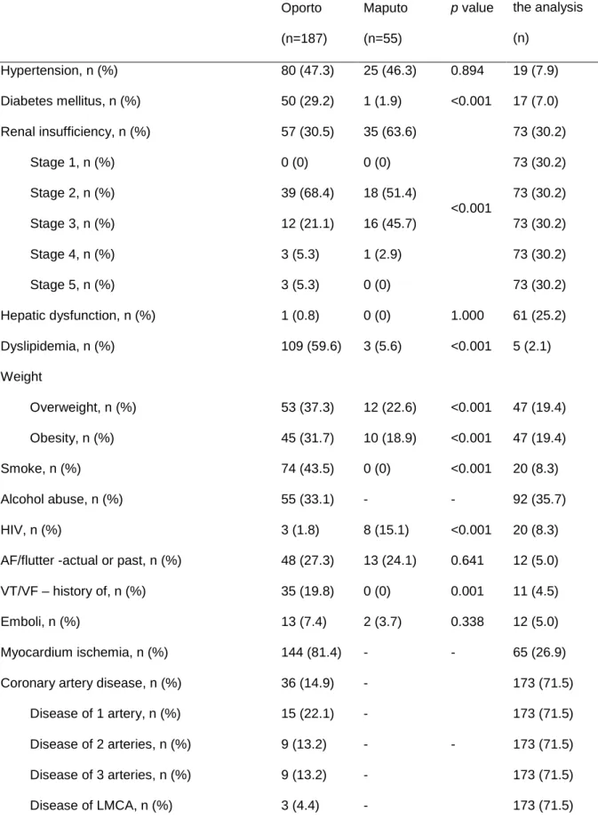
Overall, Portuguese individuals were found to have a higher proportion of co-morbidities, as shown in Table 1, except from renal insufficiency (35 – 63.6% in Mozambique vs. 50 – 30.5% in Portugal;
p<0.001) and HIV infection (8 – 15.1% in Mozambique vs. 3 – 1.8% in Portugal; p<0.001) whose
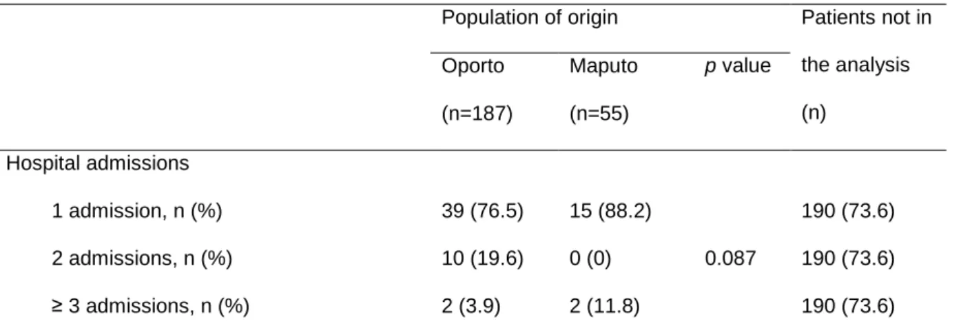
proportion was found to be greater in Mozambique. From the co-morbidities studied, no statistical difference was found on hypertension (p=0.894), hepatic dysfunction (p=1.000), presence of actual or past atrial fibrillation (AF) or flutter (p=0.641), previous episodes of emboli (p=0.338) and previous hospital admissions (p=0,087). No statistical inference was possible to be made regarding myocardium ischemia (on stress electrocardiogram or myocardium perfusion scintigraphy) and CAD
(on coronary angiography), as none of the individuals from Mozambique was submitted to the clinical test to evaluate so.
Heart failure etiology
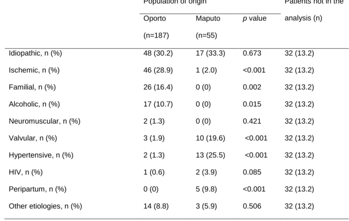
Table 2 summarizes the differences found regarding HF etiology. Ischemic (46 – 28.9% in Portugal vs. 1 – 2.0% in Mozambique; p<0.001), familial (26 – 16.4% in Portugal vs. 0 – 0% in Mozambique;
p=0.002) and alcoholic (17 – 10.7% in Portugal vs. 0 – 0% in Mozambique; p<0.015) etiologies were
more prevalent on Portuguese patients, whereas valvular (10 – 19.6% in Mozambique vs. 3 – 1.9% in Portugal; p<0.001), hypertensive (13 – 25.5% in Mozambique vs. 2 – 1.3% in Portugal; p<0.001) and peripartum (5 – 9.8% in Mozambique vs. 0 – 0% in Portugal; p<0.001) etiologies were more frequent on Mozambican patients. Regarding HIV-associated HF no significantly statistical difference was found (p=0.085).
Clinical aspects
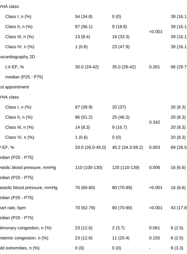
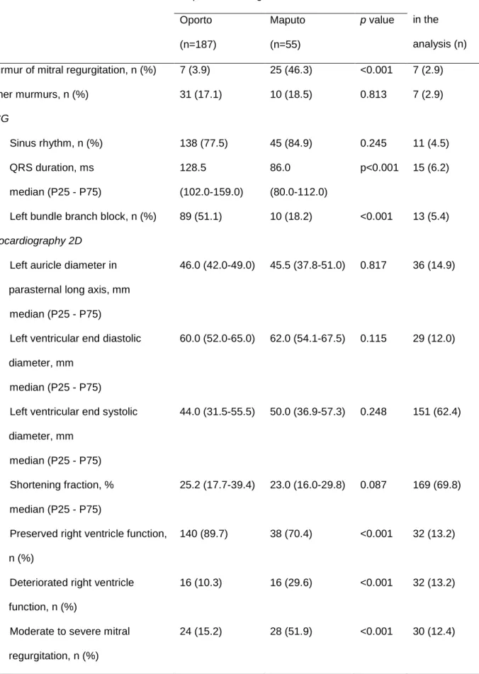
Table 3 summarizes the clinical aspects gathered from physical examination, echocardiography 2D and ECG. In what concerns New York Heart Association (NYHA) class, the most used classification for clinical categorizing of HF, we demonstrated that there were a significantly statistical difference between the populations on presentation to HF clinic (p<0.001) but that difference ceased to exist when the patients were evaluated on their last appointment (p=0.342). It is also possible to see that while in Oporto most patients were on NYHA class I and II (141 – 58.3% vs. 14 – 5.8% on classes III or IV) on their first appointment, on Maputo the majority of patients presented on NYHA class III or IV (39 – 70.9% vs. 9 – 16.4% on classes I or II).
The blood pressure profile of this groups showed that the Portuguese population has globally better control over this risk factor, as they had lower systolic (median: 110, 25-75 percentile: 100-130 in Oporto vs. median: 120, 25-75 percentile: 110-139, in Maputo; p=0.006) and diastolic (median: 70, 25-75 percentile: 60-80 in Oporto vs. median: 80, 25-75 percentile: 70-89, in Maputo; p<0.001) blood pressures.
Mitral regurgitation was also a situation more frequent in Maputo than in Oporto, both on auscultation of the respective murmur (25 – 46.3% vs. 7 – 3.9%; p<0.001, respectively) and on the evaluation by echocardiogram (28 – 51.9% vs. 24 – 15.2%; p<0.001, respectively).
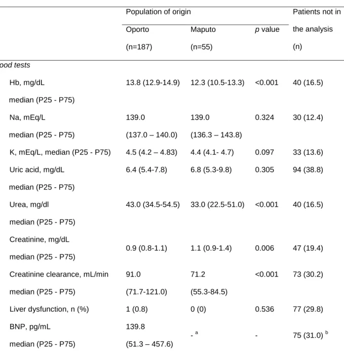
Regarding blood tests, it was not possible to establish a comparison of B-type natriuretic peptide (BNP) values between the populations as this was not evaluated in none of the patients from Maputo.
Pharmacological treatment
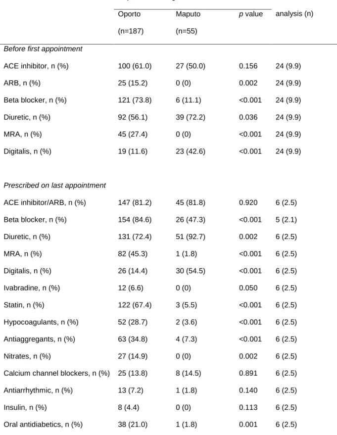
In what concerns pharmacological treatment (Table 4), on presentation to HF clinic most patients had already some treatment prescribed by other doctors, being the most prescribed drugs in Oporto beta-blockers (73.8%), angiotensin converting enzyme (ACE) inhibitors (61.0%) and diuretics (56.1%), in opposition to Maputo where the most prescribed drugs before presentation to HF clinic were diuretics (72.2%), ACE inhibitors (50.0%) and digitalis (42.6%).
On the last appointment, we were able to show a different pattern on drugs prescribed, with beta-blockers (84.6%), ACE inhibitor/angiotensin receptor blocker (ARB) (81.2%), diuretics (72.4%) and statins (67.4%) being the most prescribed drugs to patients in Oporto, in opposition to diuretics (92.7%), ACE inhibitors/ARB (81.8%), digitalis (54.5%) and beta-blockers (47.3%) which were the ones most prescribed in Maputo.
Non-pharmacological treatment
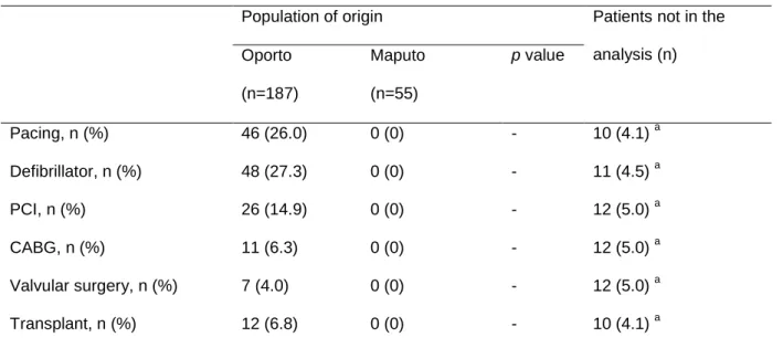
The most prominent aspect on Table 5 is that none of the patients from Maputo had undergone any of the non-pharmacological treatments studied. In Oporto, it was frequent that patients had resynchronizing devices (46 – 26.0%) or defibrillators (48 – 27.3%) implanted, and there were more patients who have been submitted to heart transplant surgery (12 – 6.8%) than the ones submitted to coronary artery surgery (11 – 6.3%) and valvular surgery (7 – 4.0%).
Discussion
In an unprecedented cross-sectional study, we aimed to compare the characteristics of HF patients between an African and an European HF clinic, considering characteristics of the disease itself, the patient’s co-morbidities and clinical status and the approach used on diagnosis and treatment of these patients. A total of 242 patients were studied, 187 from Oporto and 55 from Maputo. In Portugal the patients were older and 70.1% were man, while in Maputo there was a more homogenous distribution regarding gender.
We also found a higher prevalence within the Portuguese population of risk factors and co-morbidities (with the exception of HIV infection and renal insufficiency that are more prevalent in Maputo), which may be related to economic and social development of Mozambique, one of the poorer countries in the world.
While our data did not provide a complete picture of the etiology of the HF, it is conceivable that we made clear some differences between these populations. A similar amount of patients was categorized as having idiopathic HF, but there was a clear preponderance of ischemic, familial and alcoholic causes on Portuguese population in opposition with valvular and hypertensive heart diseases in Mozambicans, as expected27, and peripartum etiology was also more common among Mozambicans. HIV-related HF was more prevalent in Mozambique but this result lacks statistical significance.
Differences have also been made clear when it comes to the first appointment on the HF clinic, as only 1 (0.6%) Portuguese patient presented with NYHA class IV in opposition to 23 (47.9%) Mozambican patients and 54 (34.8%) Portuguese patients presented with NYHA class I in opposition to Mozambique where none of the patients presented with such NYHA class.This may be the reflex of both clinical follow-up for patients with risk factors and co-morbidities, and to a better access to health care services in Portugal. On the other hand no significant difference was found on the NYHA class on the last appointment, meaning that even with limited resources Mozambican patients become as little symptomatic as the Portuguese ones.
However, Mozambican patients showed a more accentuated left ventricle ejection fraction (LV-EF) recovery. This may be related to the institution of the correct treatment for HF, namely increased use of beta-blockers and also to the fact that peripartum cardiomyopathy frequently shows EF recovery. However, in the absence of a central validation of the echocardiograms performed at
each center, caution is needed in the interpretation of this parameter. Even so, other variables suggest that the population from Portugal has a more severe disease, as the more prolonged QRS interval, and higher percentage of patients with left bundle brunch block.
A curious finding was that even though there were a similar percentage of patients with actual or past AF/flutter, with higher usage of hypocoagulants and antiaggregants on the Portuguese population, there were a similar number of embolic events. Probably, that can be related with a lower CHA2DS2-VASc Score for Atrial Fibrillation Stroke Risk, with lower embolic risk, as the population is younger, with lower incidence of DM and CAD.
It also became clear the difference of approaches used with patients before and after being evaluated on a HF clinic. We observed that prior to first appointment on HF clinic both populations were on a drug therapy with similar proportion of ACE inhibitors prescribed but with ARB, beta-blocker and mineralocorticoid receptor antagonists (MRA) being more used in Portugal and diuretic and digitalis being more used in Maputo (all differences are statistically significant). When it comes to the drugs prescribed on last appointment, the proportion of ACE inhibitor/ARB was high and similar on both clinics, but there were significantly differences on other drugs: diuretics and digitalis were still more prescribed to Mozambican patients, while beta-blockers, MRA, statins, hypocoagulants, antiaggregants and nitrates were more prescribed to Portuguese patients. The higher prevalence of ischemic heart disease among the Portuguese patients may explain some of these differences. Oral antidiabetics were also more prescribed on Oporto, but we must have in consideration that in our study the prevalence of DM on Oporto was superior to Maputo’s prevalence. No difference was found on the use of calcium channel blockers, antiarrhythmic or insulin, with low percentages of use of these drugs.
Besides pharmacological treatment, when it comes to HF there is also the possibility to use non-pharmacological approaches, and in the setting of this study the use of these other approaches revealed that none of the Mozambican patients studied have ever been submitted to a non-pharmacological way of treatment, as these techniques are expensive and therefore are not available for Mozambican patients, who live in one of the poorer countries in the world.
This study has several limitations, starting with the size of the samples, especially the one from Maputo that may not be representative of the populations. Both clinics are situated on central
hospitals which, especially on Mozambique may lead to selection bias on the patients involved. The absence of a core lab for validation of the Echocardiograms is also a significant limitation.
Conclusion
We consider that the purpose of this study was achieved as it was possible to make clear the differences between these two populations.
Mozambican patients generally presented to the HF clinic with more severe NYHA class, usually III and IV, while the Portuguese patients usually presented with NYHA class I and II. Despite this, Mozambican patients were able to become as little symptomatic as the Portuguese ones with fewer resources, which may be explained by the fact that they had higher EF and better recovery of the LV-EF.
The difference between HIV-related HF on both populations did not prove to be as higher as expected, as no statistical difference was found.
Different approaches are used regarding pharmacological treatment, as even though the similar proportion of ACE inhibitors/ARB prescribed by both clinics, in Portugal beta-blockers, MRA, statins, hypocoagulants, antiaggregants and nitrates were more prescribed, whereas on Maputo there was a higher percentage of prescription of diuretics and digitalis. We also noticed that non-pharmacological approaches are still unavailable for Mozambican patients.
However, considering the limitations mentioned, it would be of greater interest to repeat this study, with bigger samples of the population for a longer period time, as it may help to clarify if some results are real or derived from biases.
Bibliography
1. Khand A, Gemmel I, Clark AL et al. Is the prognosis of heart failure improving?. J Am Coll Cardiol, 2000; 36:2284-6
2. Ho KK, Pinsky JL, Kannel WB et al. The epidemiology of heart failure: the Framingham Study. J Am Coll Cardiol, 1993; 22:6A-13A
3. Cleland JGF, Swedberg K, Cohen-Solal A et al. A survey on the quality of care among patients with heart failure in Europe. Eur J Heart Fail, 2000; 2:123-32
4. Dickstein K, Cohen-Solal A, Filippatos G et al. ESC guidelines for the diagnosis and treatment of acute and chronic heart failure 2008: the Task Force for the diagnosis and treatment of acute and chronic heart failure 2008 of the European Society of Cardiology. Developed in collaboration with the Heart Failure Association of the ESC (HFA) and endorsed by the European Society of Intensive Care Medicine (ESICM). Eur J Heart Fail, 2008; 10:933-89
5. Liu SS, Monti J, Kargbo HM et al. Frontiers of Therapy for Patients With Heart Failure. Am J Med, 2013; 126:6-12
6. McMurray JJV, Adamopoulos S, Anker SD et al. ESC Guidelines for the diagnosis and treatment of acute and chronic heart failure 2012. Eur Heart J, 2012; 33:1787-1847
7. Velagaleti RS, Pencina MJ, Murabito JM et al. Long-term trends in the incidence of heart failure after myocardial infarction. Circulation, 2008; 118:2057-62
8. Ezekowitz J, Kaul P, Bakal J et al. Declining in-hospital mortality and increasing heart failure incidence in elderly patients with first myocardial infarction. J Am Coll Cardiol, 2009; 53:13-20
9. Felker GM, Pang PS, Adams KF et al. Clinical trials of pharmacological therapies in acute heart failure syndromes: lessons learned and directions forward. Circ Heart Fail, 2010; 3:314-325
10. Lloyd-Jones D, Adams RJ, Brown TM et al. Executive summary: heart disease and stroke statistics - 2010 update: a report from the American Heart Association. Circulation, 2010; 121:948-954 11. Davie AP, Francis CM, Caruana L et al. Assessing diagnosis in heart failure: which features are any use?. QJM, 1997; 90:335-9
12. Mant J, Doust J, Roalfe A et al. Systematic review and individual patient data meta-analysis of diagnosis of heart failure, with modelling of implications of different diagnostic strategies in primary
13. Oudejans I, Mosterd A, Bloemen JA et al. Clinical evaluation of geriatric outpatients with suspected heart failure: value of symptoms, signs, and additional tests. Eur J Heart Fail, 2011; 13:518-27
14. Fonseca C. Diagnosis of heart failure in primary care. Heart Fail Rev, 2006; 11:95-107
15. Kelder JC, Cramer MJ, van Wijngaarden J et al. The diagnostic value of physical examination and additional testing in primary care patients with suspected heart failure. Circulation, 2011; 124:2865–73 16. Clarke KW, Gray D, Hampton JR. Evidence of inadequate investigation and treatment of patients with heart failure. Br Heart J, 1994; 71:584-87
17. Cowie MR, Wood DA, Coats AJS et al. Incidence and aetiology of heart failure: a population based study. Eur Heart J, 1999; 20:421-28
18. McMurray JM, McDonagh T, Morrison CE et al. Trends in hospitalization for heart failure in Scotland 1980-1990. Eur Heart J, 1993; 14:1158-62
19. Cleland JGF, Gemmel I, Khand A et al. Is the prognosis of heart failure improving?. Eur J Heart Failure, 1999; 1:229-41
20. Mayosi BM. Contemporary trends in the epidemiology and management of cardiomyopathy and pericarditis in sub-Saharan Africa. Heart, 2007; 93(10):1176-83
21. Sliwa K, Damasceno A and Mayosi BM. Epidemiology and Etiology of Cardiomyopathy in Africa. Circulation, 2005; 112:3577-83
22. Akinkugbe OO, Nicholson GD, Cruickshank JK. Heart disease in blacks of Africa and the Caribbean. Cardiovasc Clin, 1991; 21:377-91
23. Commerford P, Mayosi B. An appropriate research agenda for heart disease in Africa. Lancet, 2006; 367(9526):1884–6
24. Antony KK. Pattern of cardiac failure in Northern Savanna Nigeria. Trop Geogr Med, 1980; 32:118–25
25. Oyoo GO, Ogola EN. Clinical and socio demographic aspects of congestive heart failure patients at Kenyatta National Hospital, Nairobi. East Afr Med J, 1999; 76:23-7
26. Ceia F, Fonseca C, Mota T et al. Aetiology, comorbidity and drug therapy of chronic heart failure in the real world: the EPICA substudy. Eur J Heart Fail, 2004; 6:801-6
27. Damasceno A, Cotter G, Dzudie A et al. Heart Failure in Sub-Saharan Africa: Time for Action. J Am Coll Cardiol, 2007; 50(17):1688-93
Tables
Table 1 – Co-morbidities and risk factors
Population of origin Patients not in the analysis (n) Oporto (n=187) Maputo (n=55) p value Hypertension, n (%) 80 (47.3) 25 (46.3) 0.894 19 (7.9) Diabetes mellitus, n (%) 50 (29.2) 1 (1.9) <0.001 17 (7.0) Renal insufficiency, n (%) 57 (30.5) 35 (63.6) <0.001 73 (30.2) Stage 1, n (%) 0 (0) 0 (0) 73 (30.2) Stage 2, n (%) 39 (68.4) 18 (51.4) 73 (30.2) Stage 3, n (%) 12 (21.1) 16 (45.7) 73 (30.2) Stage 4, n (%) 3 (5.3) 1 (2.9) 73 (30.2) Stage 5, n (%) 3 (5.3) 0 (0) 73 (30.2) Hepatic dysfunction, n (%) 1 (0.8) 0 (0) 1.000 61 (25.2) Dyslipidemia, n (%) 109 (59.6) 3 (5.6) <0.001 5 (2.1) Weight Overweight, n (%) 53 (37.3) 12 (22.6) <0.001 47 (19.4) Obesity, n (%) 45 (31.7) 10 (18.9) <0.001 47 (19.4) Smoke, n (%) 74 (43.5) 0 (0) <0.001 20 (8.3) Alcohol abuse, n (%) 55 (33.1) - - 92 (35.7) HIV, n (%) 3 (1.8) 8 (15.1) <0.001 20 (8.3) AF/flutter -actual or past, n (%) 48 (27.3) 13 (24.1) 0.641 12 (5.0) VT/VF – history of, n (%) 35 (19.8) 0 (0) 0.001 11 (4.5) Emboli, n (%) 13 (7.4) 2 (3.7) 0.338 12 (5.0) Myocardium ischemia, n (%) 144 (81.4) - - 65 (26.9) Coronary artery disease, n (%) 36 (14.9) -
-
173 (71.5) Disease of 1 artery, n (%) 15 (22.1) - 173 (71.5) Disease of 2 arteries, n (%) 9 (13.2) - 173 (71.5) Disease of 3 arteries, n (%) 9 (13.2) - 173 (71.5)
Table 1 – Co-morbidities and risk factors (cont.)
Population of origin Patients not in the analysis (n) Oporto (n=187) Maputo (n=55) p value Hospital admissions 1 admission, n (%) 39 (76.5) 15 (88.2) 0.087 190 (73.6) 2 admissions, n (%) 10 (19.6) 0 (0) 190 (73.6) ≥ 3 admissions, n (%) 2 (3.9) 2 (11.8) 190 (73.6) AF: atrial fibrillation; LMCA: Left main coronary artery; VF: ventricular fibrillation; VT: ventricular tachycardia.
Renal insufficiency stages are defined by the Glomerular Filtration Rate (GFR): stage 1 – GFR ≥
90mL/min/1.73m2; stage 2 – GFR between 60-89mL/min/1.73m2; stage 3 – GFR between 30-59mL/min/1.73m2; stage 4 – GFR between 15-29 mL/min/1.73m2; stage 5 – GFR < 15mL/min/1.73m2 or patient on dialysis.
Overweight is defined by the World Health Organization as having a BMI ≥ 25,00kg/m2. Obesity is defined by the World Health Organization as having a BMI ≥ 30,00kg/m2.
Table 2 – Heart failure etiology
Population of origin Patients not in the analysis (n) Oporto (n=187) Maputo (n=55) p value Idiopathic, n (%) 48 (30.2) 17 (33.3) 0.673 32 (13.2) Ischemic, n (%) 46 (28.9) 1 (2.0) <0.001 32 (13.2) Familial, n (%) 26 (16.4) 0 (0) 0.002 32 (13.2) Alcoholic, n (%) 17 (10.7) 0 (0) 0.015 32 (13.2) Neuromuscular, n (%) 2 (1.3) 0 (0) 0.421 32 (13.2) Valvular, n (%) 3 (1.9) 10 (19.6) <0.001 32 (13.2) Hypertensive, n (%) 2 (1.3) 13 (25.5) <0.001 32 (13.2) HIV, n (%) 1 (0.6) 2 (3.9) 0.085 32 (13.2) Peripartum, n (%) 0 (0) 5 (9.8) <0.001 32 (13.2) Other etiologies, n (%) 14 (8.8) 3 (5.9) 0.506 32 (13.2)
Table 3 – Clinical aspects
Population of origin Patients not in the analysis (n) Oporto (n=187) Maputo (n=55) p value First appointment NYHA class Class I, n (%) 54 (34.8) 0 (0) <0.001 39 (16.1) Class II, n (%) 87 (56.1) 9 (18.8) 39 (16.1) Class III, n (%) 13 (8.4) 16 (33.3) 39 (16.1) Class IV, n (%) 1 (0.6) 23 (47.9) 39 (16.1) Ecocardiography 2D LV-EF, % median (P25 - P75) 30.0 (24-42) 35.0 (28-42) 0.261 66 (29.7) Last appointment NYHA class Class I, n (%) 67 (39.9) 20 (37) 0.342 20 (8.3) Class II, n (%) 86 (51.2) 25 (46.3) 20 (8.3) Class III, n (%) 14 (8.3) 9 (16.7) 20 (8.3) Class IV, n (%) 1 (0.6) 0 (0) 20 (8.3) LV-EF, % median (P25 - P75) 33.0 (26.0-45.0) 45.2 (34.3-59.2) 0.003 69 (28.5)
Systolic blood pressure, mmHg median (P25 - P75)
110 (100-130) 120 (110-139) 0.006 16 (6.6)
Diastolic blood pressure, mmHg median (P25 - P75) 70 (60-80) 80 (70-89) <0.001 16 (6.6) Heart rate, bpm median (P25 - P75) 70 (62-79) 80 (70-90) <0.001 43 (17.8) Pulmonary congestion, n (%) 23 (12.6) 2 (3.7) 0.061 6 (2.5) Systemic congestion, n (%) 23 (12.6) 11 (20.4) 0.155 6 (2.5) Cold extremities, n (%) 0 (0) 0 (0) - 8 (3.3)
Table 3 – Clinical aspects (cont.)
Population of origin Patients not in the analysis (n) Oporto (n=187) Maputo (n=55) p value
Murmur of mitral regurgitation, n (%) 7 (3.9) 25 (46.3) <0.001 7 (2.9) Other murmurs, n (%) 31 (17.1) 10 (18.5) 0.813 7 (2.9) ECG Sinus rhythm, n (%) 138 (77.5) 45 (84.9) 0.245 11 (4.5) QRS duration, ms median (P25 - P75) 128.5 (102.0-159.0) 86.0 (80.0-112.0) p<0.001 15 (6.2)
Left bundle branch block, n (%) 89 (51.1) 10 (18.2) <0.001 13 (5.4)
Ecocardiography 2D
Left auricle diameter in parasternal long axis, mm
median (P25 - P75)
46.0 (42.0-49.0) 45.5 (37.8-51.0) 0.817 36 (14.9)
Left ventricular end diastolic diameter, mm
median (P25 - P75)
60.0 (52.0-65.0) 62.0 (54.1-67.5) 0.115 29 (12.0)
Left ventricular end systolic diameter, mm median (P25 - P75) 44.0 (31.5-55.5) 50.0 (36.9-57.3) 0.248 151 (62.4) Shortening fraction, % median (P25 - P75) 25.2 (17.7-39.4) 23.0 (16.0-29.8) 0.087 169 (69.8)
Preserved right ventricle function, n (%)
140 (89.7) 38 (70.4) <0.001 32 (13.2)
Deteriorated right ventricle function, n (%)
16 (10.3) 16 (29.6) <0.001 32 (13.2)
Moderate to severe mitral regurgitation, n (%)
Table 3 – Clinical aspects (cont.)
Population of origin Patients not in the analysis (n) Oporto (n=187) Maputo (n=55) p value Blood tests Hb, mg/dL median (P25 - P75) 13.8 (12.9-14.9) 12.3 (10.5-13.3) <0.001 40 (16.5) Na, mEq/L median (P25 - P75) 139.0 (137.0 – 140.0) 139.0 (136.3 – 143.8) 0.324 30 (12.4) K, mEq/L, median (P25 - P75) 4.5 (4.2 – 4.83) 4.4 (4.1- 4.7) 0.097 33 (13.6) Uric acid, mg/dL median (P25 - P75) 6.4 (5.4-7.8) 6.8 (5.3-9.8) 0.305 94 (38.8) Urea, mg/dl median (P25 - P75) 43.0 (34.5-54.5) 33.0 (22.5-51.0) <0.001 40 (16.5) Creatinine, mg/dL median (P25 - P75) 0.9 (0.8-1.1) 1.1 (0.9-1.4) 0.006 47 (19.4) Creatinine clearance, mL/min
median (P25 - P75) 91.0 (71.7-121.0) 71.2 (55.3-84.5) <0.001 73 (30.2) Liver dysfunction, n (%) 1 (0.8) 0 (0) 0.536 77 (29.8) BNP, pg/mL median (P25 - P75) 139.8 (51.3 – 457.6) - a - 75 (31.0) b BNP: B-type natriuretic peptide; ECG: Electrocardiogram; LV-EF: Left ventricle ejection fraction; NYHA: New York Heart Association.
a
Table 4 – Pharmacological treatment
Population of origin Patients not in the analysis (n) Oporto (n=187) Maputo (n=55) p value
Before first appointment
ACE inhibitor, n (%) 100 (61.0) 27 (50.0) 0.156 24 (9.9) ARB, n (%) 25 (15.2) 0 (0) 0.002 24 (9.9) Beta blocker, n (%) 121 (73.8) 6 (11.1) <0.001 24 (9.9) Diuretic, n (%) 92 (56.1) 39 (72.2) 0.036 24 (9.9) MRA, n (%) 45 (27.4) 0 (0) <0.001 24 (9.9) Digitalis, n (%) 19 (11.6) 23 (42.6) <0.001 24 (9.9)
Prescribed on last appointment
ACE inhibitor/ARB, n (%) 147 (81.2) 45 (81.8) 0.920 6 (2.5) Beta blocker, n (%) 154 (84.6) 26 (47.3) <0.001 5 (2.1) Diuretic, n (%) 131 (72.4) 51 (92.7) 0.002 6 (2.5) MRA, n (%) 82 (45.3) 1 (1.8) <0.001 6 (2.5) Digitalis, n (%) 26 (14.4) 30 (54.5) <0.001 6 (2.5) Ivabradine, n (%) 12 (6.6) 0 (0) 0.050 6 (2.5) Statin, n (%) 122 (67.4) 3 (5.5) <0.001 6 (2.5) Hypocoagulants, n (%) 52 (28.7) 2 (3.6) <0.001 6 (2.5) Antiaggregants, n (%) 63 (34.8) 4 (7.3) <0.001 6 (2.5) Nitrates, n (%) 27 (14.9) 0 (0) 0.002 6 (2.5) Calcium channel blockers, n (%) 25 (13.8) 8 (14.5) 0.891 6 (2.5) Antiarrhythmic, n (%) 13 (7.2) 1 (1.8) 0.140 6 (2.5) Insulin, n (%) 8 (4.4) 0 (0) 0.113 6 (2.5) Oral antidiabetics, n (%) 38 (21.0) 1 (1.8) 0.001 6 (2.5) ACE: angiotensin converting enzyme; ARB: angiotensin receptor blocker; MRA: mineralocorticoid receptor antagonist.
Table 5 – Non-pharmacological treatment
Population of origin Patients not in the analysis (n) Oporto (n=187) Maputo (n=55) p value Pacing, n (%) 46 (26.0) 0 (0) - 10 (4.1) a Defibrillator, n (%) 48 (27.3) 0 (0) - 11 (4.5) a PCI, n (%) 26 (14.9) 0 (0) - 12 (5.0) a CABG, n (%) 11 (6.3) 0 (0) - 12 (5.0) a Valvular surgery, n (%) 7 (4.0) 0 (0) - 12 (5.0) a Transplant, n (%) 12 (6.8) 0 (0) - 10 (4.1) a CABG – coronary artery bypass graft; PCI – percutaneous coronary intervention.
a
Agradecimentos
Ao Professor Silva Cardoso pela orientação atenta e sugestões pertinentes que
auxiliaram na execução deste artigo.
À Dra. Brenda Moura pela dedicação, disponibilidade e contribuição imprescindíveis.
Ao Professor Albertino Damasceno por apesar de distante ter estado sempre presente.
À Dra. Neusa Jessen pela colaboração a partir de Maputo.
Ao Professor Luís Azevedo pela disponibilidade e colaboração na análise estatística.
À Dra. Joana Martins, minha fiel companheira, pelo apoio que me deu nos bons e
maus momentos, tendo contribuído para a realização deste projeto com mais alegria.
A Revista Portuguesa de Cardiologia, órgão oficial da Sociedade Portuguesa de Cardiologia, é uma publicação científica internacional destinada ao estudo das doenças cardiovasculares.
Publica artigos em português na sua edição em papel e em portu-guês e inglês na sua edição online, sobre todas as áreas da Medicina Cardiovascular. Se os artigos são publicados apenas em inglês, esta versão surgirá simultaneamente em papel e online. Inclui regularmen-te artigos originais sobre investigação clínica ou básica, revisões regularmen- te-máticas, casos clínicos, imagens em cardiologia, comentários editoriais e cartas ao editor. Para consultar as edições online deverá aceder através do link www.revportcardiol.org.
Todos os artigos são avaliados antes de serem aceites para publi-cação por peritos designados pelos Editores (peer review). A sub-missão de um artigo à Revista Portuguesa de Cardiologia implica que este nunca tenha sido publicado e que não esteja a ser avaliado para publicação noutra revista.
Os trabalhos submetidos para publicação são propriedade da Re-vista Portuguesa de Cardiologia e a sua reprodução total ou parcial deverá ser convenientemente autorizada. Todos os autores deverão enviar a Declaração de Originalidade, conferindo esses direitos à RPC, na altura em que os artigos são aceites para publicação.
Envio de manuscritos
Os manuscritos para a Revista Portuguesa de Cardiologia são en-viados através do link http://www.ees.elsevier.com/repc. Para enviar um manuscrito, é apenas necessário aceder ao referido link e seguir todas as instruções que surgem.
Responsabilidades Éticas
Os autores dos artigos aceitam a responsabilidade definida pelo Comité Internacional dos Editores das Revistas Médicas (consultar www.icmje.org).
Os trabalhos submetidos para publicação na Revista Portuguesa de Cardiologia devem respeitar as recomendações internacionais sobre investigação clínica (Declaração de Helsínquia da Associação Médica Mundial, revista recentemente) e com animais de laboratório (So-ciedade Americana de Fisiologia). Os estudos aleatorizados deverão seguir as normas CONSORT.
Informação sobre autorizações
A publicação de fotografias ou de dados dos doentes não devem identificar os mesmos. Em todos os casos, os autores devem apre-sentar o consentimento escrito por parte do doente que autorize a sua publicação, reprodução e divulgação em papel e na Revista Portu-guesa de Cardiologia. Do mesmo modo os autores são responsáveis por obter as respectivas autorizações para reproduzir na Revista Portuguesa de Cardiologia todo o material (texto, tabelas ou figuras) previamente publicado. Estas autorizações devem ser solicitadas ao autor e à editora que publicou o referido material.
Conflito de interesses
Cada um dos autores deverá indicar no seu artigo se existe ou não
Declaração de originalidade
O autor deverá enviar uma declaração de originalidade. Ver anexo I Protecção de dados
Os dados de carácter pessoal que se solicitam vão ser tratados num ficheiro automatizado da Sociedade Portuguesa de Cardiologia (SPC) com a finalidade de gerir a publicação do seu artigo na Revista Portugue-sa de Cardiologia (RPC). Salvo indique o contrário ao enviar o artigo, fica expressamente autorizado que os dados referentes ao seu nome, ape-lidos, local de trabalho e correio electrónico sejam publicados na RPC, bem como no portal da SPC (www.spc.pt) e no portal online www. revportcardiol.org, com o intuito de dar a conhecer a autoria do artigo e de possibilitar que os leitores possam comunicar com os autores.
INSTRUÇÕES AOS AUTORES
Todos os manuscritos deverão ser apresentados de acordo com as normas de publicação. Pressupõe-se que o primeiro autor é o repon-sável pelo cumprimento das normas e que os restantes autores conhe-cem, participam e estão de acordo com o conteúdo do manucrito. NOTA IMPORTANTE! Para que se possa iniciar o processo de avaliação, o documento com o corpo do artigo deverá incluir todos os elementos que fazem parte do artigo: Títulos em português e em inglês; autores; proveniência; palavras-chave e keywords; Resumos em português e em inglês; Corpo do artigo, incluindo as tabelas; bibliogra-fia; legendas das figuras e das tabelas.
1. Artigos Originais Apresentação do documento:
•
Com espaço duplo, margens de 2,5 cm e páginas numeradas.•
Não deverão exceder 5.000 palavras, contadas desde a primeira à última página, excluindo as tabelas.•
Consta de dois documentos: primeira página e manuscrito•
O manuscrito deve seguir sempre a mesma ordem: a) resumo estru-turado em português e palavras-chave; b) resumo estruestru-turado em inglês e palavras-chave; c) quadro de abreviaturas em português e em inglês; d) texto; e) bibliografia; f) legendas das figuras; g) tabelas (opcional) e h) figuras(opcional)-Primeira página
Título completo (menos de 150 caracteres) em português e em inglês. Nome e apelido dos autores pela ordem seguinte: nome próprio, seguido do apelido (pode conter dois nomes)
Proveniência (Serviço, Instituição, cidade, país) e financiamento caso haja. Endereço completo do autor a quem deve ser dirigida a corres-pondência, fax e endereço electrónico.
Faz-se referência ao número total de palavras do manuscrito (ex-cluindo as tabelas).
Resumo estruturado
O resumo, com um máximo de 250 palavras, está dividido em qua-tro partes: a) Inqua-trodução e objectivos; b) Métodos; c) Resultados e
Normas de publicação da Revista
Portuguesa de Cardiologia
Deverá ser elucidativo e não inclui referências bibliográficas nem abreviaturas (excepto as referentes a unidades de medida).
Inclui no final três a dez palavras-chave em português e em inglês. Deverão ser preferencialmente seleccionadas a partir da lista publica-da na Revista Portuguesa de Cardiologia, oriunpublica-das do Medical Subject Headings (MeSH) da National Libray of Medicine, disponível em: www.nlm.nihgov/mesh/meshhome.html.
O resumo e as palavras-chave em inglês devem ser apresentados da mesma forma.
Texto
Deverá conter as seguintes partes devidamente assinaladas: a) In-trodução; b) Métodos; c) Resultados; d) Discussão e e) Conclusões. Poderá utilizar subdivisões adequadamente para organizar cada uma das secções.
As abreviaturas das unidades de medida são as recomendadas pela RPC (ver Anexo II).
Os agradecimentos situam-se no final do texto. Bibliografia
As referências bibliográficas deverão ser citadas por ordem numérica no formato ‘superscript’, de acordo com a ordem de entrada no texto.
As referências bibliográficas não incluem comunicações pessoais, manuscritos ou qualquer dado não publicado. Todavia podem estar incluídos, entre parêntesis, ao longo do texto.
São citados abstracts com menos de dois anos de publicação, identificando-os com [abstract] colocado depois do título.
As revistas médicas são referenciadas com as abreviaturas utiliza-das pelo Index Medicus: List of Journals Indexed, tal como se publi-cam no número de Janeiro de cada ano. Disponível em: http://www. ncbi.nlm.nih.gov/entrez/citmatch_help.html#JournalLists.
O estilo e a pontuação das referências deverão seguir o modelo Vancouver 3.
Revista médica: Lista de todos os autores. Se o número de autores
for superior a três, incluem-se os três primeiros, seguidos da abreviatu-ra latina et al. Exemplo:
17. Sousa PJ, Gonçalves PA, Marques H et al. Radiação na AngioTC cardíaca; preditores de maior dose utilizada e sua redução ao lon-go do tempo. Rev Port cardiol, 2010; 29:1655-65
Capítulo em livro: Autores, título do capítulo, editores, título do
livro, cidade, editora e páginas. Exemplo:
23. Nabel EG, Nabel GJ. Gene therapy for cardiovascular disease. En: Haber E, editor. Molecular cardiovascular medicine. New York: Scientific American 1995. P79-96.
Livro: Cite as páginas específicas. Exemplo:
30. Cohn PF. Silent myocardial ischemia and infarction. 3rd ed. New York: Mansel Dekker; 1993. P. 33.
Material electrónico: Artigo de revista em formato electrónico. Exemplo:
Abood S. Quality improvement initiative in nursing homes: the ANA acts it an advisory role. Am J Nurs. [serie na internet.] 2002 Jun citado 12 Ago 2002:102(6): [aprox. 3] p. Disponível em: http:// www.nursingworld.org/AJN/2002/june/Wawatch.htm
. A Bibliografia será enviada como texto regular, nunca como nota de rodapé. Não se aceitam códigos específicos dos programas de gestão bibliográfica.
1. Figuras
As figuras correspondentes a gráficos e desenhos são enviadas no for-mato TIFF ou JPEG de preferência, com uma resolução nunca inferior
ao ser reduzido, os mantenha claramente legíveis. Os detalhes especiais deverão ser assinalados com setas contrastantes com a figura.
•
As legendas das figuras devem ser incluídas numa folha aparte. No final devem ser identificadas as abreviaturas empregues por ordem alfabética.•
As figuras não podem incluir dados que dêem a conhecer a proveniência do trabalho ou a identidade do paciente. As fotogra-fias das pessoas devem ser feitas de maneira que estas não sejam identificadas ou incluir-se-á o consentimento por parte da pessoa fotografada.Tabelas
São identificadas com numeração árabe de acordo com a ordem de entrada no texto.
Cada tabela será escrita a espaço duplo numa folha aparte.
•
Incluem um título na parte superior e na parte inferior são refe-ridas as abreviaturas por ordem alfabética.•
O seu conteúdo é auto-explicativo e os dados que incluem não figuram no texto nem nas figuras.2. Artigos de Revisão
Nº máximo de palavras do artigo sem contar com o resumo e qua-dros- 5.000
Nº máximo de palavras do Resumo - 250 Nº máximo de Figuras - 10
Nº máximo de quadros - 10
Nº máximo de ref. bibliográficas - 100 3. Cartas ao Editor
Devem ser enviadas sob esta rubrica e referem-se a artigos publica-dos na Revista. Serão somente consideradas as cartas recebidas no prazo de oito semanas após a publicação do artigo em questão.
•
Com espaço duplo, com margens de 2,5 cm.•
O título (em português e em inglês), os autores (máximo quatro), proveniência, endereço e figuras devem ser especificados de acordo com as normas anteriormente referidas para os artigos originais.•
Não podem exceder as 800 palavras.•
Podem incluir um número máximo de duas figuras. As tabelas estão excluídas.4. Casos Clínicos
Devem ser enviados sob esta rubrica.
•
A espaço duplo com margens de 2,5 cm.•
O título (em português e em inglês) não deve exceder 10 palavras Os autores (máximo oito) proveniência, endereço e figuras serão especificados de acordo com as normas anteriormente referidas para os artigos originais.O texto explicativo não pode exceder 3.000 palavras e contem in-formação de maior relevância. Todos os símbolos que possam constar nas imagens serão adequadamente explicados no texto.
Contêm um número máximo de 4 figuras e pode ser enviado mate-rial suplementar, como por exemplo vídeoclips.
5. Imagens em Cardiologia
•
A espaço duplo com margens de 2,5 cm.•
O título (em português e em inglês) não deve exceder oito palavras•
Os autores (máximo seis), proveniência, endereço e figuras serão especificados de acordo com as normas anteriormente referidas pa-ra os artigos originais.•
O texto explicativo não pode exceder as 250 palavras e contem informação de maior relevância, sem referências bibliográficas. To-Normas de publicação da revista portuguesa de cardiologiaANEXO II
Símbolos, abreviaturas de medidas ou estatística
6. Material adicional na WEBA Revista Portuguesa de Cardiologia aceita o envio de material electrónico adicional para apoiar e melhorar a apresentação da sua investigação científica. Contudo, unicamente se considerará para publicação o material electrónico adicional directamente relaciona-do com o conteúrelaciona-do relaciona-do artigo e a sua aceitação final dependerá relaciona-do critério do Editor. O material adicional aceite não será traduzido e publicar-se-á electronicamente no formato da sua recepção.
Para assegurar que o material tenha o formato apropriado reco-mendamos o seguinte:
Formato Extensão
Detalhes
Texto
Word
.doc ou docx
Tamanho máximo 300 Kb
Imagem TIFF
.tif
Tamanho máximo 10MB
Audio
MP3
.mp3
Tamanho máximo 10MB
Vídeo
WMV
.wmv
Tamanho máximo 30MB
Os autores deverão submeter o material no formato electró-nico através do EES como arquivo multimédia juntamente com o artigo e conceber um título conciso e descritivo para cada arquivo.
Do mesmo modo, este tipo de material deverá cumprir também todos os requisitos e responsabilidades éticas gerais descritas nes-sas normas.
O Corpo Redactorial reserva-se o direito de recusar o material electrónico que não julgue apropriado.
ANEXO I
DECLARAÇÃO
Declaro que autorizo a publicação do manuscrito: Ref.ª ... Título ... ... ... do qual sou autor ou c/autor.
Declaro ainda que presente manuscrito é original, não foi objecto de qualquer outro tipo de publicação e cedo a inteira propriedade à Revista Portuguesa de Cardiologia, ficando a sua reprodução, no todo ou em parte, dependente de prévia autorização dos editores.
Nome dos autores:
... ...
Assinaturas: Normas de publicação da revista portuguesa de cardiologia
Designação
Ampere Ano
Centímetro quadrado Contagens por minuto Contagens por segundo Curie Electrocardiograma Equivalente Grau Celsius Grama Hemoglobina Hertz Hora Joule Litro Metro Minuto Molar Mole Normal (concentração) Ohm Osmol Peso Pressão parcial de CO2 Pressão parcial de O2 Quilograma Segundo Semana
Sistema nervoso central Unidade Internacional Volt Milivolt Volume Watts Estatística: Coeficiente de correlação Desvio padrão (standard) Erro padrão (standard) da média Graus de liberdade Média Não significativa Número de observações Probabilidade Teste «t» de Student Português A ano cm2 cpm cps Ci ECG Eq °C g Hb Hz h J L ou L m min M mol N Ω osmol peso pCO2 pO2 kg s Sem SNC UI V mV Vol W r DP EPM gl χ NS n p teste t Inglês A yr cm2 cpm cps Ci ECG Eq °C g Hb Hz h J I ou L m min M mol N Ω osmol WT pCO2 pO2 kg sec Wk CNS IU V mV Vol W r SD SEM df χ NS n p t test