Recebido para publicação: Julho de 2008 • Aceite para publicação: Março de 2010
1253
na telerradiografia do tórax
[84]
RICARDOOLIVEIRA*, JOSÉDIOGOMARTINS**, HUGOMARQUES***, OLIVEIRASANTOS****, ISABELFREITAS**, FÁTIMAF. PINTO** * Serviço de Cardiologia, Hospital Reynaldo dos Santos, Vila Franca de Xira, Portugal
** Serviço de Cardiologia Pediátrica, Hospital de Santa Marta, Lisboa, Portugal ***Serviço de Radiologia, Hospital de Santa Marta, Lisboa, Portugal **** Unidade de Pneumologia, Hospital Dona Estefânia, Lisboa, Portugal
Rev Port Cardiol 2010; 29 (07-08): 1253-1259
RESUMO
A ausência unilateral de uma artéria pulmonar é uma anomalia congénita rara. Os autores descrevem o caso de um rapaz de dois anos, sem antecedentes patológicos prévios e que é referenciado para avaliação após a detecção na telerradiografia de tórax de assimetria dos campos pulmonares com desvio do mediastino para a direita. A tomografia axial computorizada e a cintigrafia de perfusão pulmonar entretanto efectuadas, indicavam para a ausência da artéria pulmonar direita que foi comprovada no cateterismo cardíaco e em ressonância magnética. Esta é uma patologia relevante pois o seu diagnóstico precoce e a sua correcção atempada podem evitar morbilidades no futuro. Dada a idade e o facto de o doente estar de momento assintomático, optou-se por uma atitude conservadora e vigilância em ambulatório. Palavras-Chave
Cardiopatia congénita; Ausência unilateral da artéria pulmonar; Agenesia da artéria
pulmonar; Cateterismo cardíaco
ABSTRACT
Pulmonary asymmetry on chest X-ray
The unilateral absence of one pulmonary artery is a rare congenital abnormality. The authors report a clinical case of a two-year-old boy with no previous medical history who was referred for evaluation after the detection of pulmonary asymmetry on the chest X-ray with a right mediastinal shift. The CT scan and pulmonary perfusion scintigraphy pointed to an absent right pul-monary artery, which was confirmed by right heart catheterization and cardiac magnetic resonance imaging. This is an important pathology because early diagnosis and time-ly correction can prevent future complica-tions. Since at this time the patient is asymptomatic, the authors opted for careful clinical vigilance.
Key words
Congenital heart disease; Unilateral absence of pulmonary artery; Pulmonary artery agenesis; Cardiac catheterization
INTRODUÇÃO
A
ausência unilateral da artéria pulmonar é uma entidade rara, presente em 0,39% das cardiopatias congénitas como a tetralogia de Fallot, truncus arteriosus, arco aórticoINTRODUCTION
T
he unilateral absence of one pulmonary artery is a rare entity, found in 0.39% of congenital heart defects such as tetralogy of Fallot, truncus arteriosus, right aortic arch,direito, defeitos do septo e persistência do canal arterial(1). O seu aparecimento isolado é
extremamente raro, com uma prevalência esti-mada de 1:200.000 individuos(2). Os autores
apresentam um caso clínico e fazem uma revisão da literatura existente sobre esta patologia incomum.
CASO CLÍNICO
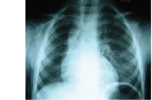
Criança de 2 anos, do sexo masculino, saudável até 4 meses antes do internamento no nosso serviço, altura em por uma traqueo-bronquite tratada com antibioterapia em am-bulatório, efectuou uma teleradiografia de tó-rax revelou uma assimetria dos campos pul-monares com predomínio do pulmão esquerdo e desvio do mediastino para a direita
(Figu-ra 1).
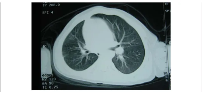
Ao exame objectivo, encontrava-se assin-tomático, apresentando apenas na auscultação pulmonar, uma ligeira diminuição do mur-múrio vesicular à direita. Perante isto, foi pedida uma tomografia computorizada do tó-rax que revelou pulmão direito hipoplásico com desvio homolateral do mediastino e au-sência de visualização da artéria pulmonar direita (Figura 2).
septal defects and persistent ductus arteriosus(1).
It is extremely rare as an isolated finding, with an estimated prevalence of 1:200,000 (2). The
authors present a clinical case and a review of the literature on this uncommon pathology.
CASE REPORT
A two-year-old boy was healthy until 4 months before admission to our department, when in the course of tracheobronchitis treat-ed with antibiotics as an outpatient a chest X-ray revealed pulmonary asymmetry with a pre-dominant left lung and a right mediastinal shift (Figure 1).
On physical examination he was asympto-matic, the only relevant finding being slightly reduced breath sounds on the right side on pulmonary auscultation. The thoracic CT scan showed a hypoplastic right lung with ipsilater-al mediastinipsilater-al shift and the right pulmonary artery could not be visualized (Figure 2).
Given the suspicion of anomalous pul-monary vascularization, pulpul-monary perfusion scintigraphy was performed, which demon-strated absence of radioisotope uptake in the right lung (Figure 3).
For further clarification, and to detect
1254
Recebido para publicação: ????????????? • Aceite para publicação: ?????????
Figura 1. Telerradiografia do torax com assimetria dos campos pulmonares e desvio do mediastino para a direita. Figure 1. Chest X-ray with pulmonary asymmetry and a right mediastinal shift.
Dada a suspeita de vascularização pulmo-nar anómala, efectuou uma cintigrafia de per-fusão pulmonar que evidenciou ausência de distribuição do radiofármaco no pulmão direi-to (Figura 3).
Para esclarecimento do quadro e despiste de outras malformações cardiovasculares con-génitas, foi referenciado ao nosso serviço para avaliação.
O electrocardiograma encontrava-se dentro dos limites da normalidade para a faixa etária e o ecocardiograma transtorácico sugeria o
dia-other congenital cardiovascular malforma-tions, the patient was referred to our depart-ment for evaluation.
The electrocardiogram was within normal limits for his age, while the right branch of the pulmonary artery could not be visualized on the transthoracic echocardiogram. This sug-gested a diagnosis of absence of the right pul-monary artery, and so cardiac catheterization was performed, which confirmed agenesis of the right pulmonary artery, with a hypoplastic right lung perfused by a collateral of the
1255
Recebido para publicação: ????????????? • Aceite para publicação: ?????????
Figura 2. TAC torácico revelando ausência da artéria pulmonar direita e hipoplasia pulmonar ipsilateral. Figure 2. Thoracic CT scan revealing absence of right pulmonary artery and ipsilateral pulmonary hypoplasia.
Figura 3. Cintigrafia de perfusão pulmonar com ausência de captação do radiofármaco à direita. Figure 3. Pulmonary perfusion scintigraphy showing absence of radioisotope uptake on the right.
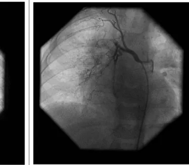
gnóstico dado não se visualizar o ramo direito da artéria pulmonar pelo que decidimos efec-tuar um cateterismo cardíaco. Este último comprovou agenésia da artéria pulmonar direita com pulmão direito hipoplásico e per-fundido por colateral da aorta descendente (diâmetro de 7 milímetros) e por múltiplos pe-quenos colaterais brônquicos (Figuras 4 e 5), sendo o retorno venoso pulmonar normal. Não se detectaram outras anomalias cardiopulmo-nares nem hipertensão pulmonar. Por
apre-descending aorta (diameter 7 mm) and by multiple small bronchial collaterals (Figures 4 and 5); pulmonary venous return was normal. No other cardiopulmonary abnormalities were detected and there was no pulmonary hyper-tension. Since the atrial septum was whole, ruling out countercurrent contrast injection in the pulmonary veins, thoracic magnetic reso-nance imaging (MRI) was requested to show any remnant of the right pulmonary artery. This exam confirmed the absence of the right
1256
Recebido para publicação: ????????????? • Aceite para publicação: ?????????
Figura 4. Angiografia da artéria pulmonar com ausência do ramo
direito.
Figure 4. Angiography of the pulmonary artery, showing absence
of the right branch.
Figura 5. Colateral aorto-pulmonar e colaterais brônquicos para o
pulmão afectado.
Figure 5. Aortopulmonary collateral and bronchial collaterals to
the affected lung.
sentar septo interauricular íntegro que impos-sibilitou injecção contra-corrente nas veias pulmonares e no intuito de demonstrar o even-tual remanescente da artéria pulmonar direita, foi solicitada uma ressonância magnética do tórax (RM). Este exame confirmou a ausência da artéria pulmonar direita sem outras altera-ções, não se tendo visualizado coto hilar da artéria pulmonar (Figura 6).
Perante o facto de o doente ser assintomá-tico e apresentar uma boa evolução estatuto-ponderal e psicomotora, decidiu-se por não intervir nesta altura e manter uma vigilância clínica cuidadosa.
pulmonary artery, with no other abnormalities, and the hilar ostium of the pulmonary artery could not be visualized (Figure 6).
Since the patient is asymptomatic and presents good height and weight and psy-chomotor development, it was decided not to intervene at this time and to maintain careful clinical vigilance.
DISCUSSION
In terms of embryological development, the pulmonary arteries are formed of two
ele-DISCUSSÃO
Do ponto de vista embrionário, as artérias pulmonares são formadas por dois componen-tes, uma porção intrapulmonar distal que tem origem no respectivo botão pulmonar e à qual se vai juntar o segmento extrapulmonar forma-do pela parte proximal forma-do 6.º arco aórtico(1,3). A
ausência unilateral da artéria pulmonar deve--se à involução do 6.º arco aórtico proximal e à respectiva não formação da porção extra-pul-monar da artéria com persistência da conexão do segmento intrapulmonar ao arco aórtico dis-tal pelo canal arterial(1). Quando o canal
arte-rial encerra, a porção hilar da artéria pulmonar regride, havendo hipoplasia e obliteração da árvore intraparenquimatosa pulmonar. A vas-cularização do pulmão ipsilateral fica então a cargo das artérias brônquicas, colaterais aorto-pulmonares e de outras artérias sistémicas como as intercostais e as pleurais(3).
No nosso doente, a artéria pulmonar direita é inexistente, sendo esta a artéria mais fre-quentemente envolvida, com cerca do dobro da prevalência, nos outros casos publicados (3-4). A
ausência da artéria pulmonar esquerda encon-tra-se muitas vezes associada a outras malfor-mações congénitas e estes doentes são geral-mente sintomáticos durante a infância(5).
A mediana da idade de diagnóstico referi-da na literatura é de 14 anos, com os limites compreendidos entre o primeiro mês de vida e os 58 anos(4). Clinicamente, a maioria dos
doentes desenvolve sintomas com o tempo e 44% apresenta hipertensão pulmonar por aumento do fluxo sanguíneo no pulmão con-tra-lateral. As manifestações clínicas mais comuns são a dispneia ou intolerância ao esforço em 40%, infecções pulmonares reco-rrentes em 37% e hemoptises devido à exten-sa rede de colaterais e presença de fístulas arterio-venosas em 20%(3-5). Cerca de 13% dos
doentes permanecem assintomáticos, podendo a doença só se revelar em situações que favo-reçam o aparecimento de hipertensão pulmo-nar, como a gravidez e a altitude, por vezes sob a forma de edema pulmonar agudo unila-teral(4,6).
Nos doentes assintomáticos, o diagnóstico,
ments, a distal intrapulmonary part originat-ing in the respective lung bud, which joins the extrapulmonary segment formed by the proxi-mal part of the sixth aortic arch(1, 3). Unilateral
absence of a pulmonary artery is caused by the involution of the proximal sixth aortic arch and the corresponding failure of the extrapul-monary part of the artery to form, with persist-ence of the connection of the intrapulmonary segment to the distal aortic arch via the duc-tus arteriosus(1). When the ductus arteriosus
closes, the hilar portion of the pulmonary artery regresses, resulting in hypoplasia and obliteration of the intraparenchymal pul-monary artery tree. Vascularization of the ipsi-lateral lung is therefore carried out by the bronchial arteries, aortopulmonary collaterals and other systemic arteries such as the inter-costal and pleural arteries(3).
In our patient, the right pulmonary artery was absent, which is more often the case than with the left, the prevalence being about dou-ble in published cases(3, 4). Absence of the left
pulmonary artery is frequently associated with other congenital malformations, and these patients are usually symptomatic during infancy(5).
The median age of diagnosis in the litera-ture is 14 years (between 1 month and 58 years)(4). Most affected individuals eventually
develop symptoms; 44% present pulmonary hypertension due to increased blood flow in the contralateral lung. The most frequent clin-ical manifestations are dyspnea or exercise intolerance in 40%, recurrent pulmonary infections in 37% and hemoptysis (due to the extensive collateral network and the presence of arteriovenous fistulas) in 20%(3-5). About
13% remain asymptomatic, and the disease may only be revealed by situations that pro-voke pulmonary hypertension such as preg-nancy or high altitude, sometimes in the form of acute unilateral pulmonary edema(4, 6).
In asymptomatic individuals, diagnosis is usually the result of an abnormal X-ray per-formed for other reasons. The most typical findings are cardiac and mediastinal displace-ment, absence of the hilar shadow, smaller hemithorax and elevation of the diaphragm on 1257
em geral, deve-se a exames radiológicos anor-mais, efectuados por outros motivos, sendo as alterações mais típicas, o desvio do coração e mediastino, ausência da sombra hilar, hemitó-rax pequeno e elevação do diagfragma no lado envolvido, com pulmão contralateral hiperin-suflado e herniado através da linha média(6).
Com base na teleradiografia do tórax (RX), o diagnóstico diferencial é feito com tromboem-bolismo pulmonar maciço, coarctação ou este-nose da artéria pulmonar, enfisema unilateral obstructivo e síndrome Swyer-James(6). Outros
exames utilizados na confirmação do diagnós-tico, são o ecocardiograma que pode docu-mentar a ausência do ramo da artéria pulmo-nar e posteriormente, o cateterismo cardíaco para despistar outras malformações cardiovas-culares e a presença de hipertensão pulmonar. Este último pode eventualmente ser substituí-do pela RM ou por uma Angio TAC de alta resolução(4).
As provas de função respiratória nestes doentes são habitualmente normais ou reve-lam um ligeiro defeito do tipo restritivo, com aumento do espaço morto fisiológico. Existe também uma diminuição da capacidade de difusão, consistente com a redução para cerca de metade dos alvéolos activos(5,6).
O tratamento destes doentes é geralmente médico e apenas cerca de 17% dos casos documentados até 2000 foram operados(4). A
cirurgia reconstrutiva da artéria pulmonar foi efectuada em 11 dos 121 casos descritos na literatura (9%), encontrando-se reservada para doentes com hipertensão pulmonar sinto-mática e insuficiência cardíaca congestiva(7).
Recentemente, Welch et al, descreveram excelentes resultados com a correcção cirúrgi-ca total através da construção de um conduto entre o tronco pulmonar ao remanescente hilar do pulmão afectado(8). Além desta
interven-ção, alguns doentes têm de ser submetidos a pneumectomias ou lobectomias por hemopti-ses recorrentes.
Como foi descrito, durante o cateterismo cardíaco do nosso doente optou-se por não se efectuar punção transeptal inter-auricular e aguardar pelo resultado da RM, que não evi-denciou a artéria pulmonar hilar
remanescen-the affected side, with hyperinflation and her-niation of the lung across the midline(6). On
the basis of the chest X-ray, differential diag-nosis is with massive pulmonary thromboem-bolism, coarctation or stenosis of the pul-monary artery, unilateral obstructive emphy-sema and Swyer-James syndrome(6). Other
exams used to confirm the diagnosis include echocardiography, which can demonstrate absence of a pulmonary artery branch, fol-lowed by cardiac catheterization to search for other cardiovascular malformations and the presence of pulmonary hypertension; the lat-ter function can also be performed by MRI or high-resolution CT angiography(4).
Respiratory function tests in these patients are usually normal or show a mild restrictive defect, with increased physiological dead space. There is also decreased diffusing capacity consistent with absence of half of the active alveoli(5, 6).
Treatment is usually medical, and only 17% of the cases documented up to 2000 were operated. Surgical reconstruction of the pul-monary artery was performed in 11 of the 121 in the literature (9%) and is reserved for patients with symptomatic pulmonary hyper-tension and congestive heart failure(7). Welch
et al. recently reported excellent results with total surgical correction by constructing a con-duit between the pulmonary trunk and the hilar remnant of the affected lung(8). Some
patients require pneumonectomy or lobectomy due to recurrent hemoptysis.
In our patient, as stated above, it was decided during cardiac catheterization not perform a transseptal puncture but to await the MRI results, which showed no evidence of a remnant hilar pulmonary artery. If the deci-sion is made to go ahead with corrective sur-gery, it could be preceded by cardiac catheter-ization to perform pulmonary venous wedge angiography.
This pathology, although rare, is of consid-erable importance, since although it can be asymptomatic in the early stages, allowing patients to reach adulthood, in the long term morbidity can be significant. The main factor for poor prognosis is the development of
pul-1258
1259
Recebido para publicação: ????????????? • Aceite para publicação: ?????????
1. Apostolopoulou S, Kelekis N, Brountzos E, Rammos S, Kelekis D. ‘Absent’ pulmonary artery in one adult, and five pediatric patients. Imaging, embryology, and therapeutic implications. Am J Roentgenol 2002;179:1253–60.
2. Bouros D, Pare P, Panagou P, Tsintiris K, Siafakas N. The varied manifestation of pulmonary artery agenesis in adult-hood. Chest 1995; 108:670–6.
3. Griffin N, Mansfield L, Redmond KC, Dusmet M, Goldstraw P, Mittal TK, Padley S. Imaging features of isolated unilateral pulmonary artery agenesis presenting in adulthood: a review of four cases. Clin Radiol. 2007 Mar; 62(3): 238-44.
4. Ten Harkel A, Blom NA, Ottenkamp J. Isolated unilateral absence of a pulmonary artery: a case report and review of the literature. Chest 2002; 1224:1471–7.
5. Mimura S, Kobayashi H, Shinkai M, Kanoh S, Motoyoshi K. A case report of congenital isolated absence of the right
pul-BIBLIOGRAFIA / REFERENCES
monary artery: Bronchofibrescopic findings and chest radio-logical tracings over nine years. Respirology 2005; 10: 250-253.
6. Werber J, Ramilo JL, London R, Harris VJ. Unilateral absence of a pulmonary artery. Chest. 1983 Dec; 84(6):729-32. 7. Atik E, Tanamati C, Kajati L, Barbero-Marcial M. Isolated unilateral pulmonary artery agenesis. Evaluation of natural and long-term evolution after corrective surgery. Arq Bras Cardiol 2006; 87: 381-385.
8. Welch K, Hanley F, Johnston T, Cailes C, Shah MJ. Isolated unilateral absence of right proximal pulmonary artery: surgical repair and follow-up. Ann Thorac Surg. 2005 Apr; 79(4): 1399-402.
9. Heper G, Korkmaz M. High-pressure pulmonary artery aneurysm and unilateral pulmonary artery agenesis in an adult. Tex Heart Inst J 2007; 34: 425-430.
monary hypertension and right heart failure(9).
In the largest published series (108 patients), overall mortality was 7%(4).
Early detection allows the possibility of surgical correction to provide anterograde flow to the hilar segment of the pulmonary artery, thus preventing obliteration of the pulmonary vasculature and the development of pul-monary hypertension.
Pedido de separatas para: Address for Reprints: Ricardo Gil Oliveira Serviço de Cardiologia Hospital Reynaldo dos Santos R Dr. Luís César Pereira 2600-178 Vila Franca De Xira [email protected]
te. No caso de se decidir por uma cirurgia correctiva, o doente poderá ser submetido a novo caterismo cardíaco para realização de uma angiografia venosa pulmonar encravada. Esta patologia, apesar de muito rara, tem uma grande relevância clínica pois no seu estádio inicial pode ser assintomática, permitindo aos doentes chegarem à idade adulta, mas a longo prazo, pode ter uma morbilidade significativa. O principal factor de mau prognóstico é o des-envolvimento de hipertensão pulmonar com falência ventricular direita(9). Na maior série
de revisão descrita na literatura (108 doentes), a mortalidade total foi de 7%(4).
A sua detecção precoce possibilita a correcção cirúrgica fornecendo fluxo anteró-grado ao segmento hilar da artéria pulmonar evitando a obliteração da vasculatura pulmo-nar e por consequência, prevenindo o apareci-mento de hipertensão pulmonar.