2014/2015
Mafalda Sofia Barros Gomes
ABOUT THE FETAL RISKS FROM
DIAGNOSTIC USE OF RADIATION
DURING PREGNANCY: A
SYSTEMATIC REVIEW AND
PROPOSAL OF A CLINICAL
PROTOCOL
Mestrado Integrado em Medicina
Área: Obstetrícia
Tipologia: Monografia
Trabalho efetuado sob a Orientação de:
Doutora Alexandra Matias
Trabalho organizado de acordo com as normas da revista:
Pediatric Radiology
Mafalda Sofia Barros Gomes
ABOUT THE FETAL RISKS FROM
DIAGNOSTIC USE OF RADIATION
DURING PREGNANCY: A
SYSTEMATIC REVIEW AND
PROPOSAL OF A CLINICAL
PROTOCOL
Dedico esta monografia aos meus pais
Madalena Sofia Rodrigues de Carvalho Barros e
António Rui Flores Gomes,
e à minha irmã
Diana Raquel Barros Gomes.
ABOUT THE FETAL RISKS FROM DIAGNOSTIC USE OF RADIATION DURING PREGNANCY: A SYSTEMATIC REVIEW AND PROPOSAL OF A CLINICAL PROTOCOL
Mafalda Gomes, Degree in Basic Health Sciences, Master Degree in Medicine, Faculty of Medicine, University of Porto
Alexandra Matias, MD PhD, Senior Consultant of OB/GYN, S. João, Faculty of Medicine, University of Porto
Filipe Macedo, MD, Senior Consultant of Radiology, SMIC, Porto, Portugal
CORRESPONDENCE:
Mafalda Sofia Barros Gomes
Rua Sacra Família, nº70, 4490-548, Póvoa de Varzim, Porto, Portugal 910036474
[email protected]
ACKNOWLEDGEMENTS
We would like to thank Anabela Rocha, MD, Resident in OB/GYN, S. João, Faculty of Medicine, University of Porto, for taking interest in this systematic review.
1
ABOUT THE FETAL RISKS FROM DIAGNOSTIC USE OF RADIATION DURING PREGNANCY: A SYSTEMATIC REVIEW AND PROPOSAL OF A CLINICAL PROTOCOLABSTRACT
Aim: Analyze existing literature about the fetal risks of radiation exposure, producing a clinical
protocol to guide radiation exposure in a clinical setting.
Methods: An initial query was made on PubMed: “Diagnostic radiography in pregnancy AND
radiation”, with the limits “published from January 1st
1993 to December 31st 2013, in English or Portuguese”. The articles that presented our aim were analyzed according to their MESH terms and created the final query: “((radiation) AND pregnancy) AND diagnostic imaging”. Research on April 15th
of 2014, with the same limits, on PubMed gathered 688 articles; on SCOPUS 245 additional articles. After reading the title and abstract 298 articles remained. 179 allowed access to full text and were analyzed according to inclusion and exclusion criteria. A total of 103 articles were used and an additional one regarding In utero radiation exposure from atomic bombs. The PRISMA statement was followed.
Results: Deterministic effects like pregnancy loss, congenital malformations, growth retardation
and neurobehavioral abnormalities have threshold doses greater 100-200 mGy, being the risk considered negligible at 50 mGy. No diagnostic exam exceeds this limit.The most crucial time to avoid radiation exposure is from the 8th to the 15th week of gestation. The risk of carcinogenesis is slightly higher than the general population, although very similar.Intravenous contrast is discouraged, except in highly-selected patients.
Conclusion: Measures to diminish radiation are essential and affect the fetal outcome.
Nonionizing procedures should be considered whenever possible and every radiology center should have its own data on fetal radiation exposure.
2
INTRODUCTIONEveryday medical practitioners face the dilemma of exposing pregnant or presumably pregnant patients to radiation from complementary diagnostic exams [1-3]. In fact, irradiation of the fetus is a very common phenomenon [1], but one should be aware of the implicated risks [4].
There are many circumstances for fetal exposure to radiation. The most common one, especially during the first trimester, is accidental as the patient was not aware of the pregnancy [5-11]. To this we add the need for medical diagnosis of the mother (at any given time during gestation) or the fetus (to confirm an abnormality or provide further information, usually after ultrasound during the 2nd and 3rd trimesters). More frequently irradiation during pregnancy derives from diagnostic need for both mother and fetus, if no alternative to ionizing radiation is available [1]. Finally, special consideration should be granted to pregnant radiology staff [6,7,11].
Much of the information regarding radiation exposure of the fetus comes from “opportunistic” accidents in the world’s history. Survivors of the atomic bombs of Hiroshima and Nagasaki have shown
risks of fetal exposure to radiation, the most common one being microcephaly at 100-200 mSv [5,12-14]. Mental retardation was also observed among survivors (20-30 points per 100 rad; 25-31 points per Gy above 0.1 Gy) [12,13,15,16], as well as growth retardation (permanent above 250 mSv, 25 rad or 0.25 Gy) [5,13], teratogenesis (above 1Gy) and cancer (increased rate of leukemia) [13]. Studies on cancer after intrauterine exposure to the atomic bomb are inconsistent [17]. The Chernobyl reactor accident was also associated with increased rate of cancer [13]. Studies on children exposed to radiation before 15 weeks of gestational age showed a higher susceptibility to these effects [12].
Ionizing radiation is frequently used with the purpose of achieving a medical diagnosis since the discovery of X rays [18] and is still a helpful tool. In recent years there has been a great concern in developing new techniques and methods to decrease the risk of radiation for pregnant women and, just as important, to their fetus [4,19].
Both doctors and patients often have questions about the risks of radiation. Therefore, creating a guideline is not only a useful tool for every medical practitioner but also a necessity [1]. The main objectives of this systematic review are to analyze the existing literature about the risks of radiation exposure and safety of contrast agents. Additionally, a clinical protocol is proposed to guide radiation exposure in a clinical setting.
3
METHODSThe present article is a systematic review that aims to analyze the existing literature about the fetal risks from radiation exposure during pregnancy. An initial query was made on PubMed: “Diagnostic radiography in pregnancy AND radiation”, with the limits “published from January 1st 1993 to December 31st 2013, in English or Portuguese”. This research yielded a total of 381 articles. Those who presented the same objective as intended in this systematic review where analyzed according to their MESH terms. Gathering the most frequent MESH terms the final query was created: “((radiation) AND pregnancy) AND diagnostic imaging”. On April 15th 2014, the total of articles retrieved from this research on PubMed was 1462. After applying the same restrictions to publication date and language, 688 articles remained, 261 of them reviews. The same query and research limits were applied on SCOPUS, gathering an additional 245 articles (Fig. 1 – Flowchart of the methods). After reading the title and abstract, when available, 635 were excluded.
The main inclusion criteria considered: Radiation doses absorbed by the fetus;
Risks of radiation from diagnostic exams to the fetus;
Protection measures for diagnostic radiology exams in pregnant women.
The following excluding criteria were also used: o Studies on radiotherapy;
o Studies on occupational hazards of radiation; o Risks of ultrasound;
o Discussion of ethical problems regarding radiation usage; o Molecular studies of radiation rather than clinical ones; o Articles with an iconographic purpose;
o Studies on animals other than Humans;
o Studies with the objective of comparing diagnostic exams for specific pathologies regardless of the risks for the fetus (for example: comparison of sensitivity and specificity of two different diagnostic exams).
4
From the 298 final articles, 179 allowed access to full text and were analyzed according to different variables: dosages of radiation absorbed by the fetus according to the irradiated area of the pregnant woman, effects and safety limits of radiation. A total of 103 articles were used and an additional on regarding in utero exposure from atomic bombs (Fig. 1 – Flowchart of the methods). The PRISMA
statement was followed for the construction of this systematic review. As a result of our research from the literature, a protocol for medical use was designed.
DOSAGE OF RADIATION TO THE FETUS
Background radiation is considered to vary across the globe [4] between 1.3-5.8 mSv/year [20], being that the average annual effective dose from it is about 3.6 mSv (0.36 rem) for an adult [3,15,21,22] and 0.5-1 mSv or 1.1-2.5 mGy [23,24] for a fetus during the entire period of gestation [3,25-28]. The fetus is more radiosensitive than the mother [28,29].
If a pregnant woman is in need of medical care and, to achieve diagnosis, requires the use of a diagnostic procedure that will expose her unborn child to radiation, we need to take into account not only the type of energy but also the quantity of photons, size of the patient and vulnerability of irradiated tissues. However, quantifying the dosage delivered to the fetus is not an easy task [21,30].
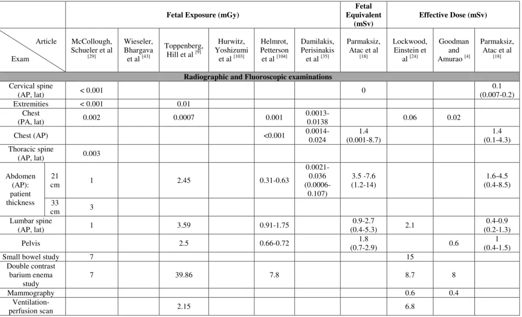
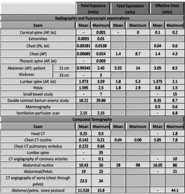
In radiographic and fluoroscopic examinations, if the uterus is outside the field of view, the fetus is only exposed to scattered radiation in minimal doses [31,32]. Therefore, the fetal exposure increases if the uterus is within the field of view (Table 1). It appears that posteroanterior chest x-rays exposes the fetus to less radiation than the anteroposterior projection [31]. The dosage applied to the fetus in radiography depends on the patient thickness, the direction of projection, the depth of the fetus from the skin surface and x-ray technique factors [25,33] (Table 1).
Maximum exposure of the fetus to radiation comes from abdominal computed tomography (CT) [18,25]. However, the dosage is minimal and the patient can benefit significantly from the exam [25] (Table 1). If the abdomen is not in the field of view, the fetus is only exposed to scatter radiation [24]. The fetal radiation dose from a CT depends on kilovolt peak, milliamperes, slice thickness [34], gestational age, the depth of the fetus and proximity of the uterus to the field of interest [25,35] (Table 1).
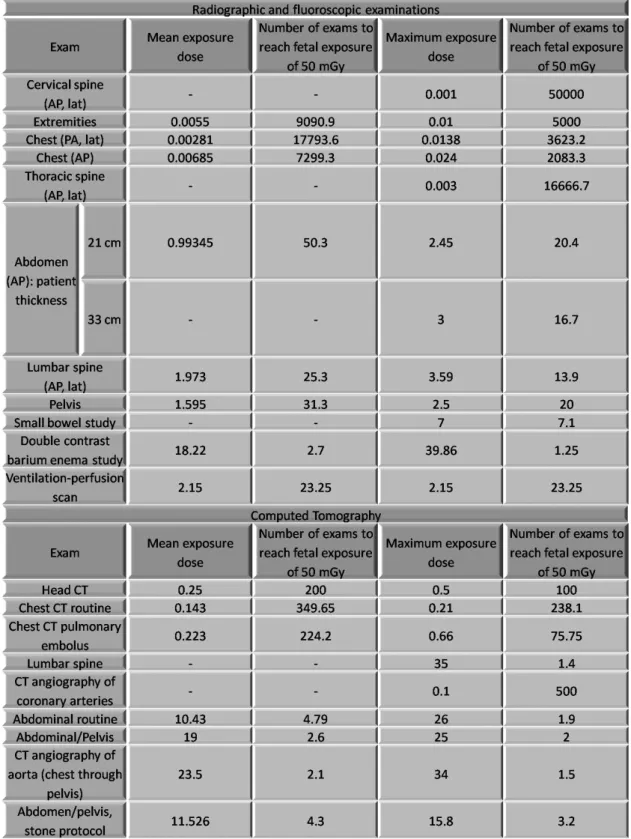
The mean effective dose of radiation for each procedure to the mother, the fetal exposure and the fetal equivalent dose (Table 2) and the number of exams needed to reach the accepted cumulative dose of
5
fetal exposure (Table 3) are presented. The measurements vary extensively, requiring each radiology department to have access to its own data.
RISKS TO THE FETUS FROM RADIATION OF DIAGNOSTIC EXAMS
When using radiation we have to consider two kinds of effects: deterministic and stochastic. Deterministic effects are those whose severity increases with the dose of radiation, having a threshold dose below which its effect is clinically irrelevant. For them to have an effect on the fetus, the threshold dose must be reached. After this limit, the severity of the effect increases with the dose [3,5,13,16,32,34,36-45]. Stochastic effects are those whose probability of occurring increases with the dose, not caring for a threshold dose because the result is the same (acting on one single cell or a group of them). The severity of the effect is dose-independent [3,5,13,16,21,32,34,37-45].
The effects of radiation on the fetus depend on the stage of the pregnancy, radiation dose [5,8,11,13,15,23,32,46-48] and fetal cellular repair mechanisms [25]; demographic factors (patient age and weight), medical history factors (coexisting diseases, genetic factors, medication use and radiation history) and procedure factors influence as well [3,16,23,28,40,42,49]. We can divide the fetal effects of radiation in:
1. Pregnancy loss;
2. Congenital malformations (teratogenesis) [21,34]; 3. Neurobehavioral abnormalities [13];
4. Fetal growth retardation [9,36,50]; 5. Carcinogenesis [9,21,34,36,50,51].
1. Pregnancy Loss
At the beginning of every pregnancy the risk of spontaneous miscarriage is about 15% [3,16,24,32,36]. After conception and during preimplantation and preorganogenesis, the embryo cells are omnipotential. This means that it is unlikely for malformations to occur by the effects of ionizing radiation during these stages. Other cells can replace adjacent cells that have been deleteriously affected. This period is called “the all-or-none period” [11,13,14,36,49].
6
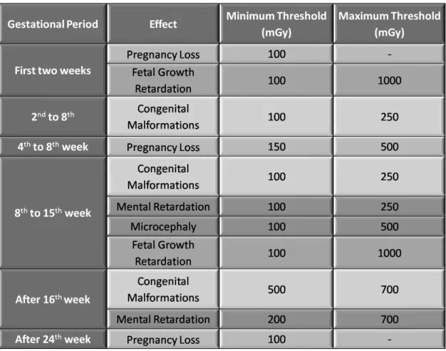
If the exposure to radiation exceeds 100 mGy or 100 mSv during the first 2 weeks after conception, the “all-or-none” phenomena can result in spontaneous abortion instead of a completely
unaffected embryo [3,5,11,14,16,20,28,34,38,44,45,48,49]. From the fourth to eight week of gestation, the threshold goes up to 150mGy [42], 200 mGy [45] or 250 mGy and 500 mGy [36,52]. After 26 weeks the risk of neonatal or fetal death rises with doses above 1Gy, with a threshold of 100 mGy [34,53,54].
Exposure to less than 5 rad (50 mGy) has not been associated with increased fetal anomalies or pregnancy loss [25,45,46,55]. The exposure to radiation on its own is not an indication for terminating the pregnancy [5,9,13,25,46] and should only be considered if the exposure dose is higher than 100 mGy – “Danish rule” [7,20,24,31,39,49,56]. Some propose a limit dose of 150 mGy [8].
2. Congenital Malformations
In every pregnancy the background risk for birth defects is about 3% [3,13,16,24,32,36]. The most sensitive period for malformations is from the 2nd to 8th week of gestation, during organogenesis [13,21,34,50] and during the early fetal period (up to 15th week) [11,14], with a threshold of 100 mGy [15,18,23,28,31,38,48,56-58]. A threshold of 150 mGy [3,8,10,25,41,59,60], 200 mGy [3,11,15,20,36] or 250 mGy [25,45] has been suggested. After 16 weeks, the threshold is about 500 mGy to 700 mGy [11,45,52]. During the last trimester major organ malformations and functional anomalies are unlikely [13,14]. There has been no evidence of congenital malformations at doses below 50 mGy or 5 rad being this value the accepted cumulative dose of ionizing radiation for the entire gestational period [11,16,20,25,31,55,56,61]. No diagnostic exam exceeds this limit [9,13]. The risk of malformations is significantly increased above 150 mGy (15 rad) [13,16,26,46]. When the dose of exposure exceeds 100 mGy the probability of congenital birth defects increases 10% [62].
In the light of current knowledge, diagnostic x-rays, CTs or nuclear medicine procedures cannot be considered a risk for malformations [11,20,25,26,53].
3. Neurobehavioral Abnormalities
The background risk for neurological development problems is about 1% [3,24,32] up to 6% [5] The most sensitive stage for mental retardation and microcephaly is from the 8th week to 15th week of the gestational period [3,9,11,13,15,16,36,47,58,63]. Exposure up to 20 weeks of development increases the risk of microcephaly and mental retardation [13,21]. However, from the 16th to the 25th week the
7
central nervous system is less radiosensitive [3,13-15,63]. After the 25th week it becomes radioresistent [13].
Mental retardation has a threshold of 100 mGy to 250 mGy [3,13,15,16,20,25,28,38,44,45,48,55] or 120 mSv [18] and is not directly linked to microcephaly [13,48].Severe cases occur with higher doses: 350-500 mGy [16,20,36,45] or even 1 Gy [20,34], 120-230 mGy between 8th and 15th weeks, 210 mGy between 16th and 25th weeks [13]. The IQ loss is about 25 to 31 points per 1 Gy beyond 100 mGy of radiation [11,15,41,45,63] or 21-29 IQ points per Gy, 30 points for every Sv [13]. Eight weeks after conception, intellectual damage has not been demonstrated [58]. But others find that at 8 to 15 weeks the incidence of severe mental retardation establishes a linear connection without a threshold dose, with an increased risk of 40% per gray of radiation [9,14,15,50,63] or 40% per 100 mSv to 200 mSv (200 mGy) [5]. After this period, the incidence is lower and doses from 20 mGy to 250 mGy may show cognitive loss [50], although it is more common at high doses (>=200 mGy) [64]. In the 16 to 25 week stage the average IQ loss is approximately 13-21 points per Gy (per 100 rads) at doses above 700 mGy [13,15,63]. Microcephaly occurs at a threshold of 100 mGy [28], 200 mGy [48] or 350 mGy to 500 mGy [31,36].
Based on the evidence seen so far, no diagnostic exam (x-ray, CT or nuclear medicine procedures) can cause neurodevelopment effects [20].
4. Fetal Growth Retardation
Growth retardation due to radiation exposure has a risk of 4% in all pregnancies [3,16,24,32]. It occurs mainly during the first trimester, after 14 days of conception [34]. Exposure to radiation up to 20 weeks of development increases the risk of growth retardation [21]. It shows a dose threshold of 100 mGy to 250 mGy [16,25,44,55], in some studies up to 500 mGy [20,36,45,52], 1 Gy [15] or 50-100 mSv [1,15]. Growth retardation usually is not permanent and the fetus will recover [5].
8
5. CarcinogenesisCancer and hereditary effects after radiation exposure occur without a threshold dose [9,17,24,25,31,49,52,60,66] and appear at the same age as spontaneous ones [50]. The risk of this occurrence is constant throughout the whole pregnancy [15,28,34,48] except for the first two/three weeks of pregnancy when the risk is low [51,58]. After radiation exposure to the fetus there is an increase in risk for all cancers [14,26,30,32,35,67] (including solid tumors [11,16]) and leukemia, especially acute myeloid leukemia. However, this is not statistically significant [67].
After pelvic procedures like barium enema or CT, the carcinogenic risk is similar to the natural incidence of fatal carcinogenic risk before age 15 [14]. If the absorbed dose is 5 rad, the risk of childhood cancer is 0,3% (0.2-0.8%) - the same value as the natural risk for fatal childhood cancer [14-16,47,58]. The risk can be of 0.06% per 10 mSv or 10 mGy [1,28,39] or 0.06% per 1 rad [47], 5% per Sv (100 rem) [5] (Table 3). Others say that 100 mGy of radiation increases the risk for childhood cancer by 0.1% [52]; a dose of 10 mSv during the last trimester increases the risk of leukemia by 40%. 10 mSv at any stage of the pregnancy increases the risk of leukemia by a multiple of 1.5. Doses above 10 mSv increases the risk coefficient 6% per Sv [5]. The most consensual attitude is to consider a risk slightly higher than the general population, but still very similar [45].
Most of the articles included in this review mention leukemia as the most common carcinogenic phenomenon associated with in utero radiation [9,32,37,42]. However, leukemia associated with high exposure to radiation is not more severe than a spontaneously occurring leukemia [36]. The background risk is about 3.6 per 10000; after exposure increases to 5 per 10000 [9]. In utero exposure to 0.01 Gy increases the risk of cancer in the first and second decades of life from 0.03% to 0.04% [37].
Some studies report that radiation exposure at all gestational ages increase the risk of childhood leukemia [21,68] but others find that there is little evidence of any increased risk of childhood acute lymphoblastic leukemia associated with maternal x-rays during the pregnancy [69]. In some cases it has been recorded an excess of maternal x-ray exposure among children with acute lymphoblastic leukemia but the statistical analyses and experimental data were reassuring and do not support this connection [68]. Although perfusion scanning exams do not pose a risk for deterministic effects they can be linked to cancer or genetic effects, regardless of the dose [62]. Carcinogenesis associated with diagnostic radiation is a dose independent event but the risk seems relatively low with doses less than 10 rad (100 mGy) [3,14,16,37,41,42,70] or 10 mSv [22,70]. A cutoff of 50 mGy has also been proposed [41].
9
Complementary use of contrastsIntravenous contrast is discouraged during gestation, except in highly-selected patients where there is no other alternative to obtain important diagnostic information [71]. These contrast agents are used in CT and MRI to detect, characterize and stage diseases [72]. There are two main contrast agents: iodine or gadolinium-based.
Radioiodine crosses the placenta and starts to accumulate in the fetal thyroid since the 12th week of gestation, not exceeding the 100 mGy limit [20,23,41,46,66,73]. We have to consider the possible risk of hypothyroidism and thyroid cancer induction to the fetus, so radioiodine is contraindicated during pregnancy [3,20,23,24,26,34,49,74,75]. Internal uptake of iodine occurs mostly during the 16th to 25th week stage [15]. However, there aren’t sufficient human studies on fetal thyroid depression due to iodine
[46,53] and it has not been observed with the administration during pregnancy [28,39]. It is considered generally safe during pregnancy and therefore iodinated contrast could be used during pregnancy after assessing the risk-benefit ratio [7,8,24,39,49,74,76,77]. If the mother received iodinated contrast material during her pregnancy, the thyroid function of the newborn should be evaluated in his first week of life [23,26,37,41,49,73,75]. Evidence of mutagenic or teratogenic risk does not exist, but there is a lack of human studies [3,23,24,37,48,66,75].
Gadolinium-based contrast cross the placenta, enter the fetal circulation and are excreted into the amniotic fluid, where they remain for some time [3,39,41,66,78,79]. It appears there are no teratogenic or mutagenic effects in humans when using these agents [3,39,41,46,66,72,75,76] but gadolinium’s safety has not been established [3,23,34,37,74,79]. Apparently nephrogenic systemic fibrosis and dissociation of toxic-free gadolinium are some of the effects in discussion [46]. At higher doses than the ones used in human studies gadolinium has been associated with growth retardation and congenital anomalies [26]. Gadolinium should be contraindicated during pregnancy, only used when the benefits outweigh the risks, with extreme caution [8,27,28,39,47,49,74,76,80].
Barium sulphate is used during fluoroscopic exams and appears to be safe for the fetus [81].
Computed Tomography
Computed Tomography (CT) examinations on pregnant women are usually in areas away from the uterus, so the fetus is not directly exposed to radiation. The risk in these cases is scatter radiation that only hits low levels of radiation, thus carrying a small risk for the fetus [29].
10
CT of maternal head and chest have negligible fetal exposure. Maternal pelvic CT may increase the risk of cancer [53]. Computed tomography pulmonary angiogram exposes the fetus to similar or lower doses of radiation as V/Q scans [82]. Helical CT has an average fetal exposure dose smaller than ventilation-perfusion lung scanning [83].
Magnetic Resonance Imaging
Non ionizing procedures should be considered whenever possible [26,42,43,84-86]. In fact, Magnetic Resonance Imaging (MRI) should be the second line examination, after ultrasound, because it is an expensive, complex and less available exam [6,46,76,80,87-89].
MRI can be performed at any stage of the gestational period, but safety during the first trimester is not yet established [16,46,49,78,79]. The major concerns are heating effects of radiofrequency pulses and effects of acoustic noise on the fetus [6,26,41,47,49,74,89-91]. Thermal heating can cause biologic damage, related to cell migration, proliferation and differentiation, up to and including miscarriage [74,87,91]. The central nervous system is especially sensitive to heat rising. A 2°C rise over 24h can result in abnormalities like neural tube and cranio-facial defects [6,90,91]. Some say that MRI should be avoided in the first trimester to avoid excessive heating and high fetal exposure; however, after 24 weeks (when the fetal hearing is developing) is not easy to give additional protection from acoustic noise to the fetus [3,41,92]. Acoustic damage appears to be a more theoretical risk and not a significant practical issue [8,49].
MRI shows no harmful effects on the fetus under 1.5 Tesla [7,18,26,28,39,41,49,93], considered generally safe for use in pregnant women [46,76,84]. In some radiology centers higher field strengths are used with no apparent risk to the fetus. The use of 3 Tesla equipments is gradually being introduced in clinical practice. Field strengths above 2.5 Tesla should be avoided [3,26,41,92]. Safety of the fetus is overestimated because the effect of heat dissipation by convection in the amniotic fluid is overlooked. There should be more studies on this matter [93].
Until today no evidence of conclusive harmful effects to the fetus from MRI exists [3,8,41,47,78-80,87,89,91].
The risk is considered to be negligible at 50 mGy or less [3,5,8,13,15,16,23,28,38,43,52] and diagnostic exams have lower doses [3,23,24,37,45,48,52,75]. Deterministic effects have thresholds
11
greater than 100-200 mGy (below are considered safe) [14,32,45,56,64] and the most crucial time to avoid radiation exposure is from the 8th to the 15th week of gestation [86]. Measuring the dosage of exposure is important to determine the risk to the fetus [28,61].
MEASURES TO DIMINISH THE RISKS OF RADIATION
Accurate imaging helps to achieve a definitive diagnosis, deciding proper treatment, avoid complications and unnecessary interventions [76,80]. Withholding proper diagnostic imaging care can result in significant harm for the mother and therefore to the fetus, considered an irresponsible medical action [64]. Protection in radiology follows some basic principles: there should be no risk without benefit, prescribed limits should not be exceeded and, at all times, the “ALARA” concept (as low as reasonably achievable) should be kept [21,23,26,38,40-42,46,52,77]. Therefore, measures to reduce the dosage to the fetus should be implemented.
Screening for pregnancy
The first step to take is screening for pregnancy [2,38,40,45,78,79]. The “10 day rule” states that,
in women of childbearing potential, non urgent radiography examinations that involve pelvic irradiation should be restricted to the first 10 days of the menstrual cycle [51,54,57,58]. Hence avoiding irradiating the fetus before the mother knows that she is pregnant [51] and the risk of pregnancy loss [57].
Recently, the accepted interpretation is that if the patient’s menstruation started less than 10 days, the chance of pregnancy is very low and no cause of concern [54]. Most radiology departments no longer follows this principle [57].
In all situations, informed consent should be acquired, if the patient is stable [38,40,79].
General measures
Ionizing radiation should be avoided especially during the first trimester but, whenever possible, through the whole pregnancy ultrasound and MRI should be preferred [26,41,76,85,86]. Special care is advised between 10 and 17 weeks because of the risk for central nervous system teratogenesis. In this period, non urgent exams should be postponed [9,47].
12
Additionally, all radiologic equipment should be well-maintained and periodically inspected for radiation safety [2]. It is important to monitor the radiation dose of every exam [5,40].
For all diagnostic exams is important to minimize exposure time [2,35,44,46,58,61,77,94-96]. In a general way, protraction and fractionation of exposures of ionizing radiation to the embryo decrease the magnitude of the deleterious effects of deterministic effects [36]. Radiography, fluoroscopy and computed tomography share the following measures:
Lead shielding whenever possible [5,8,14,16,23,24,28,38,40,41,43,61,76,77]; Collimators [3,5,23,28,41,46,48,61,77,97,98];
Minimize the number of acquisitions [2,23,41,42,48,55,61];
Scan the minimum body area needed to provide sufficient guidance [3,24,32,41,42,61,99,100]. Specific technicalities adopted in radiographic, fluoroscopic and CT examinations are detailed in the clinical protocol section.
CLINICAL PROTOCOL
Every female patient in reproductive age should be screened for pregnancy before undergoing diagnostic radiation exams. If the pregnancy is a possibility or confirmed the risks of radiation to the mother and fetus need to balance with the benefit of the exams.
Deterministic effects like pregnancy loss, congenital malformations, growth retardation and neurobehavioral abnormalities have threshold doses greater 100-200 mGy [14,32,45,56,64] (Table 1 Protocol), being that the is considered to be negligible at 50 mGy or less [3,5,8,13,15,16,23,28,38,43,52]. No diagnostic exam exceeds these values [3,23,24,37,45,48,52,75](Fig. 2 – Comparison of the minimal threshold doses for the deterministic effects of radiation with the accepted cumulative radiation during pregnancy). Moreover, the most crucial time to avoid radiation exposure is from the 8th to the 15th week of gestation [86]. The risk of carcinogenesis is slightly higher than the general population, but still very similar and should be considered during the entire gestational period [45].
Intravenous contrast is discouraged during gestation, except in highly-selected patients where there is no other alternative to obtain important diagnostic information [71].
13
For radiography and fluoroscopy: Highest peak kilovoltage possible that results in acceptable image contrast [33];
Lead shielding whenever the abdomen or pelvis is not being imaged to protect the uterus from external scattered radiation [5,14,16,28,38,40,43,61,76,77]. If a specifically designed shield is not available, lead aprons should be reserved specifically for this task [101];
Minimize fluoroscopy time [2,44,58,61,77,94,95] and the number of images acquired during digital subtraction angiography and cinematic acquisitions [2,55,61];
Magnification only if necessary [41,61];
Perform pulsed fluoroscopy at the lowest pulse rate that provides sufficient image quality [41,61,77];
Maximize the distance between the x-ray source and the receptor and the distance between the patient and the receptor [40,61,77];
Collimators [5,28,41,48,61,77]; Decrease Filtration[5] with copper [77];
Avoid taking radiographs during fluoroscopy [77]; Increase tube voltage [77];
Posterior-anterior projection should be preferred to anterior-posterior projection [18,28].
In a general way, protraction and fractionation of exposures of ionizing radiation to the embryo decrease the magnitude of the deleterious effects of deterministic effects [36].
For Computed Tomography:
Lead shielding if it does not affect the image result, best with circumferential shielding
[5,8,23-25,41,43,98,99,102];
Reduce kilovoltage peak [3,4,23,24,35,41,42,52,96,98,100], milliampere-second setting
[4,41,42,96,100], the length of the scan [35,46,96] and the number of acquisitions [23,41,42,48,61];
Center the patient in the CT Gantry [99];
Use a low tube current-time product for all acquisitions after the preliminary scan [3,23,24,42,43,48,52,61,73,98];
14
Increase the pitch [3,23,41,42,61,97,98]; Limit Z axis [23,35,98];
Internal barium shielding with use of oral 30% barium sulfate solution [23,41,43,99]; Customize protocols to patient size and clinical indication [98].
Since CT scans are associated with higher radiation exposure dosage than other medical exams, its use should be restrained [21,76]. Here the alternatives (ultrasound and MRI) have to be considered and offered to the patient if the benefit is higher than the risk [9,21,38].
Every radiology center should have its own data on fetal radiation exposure in order to determine the risks [28,61].
CONCLUSION
With the increase of technology and availability of diagnostic exams, more and more pregnant women are irradiated unaware of their current state. The risks of fetal exposure to radiation are still very misunderstood by the general population and, to some degree, even by medical professionals. When using radiation to achieve a diagnosis, one has to balance the welfare of the mother and of her unborn child, weighing the risks and benefits. [21,64]
Deterministic effects like pregnancy loss, congenital malformations, growth retardation and neurobehavioral abnormalities have threshold doses greater 100-200 mGy [14,32,45,56,64], being that the risk is considered to be negligible at 50 mGy or less [3,5,8,13,15,16,23,28,38,43,52]. No diagnostic exam exceeds these values [3,23,24,37,45,48,52,75]. Moreover, the most crucial time to avoid radiation exposure is from the from the 8th to the 15th week of gestation [86]. The risk of carcinogenesis is slightly higher than the general population, but still very similar [45] and should be considered during the entire gestational period [45]. Intravenous contrast is discouraged during gestation, except in highly-selected patients [71].
Non ionizing procedures like ultrasound and MRI should be considered whenever possible [26,42,43,84-86]. Ideally, every radiology center should have their own data on fetal radiation exposure in order to determine the risks [28,61].
15
LIMITATIONSDuring the construction of this systematic review the most hindering obstacle found was the conflicting data. An attempt to present the most information was made. Additionally, there were few original articles on fetal doses of exposure to radiation and absorbed values. More studies are needed in order to warrant the safety of diagnostic exams using radiation.
REFERENCES
1. El-Khoury GY, Madsen MT et al. (2003) A new pregnancy policy for a new era. Am J
Roentgenol 181 (2):335-340.
2. Berlin L (1996) Radiation exposure and the pregnant patient. Am J Roentgenol 167
(6):1377-1379.
3. Wang PI, Chong ST et al. (2012) Imaging of pregnant and lactating patients: part 1,
evidence-based review and recommendations. Am J Roentgenol 198 (4):778-784.
4. Goodman TR, Amurao M (2012) Medical imaging radiation safety for the female
patient: rationale and implementation. Radiographics 32 (6):1829-1837.
5. Ursprung WM, Howe JW et al. (2006) Plain film radiography, pregnancy, and
therapeutic abortion revisited. J Manipulative Physiol Ther 29 (1):83-87.
6. De Wilde JP, Rivers AW et al. (2005) A review of the current use of magnetic resonance
imaging in pregnancy and safety implications for the fetus. Prog Biophys Mol Biol 87
(2-3):335-353.
7. Shin DS, Poder L et al. (2011) CT and MRI of early intrauterine pregnancy. Am J of
Roentgenol 196 (2):325-330.
8. Colletti PM, Lee KH et al. (2013) Cardiovascular imaging of the pregnant patient. Am J
Roentgenol 200 (3):515-521.
9. Toppenberg KS, Hill DA et al. (1999) Safety of radiographic imaging during pregnancy.
Am Fam Physician 59 (7):1813-1818, 1820.
10. Jaffe TA, Yoshizumi TT et al. (2008) Early first-trimester fetal radiation dose estimation
in 16-MDCT without and with automated tube current modulation. Am J Roentgenol 190
(4):860-864.
11. Williams PM, Fletcher S (2010) Health effects of prenatal radiation exposure. Am Fam
Physician 82 (5):488-493.
12. Wood JW, Johnson KG et al. (1967) In utero exposure to the Hiroshima atomic bomb.
An evaluation of head size and mental retardation: twenty years later. Pediatrics 39
(3):385-392.
13. De Santis M, Di Gianantonio E et al. (2005) Ionizing radiations in pregnancy and
teratogenesis: a review of literature. Reprod Toxicol 20 (3):323-329.
14. Strzelczyk J, Damilakis J et al. (2007) Facts and Controversies About Radiation
Exposure, Part 2: Low-Level Exposures and Cancer Risk. Journal of the American College of
Radiology 4 (1):32-39.
15. Donnelly EH, Smith JM et al. (2011) Prenatal radiation exposure: background material
for counseling pregnant patients following exposure to radiation. Disaster Med Public Health
Prep 5 (1):62-68.
16. Ratnapalan S, Bentur Y et al. (2008) "Doctor, will that x-ray harm my unborn child?".
CMAJ 179 (12):1293-1296.
17. Ray JG, Schull MJ et al. (2010) Major radiodiagnostic imaging in pregnancy and the risk
of childhood malignancy: a population-based cohort study in Ontario. PLoS Med 7
(9):e1000337.
16
18. Parmaksiz A, Atac GK et al. (2013) UNINTENTIONAL IRRADIATION OF CONCEPTUS BY
DIAGNOSTIC IMAGING EXAMINATIONS IN TURKEY. Radiat Prot Dosimetry.
19. Gu J, Xu XG et al. (2013) Fetal doses to pregnant patients from CT with tube current
modulation calculated using Monte Carlo simulations and realistic phantoms. Radiat Prot
Dosimetry 155 (1):64-72.
20. Lowe SA (2004) Diagnostic radiography in pregnancy: risks and reality. Aust N Z J
Obstet Gynaecol 44 (3):191-196.
21. Lockwood D, Einstein D et al. (2007) Diagnostic Imaging: Radiation Dose and Patients'
Concerns. Journal of Radiology Nursing 26 (4):121-124.
22. Jones JGA, Mills CN et al. (2012) Radiation dose from medical imaging: A primer for
emergency physicians. Western Journal of Emergency Medicine 13 (2):202-210.
23. Pahade JK, Litmanovich D et al. (2009) Quality initiatives: imaging pregnant patients
with suspected pulmonary embolism: what the radiologist needs to know. Radiographics 29
(3):639-654.
24. Cogley JR, Ghobrial PM et al. (2012) Pulmonary embolism evaluation in the pregnant
patient: a review of current imaging approaches. Semin Ultrasound CT MR 33 (1):11-17.
25. McCollough CH, Schueler BA et al. (2007) Radiation exposure and pregnancy: when
should we be concerned? Radiographics 27 (4):909-917; discussion 917-908.
26. Patel SJ, Reede DL et al. (2007) Imaging the pregnant patient for nonobstetric
conditions: algorithms and radiation dose considerations. Radiographics 27 (6):1705-1722.
27. Disher AC, Geary FH, Jr. (2010) Pulmonary imaging during pregnancy. Clin Obstet
Gynecol 53 (2):337-344.
28. Sadro C, Bernstein MP et al. (2012) Imaging of trauma: Part 2, Abdominal trauma and
pregnancy--a radiologist's guide to doing what is best for the mother and baby. Am J
Roentgenol 199 (6):1207-1219.
29. Gu J, Bednarz B et al. (2009) The development, validation and application of a
multi-detector CT (MDCT) scanner model for assessing organ doses to the pregnant patient and the
fetus using Monte Carlo simulations. Phys Med Biol 54 (9):2699-2717.
30. Duran-Mendicuti A, Sodickson A (2011) Imaging evaluation of the pregnant patient
with suspected pulmonary embolism. Int J Obstet Anesth 20 (1):51-59.
31. Damilakis J, Perisinakis K et al. (2003) Conceptus radiation dose and risk from chest
screen-film radiography. Eur Radiol 13 (2):406-412.
32. Shetty MK (2010) Abdominal computed tomography during pregnancy: a review of
indications and fetal radiation exposure issues. Semin Ultrasound CT MR 31 (1):3-7.
33. Parry RA, Glaze SA et al. (1999) The AAPM/RSNA physics tutorial for residents. Typical
patient radiation doses in diagnostic radiology. Radiographics 19 (5):1289-1302.
34. Nguyen CP, Goodman LH (2012) Fetal risk in diagnostic radiology. Semin Ultrasound
CT MR 33 (1):4-10.
35. Chatterson LC, Leswick DA et al. (2011) Lead versus bismuth-antimony shield for fetal
dose reduction at different gestational ages at CT pulmonary angiography. Radiology 260
(2):560-567.
36. Brent RL (2009) Saving lives and changing family histories: appropriate counseling of
pregnant women and men and women of reproductive age, concerning the risk of diagnostic
radiation exposures during and before pregnancy. Am J Obstet Gynecol 200 (1):4-24.
37. Moradi M (2013) Pulmonary thromboembolism in pregnancy: Diagnostic imaging and
related consideration. J Res Med Sci 18 (3):255-259.
38. Puri A, Khadem P et al. (2012) Imaging of trauma in a pregnant patient. Semin
Ultrasound CT MR 33 (1):37-45.
39. Sadro CT, Dubinsky TJ (2013) CT in pregnancy: Risks and benefits. Applied Radiology 42
(10):6-16.
40. Miller DL, Balter S et al. (2010) Clinical radiation management for fluoroscopically
guided interventional procedures. Radiology 257 (2):321-332.
17
41. Wieseler KM, Bhargava P et al. (2010) Imaging in pregnant patients: examination
appropriateness. Radiographics 30 (5):1215-1229; discussion 1230-1213.
42. Goldberg-Stein SA, Liu B et al. (2012) Radiation dose management: part 2, estimating
fetal radiation risk from CT during pregnancy. Am J Roentgenol 198 (4):W352-356.
43. Gilet AG, Dunkin JM et al. (2011) Fetal radiation dose during gestation estimated on an
anthropomorphic phantom for three generations of CT scanners. Am J Roentgenol 196
(5):1133-1137.
44. Samara ET, Stratakis J et al. (2009) Therapeutic ERCP and pregnancy: is the radiation
risk for the conceptus trivial? Gastrointest Endosc 69 (4):824-831.
45. Dauer LT, Thornton RH et al. (2012) Radiation management for interventions using
fluoroscopic or computed tomographic guidance during pregnancy: a joint guideline of the
Society of Interventional Radiology and the Cardiovascular and Interventional Radiological
Society of Europe with Endorsement by the Canadian Interventional Radiology Association. J
Vasc Interv Radiol 23 (1):19-32.
46. Masselli G, Derme M et al. (2013) Imaging of stone disease in pregnancy. Abdom
Imaging 38 (6):1409-1414.
47. Pearl J, Price R et al. (2011) Guidelines for diagnosis, treatment, and use of
laparoscopy for surgical problems during pregnancy. Surgical Endoscopy and Other
Interventional Techniques 25 (11):3479-3492.
48. Goldman SM, Wagner LK (1996) Radiologic management of abdominal trauma in
pregnancy. Am J Roentgenol 166 (4):763-767.
49. Chen MM, Coakley FV et al. (2008) Guidelines for computed tomography and
magnetic resonance imaging use during pregnancy and lactation. Obstet Gynecol 112 (2 Pt
1):333-340.
50. Hall EJ (2009) Radiation biology for pediatric radiologists. Pediatr Radiol 39 Suppl
1:S57-64.
51. Bury B, Hufton A et al. (1995) Radiation and women of child bearing potential. BMJ
310 (6986):1022-1023.
52. Goldberg-Stein S, Liu B et al. (2011) Body CT during pregnancy: utilization trends,
examination indications, and fetal radiation doses. Am J Roentgenol 196 (1):146-151.
53. Baysinger CL (2010) Imaging during pregnancy. Anesth Analg 110 (3):863-867.
54. Schuck AD (2008) A patient might be pregnant: now what? Radiol Technol 79
(3):270-272.
55. Baron TH, Schueler BA (2009) Pregnancy and radiation exposure during therapeutic
ERCP: time to put the baby to bed? Gastrointest Endosc 69 (4):832-834.
56. Choi JS, Han JY et al. (2013) Foetal and neonatal outcomes in first-trimester pregnant
women exposed to abdominal or lumbar radiodiagnostic procedures without administration of
radionucleotides. Intern Med J 43 (5):513-518.
57. Applegate K (2007) Pregnancy screening of adolescents and women before radiologic
testing: does radiology need a national guideline? J Am Coll Radiol 4 (8):533-536.
58. Goldman SM (2000) Overview of emergency radiological management of the pregnant
patient, especially the traumatized pregnant patient. Emergency Radiology 7 (4):198-205.
59. Devine CE, Mawlawi O (2010) Radiation safety with positron emission tomography
and computed tomography. Semin Ultrasound CT MR 31 (1):39-45.
60. Jaffe TA, Neville AM et al. (2009) Early first trimester fetal dose estimation method in a
multivendor study of 16- and 64-MDCT scanners and low-dose imaging protocols. Am J
Roentgenol 193 (4):1019-1024.
61. Thabet A, Kalva SP et al. (2012) Interventional radiology in pregnancy complications:
indications, technique, and methods for minimizing radiation exposure. Radiographics 32
(1):255-274.
62. Tutty L (2001) Risk/benefit ratios of perfusion scanning in the diagnosis of pulmonary
embolus in pregnant patients. Radiography 7 (4):227-234.
18
63. Bural GG, Laymon CM et al. (2012) Nuclear imaging of a pregnant patient: should we
perform nuclear medicine procedures during pregnancy? Mol Imaging Radionucl Ther 21
(1):1-5.
64. Austin LM, Frush DP (2011) Compendium of national guidelines for imaging the
pregnant patient. Am J Roentgenol 197 (4):W737-746.
65. Hujoel PP, Bollen AM et al. (2004) Antepartum dental radiography and infant low birth
weight. JAMA 291 (16):1987-1993.
66. Leung AN, Bull TM et al. (2011) An official American Thoracic Society/Society of
Thoracic Radiology clinical practice guideline: evaluation of suspected pulmonary embolism in
pregnancy. Am J Respir Crit Care Med 184 (10):1200-1208.
67. Rajaraman P, Simpson J et al. (2011) Early life exposure to diagnostic radiation and
ultrasound scans and risk of childhood cancer: case-control study. BMJ 342:d472.
68. Shu XO, Potter JD et al. (2002) Diagnostic X-rays and ultrasound exposure and risk of
childhood acute lymphoblastic leukemia by immunophenotype. Cancer Epidemiol Biomarkers
Prev 11 (2):177-185.
69. Bailey HD, Armstrong BK et al. (2010) Exposure to diagnostic radiological procedures
and the risk of childhood acute lymphoblastic leukemia. Cancer Epidemiol Biomarkers Prev 19
(11):2897-2909.
70. Hui CM, MacGregor JH et al. (2009) Radiation dose from initial trauma assessment and
resuscitation: review of the literature. Can J Surg 52 (2):147-152.
71. Wallace GW, Davis MA et al. (2012) Imaging the pregnant patient with abdominal
pain. Abdom Imaging 37 (5):849-860.
72. Weinreb JC (2008) Which Study When? Is Gadolinium-enhanced MR Imaging Safer
than Iodine-enhanced CT? 1. Radiology 249 (1):3-8.
73. Niemann T, Nicolas G et al. (2010) Imaging for suspected pulmonary embolism in
pregnancy-what about the fetal dose? A comprehensive review of the literature. Insights
Imaging 1 (5-6):361-372.
74. Tremblay E, Therasse E et al. (2012) Quality initiatives: guidelines for use of medical
imaging during pregnancy and lactation. Radiographics 32 (3):897-911.
75. Scarsbrook AF, Evans AL et al. (2006) Diagnosis of suspected venous thromboembolic
disease in pregnancy. Clin Radiol 61 (1):1-12.
76. Hodnett PA, Maher MM (2007) Imaging of gastrointestinal and hepatic diseases during
pregnancy. Best Pract Res Clin Gastroenterol 21 (5):901-917.
77. Dumonceau JM, Garcia-Fernandez FJ et al. (2012) Radiation protection in digestive
endoscopy: European Society of Digestive Endoscopy (ESGE) guideline. Endoscopy 44
(4):408-421.
78. Kanal E, Barkovich AJ et al. (2013) ACR guidance document on MR safe practices:
2013. Journal of Magnetic Resonance Imaging 37 (3):501-530.
79. Kanal E, Barkovich AJ et al. (2007) ACR guidance document for safe MR practices:
2007. Am J of Roentgenol 188 (6):1447-1474.
8 . Köşüş A, Köşüş N et al.
Fetal ag eti reso a e i agi g i o stetri pra ti e. J
Turkish German Gynecology Association 12 (1):39-46.
81. Han BH, Lee KS et al. (2011) Pregnancy outcome after 1st-trimester inadvertent
exposure to barium sulphate as a contrast media for upper gastrointestinal tract radiography. J
Obstet Gynaecol 31 (7):586-588.
82. Ong SJ, Clarke L et al. (2011) Imaging the patient with suspected pulmonary venous
thromboembolism. Br J Hosp Med (Lond) 72 (9):M134-137.
83. Winer-Muram HT, Boone JM et al. (2002) Pulmonary embolism in pregnant patients:
fetal radiation dose with helical CT. Radiology 224 (2):487-492.
84. Mullins JK, Semins MJ et al. (2012) Half Fourier single-shot turbo spin-echo magnetic
resonance urography for the evaluation of suspected renal colic in pregnancy. Urology 79
(6):1252-1255.
19
85. Kahaleh M, Hartwell GD et al. (2004) Safety and efficacy of ERCP in pregnancy.
Gastrointest Endosc 60 (2):287-292.
86. Whitt CK (2010) Protecting pregnant women. Radiol Technol 81 (4):387-389.
87. Casele H, Meyer J (2004) The selective use of magnetic resonance imaging in prenatal
diagnosis. Ultrasound in Obstetrics and Gynecology 23 (2):105-110.
88. Levine D (2001) Ultrasound versus magnetic resonance imaging in fetal evaluation.
Top Magn Reson Imaging 12 (1):25-38.
89. Forstner R, Kalbhen CL et al. (1996) Abdominopelvic MR imaging in the nonobstetric
evaluation of pregnant patients. American Journal of Roentgenology 166 (5):1139-1144.
90. Elster AD (1994) Does MR imaging have any known effects on the developing fetus?
Am J Roentgenol 162 (6):1493.
91. Hand JW, Li Y et al. (2006) Prediction of specific absorption rate in mother and fetus
associated with MRI examinations during pregnancy. Magn Reson Med 55 (4):883-893.
92. Crook N, Robinson L (2009) A review of the safety implications of magnetic resonance
imaging at field strengths of 3 Tesla and above. Radiography 15 (4):351-356.
93. Kikuchi S, Saito K et al. (2010) Temperature elevation in the fetus from
electromagnetic exposure during magnetic resonance imaging. Phys Med Biol 55
(8):2411-2426.
94. Al-Hashem H, Muralidharan V et al. (2009) Biliary disease in pregnancy with an
emphasis on the role of ERCP. J Clin Gastroenterol 43 (1):58-62.
95. Bates SM, Ginsberg JS (2002) How we manage venous thromboembolism during
pregnancy. Blood 100 (10):3470-3478.
96. Litmanovich D, Boiselle PM et al. (2009) Dose reduction in computed tomographic
angiography of pregnant patients with suspected acute pulmonary embolism. J Comput Assist
Tomogr 33 (6):961-966.
97. McCollough CH, Guimarães L et al. (2009) In defense of body CT. Am J Roentgenol 193
(1):28-39.
98. Cook TS, Hilton S et al. (2013) Perspectives on radiation dose in abdominal imaging.
Abdominal Imaging 38 (6):1190-1196.
99. Birnbaum S (2010) Radiation protection in the era of helical CT: practical patient based
programs for decreasing patient exposure. Semin Ultrasound CT MR 31 (1):46-52.
100. Tan M, Huisman MV (2011) The diagnostic management of acute venous
thromboembolism during pregnancy: recent advancements and unresolved issues. Thromb
Res 127 Suppl 3:S13-16.
101. Iball GR, Kennedy EV et al. (2008) Modelling the effect of lead and other materials for
shielding of the fetus in CT pulmonary angiography. Br J Radiol 81 (966):499-503.
102. Doshi SK, Negus IS et al. (2008) Fetal radiation dose from CT pulmonary angiography
in late pregnancy: a phantom study. Br J Radiol 81 (968):653-658.
103. Hurwitz LM, Yoshizumi T et al. (2006) Radiation dose to the fetus from body MDCT
during early gestation. Am J Roentgenol 186 (3):871-876.
104. Helmrot E, Pettersson H et al. (2007) Estimation of dose to the unborn child at
diagnostic X-ray examinations based on data registered in RIS/PACS. Eur Radiol 17 (1):205-209.
Gostaria de agradecer à minha Orientadora
Professora Doutora Alexandra Matias e
ao Dr. Filipe Macedo pela grande ajuda
que me prestaram na realização desta monografia.
Agradeço também à Dr.ª Anabela Rocha pelo
Instructions for Authors
General Information
Please note that the journal does not offer pre-evaluation. Therefore please directly
submit your manuscript to EditorialManager at the link below. The Editors will then
contact you.
EditorialManager
It is the Corresponding Author’s responsibility to ensure that he/she has the correct
authors’ names, affiliations, addresses and author sequence when the final corrected
proofs are submitted. Please keep in mind that corrections are no longer possible after
online first publication. All additional corrections need the approval of the Managing
Editors and would result in the publication of an erratum that will be hyperlinked to the
article.
Important Information Regarding Radiation Dosimetry
In order to adhere to the ALARA concept, authors should not submit manuscripts that
describe techniques that have used inappropriately high radiation exposures for
children. Furthermore, when CT has been used, the text should include the CTDI (as a
single value when there is one exam or as a range in multiple exams) in manuscript
submissions. This will provide significant information for appropriate dosimetry.
Types of Papers
Original article
This is the most important type of article because it provides new information based on
original research. An original report is new because of the imaging findings in a disease
or syndrome; it is new because of unique interventional processes; it is new because it
expresses new manifestations or complications or follow-up of a disease or disorder.
Original reports can be prospective or retrospective. They can be clinical or basic
research. This type of article must not exceed 18 double
–spaced typed pages excluding
tables and pictures.
Format:
Structured Abstract which should be divided into the following sections:
1) Background
– reason for study
2) Objective
– give hypothesis being tested
3) Materials and methods
– brief but specific to number of subjects, how collected, and
what was done
4) Results
– the findings of the study with statistical significance
5) Conclusion
Body of paper:
Introduction: Briefly describe the objective of the investigation and explain why it is
important.
Materials and methods: Describe the research plan, the materials (or subjects), and the
methods used, in that order.
Explain in detail how disease was confirmed and how subjectivity in observations was
controlled.
Results: Present results in a clear, logical sequence. If tables are used, do not duplicate
tabular data in text, but do describe important trends and points.
Discussion:
Describe the limitations of the research plan, materials (or subjects), and methods,
considering both the objective and the outcome of the study. When results differ from
those of previous investigators, explain the discrepancy.
Conclusion:
In one or two sentences, present the message to be remembered when all else is
forgotten. Describe the conclusion of the study, based solely on the data provided in the
body of the report. Conclusions must relate directly to the objective of the paper as
defined in the title and first paragraph of the report. Do not use abbreviations. Do not
use reference citations.
Editorial
Brief article (6 or fewer double spaced typed pages) stating the author’s personal
opinion on a contentious or timely topic. Minimum illustrations. Author will review
articles to align his/her viewpoint.
Format:
No abstract
Sections divided by topic headings
Technical innovation
A short explanation of a certain method or procedure, alteration of a method, or new
equipment of interest to radiologists. Limited to 6 double-spaced typed pages.
References limited to 8.
Format:
Abstract in paragraph form of less than 125 words
A brief, one
–paragraph introduction giving the general background
Body of report:
Introduction with general background.
Description of new technical innovation.
Discussion.
Case report
Short discussion of a single case with unique features not previously described. A case
report must be unique by imaging findings, a unique manifestation of a disease or
disorder or by making unique use of imaging to reveal a disease or disorder. Images of a
second case may supplement either the discussion or the illustration of findings, but a
single case must remain the concentration. Limited to 6 double-spaced typed pages.
References limited to 8. Authors limited to 5 who are affiliated with the institution that
managed the case.
Format:
Abstract in paragraph form (<125 words) and includes:
1) Reason to report
2) What was unique
3) Ramification of this report
Body of report:
Introduction
– is a brief paragraph giving general background and specific
interest of the case.
Case report
– Stress should be on the radiologic aspects; clinical information
must be limited to that which provides a background for the radiologist.
Discussion
– Concise and focuses on the specific message and significance of
radiologic methods. A review of the literature is not appropriate.
Since we receive many case reports, we will attempt to publish those accepted as
rapidly as possible. However, priority in getting to publication will be given to original
articles and review articles.
Review
Scholarly examination of recent developments on a certain topic as reported in the
literature. No new information is described but personal experiences may be expressed.
Reviews are not encyclopedic like a chapter in a textbook; rather, they include only the
highlights. Limited to 20 double
–space typed pages.
Format:
Abstract in paragraph form introducing scope of paper.
Body of report:
Introduction
– background and scope
Headings
– used to organize text
Pictorial essay
This is a teaching exercise with the message in the figures and their legends. Text is no
more than 9 double
–spaced typed pages, and there may be as many as 30 figure parts;
however, no new information is included. The value of the paper turns on the quality of
the illustrations as well as the timeliness and utility of the message.
Format:
Abstract in paragraph form defining the goals of the essay.
Body:
Introduction
Headings
– used to organize text
Clinical image
Clinical images are no longer accepted
Letter to the Editor and Reply
Letters to the editor and replies should offer objective analysis of published articles.
Letters may also discuss matters of general interest to pediatric radiologists. Material
being submitted or published elsewhere should not be repeated in letters.
Format:
Double-spaced on non-
letterhead paper, with a salutation of ‘‘Sir’’. The title included
on the letter should be short and relevant. The title for a reply is simply
‘‘Reply.’’ Do
not use abbreviations in the title, letter, or reply.
Summary of Format for Articles
Types of articles
Maximum pages* (words) Abstract
Original article
18 (4,500)
Structured
Editorial (Opinion/Commentary) 6 (1,500)
None
Technical innovation
6 (1,500)
Paragraph
Case report
6 (1,500)
Paragraph
Review
20 (5,000)
Paragraph
Pictorial essay
9 (2,250)
Paragraph
Letters to the Editor
2 (500)
None
*Each page double
–spaced is approximately 250 words. Total pages include references
but not pictures.
Editorial procedure
Double-blind peer review
This journal follows a double-blind reviewing procedure. Authors are therefore
requested to submit:
A blinded manuscript without any author names and affiliations in the text or on
the title page. Self-identifying citations and references in the article text should
be avoided.
A separate title page, containing title, all author names, affiliations, and the
contact information of the corresponding author. Any acknowledgements,
disclosures, or funding information should also be included on this page.
Manuscript Submission
Manuscript Submission
Submission of a manuscript implies: that the work described has not been published
before; that it is not under consideration for publication anywhere else; that its
publication has been approved by all co-authors, if any, as well as by the responsible
authorities
– tacitly or explicitly – at the institute where the work has been carried out.
The publisher will not be held legally responsible should there be any claims for
compensation.
Permissions
Authors wishing to include figures, tables, or text passages that have already been
published elsewhere are required to obtain permission from the copyright owner(s) for
both the print and online format and to include evidence that such permission has been
granted when submitting their papers. Any material received without such evidence will
be assumed to originate from the authors.
Online Submission
Authors should submit their manuscripts online. Electronic submission substantially
reduces the editorial processing and reviewing times and shortens overall publication
times. Please follow the hyperlink “Submit online” on the right and upload all of your
manuscript files following the instructions given on the screen.
Title page
Title Page
The title page should include:
The name(s) of the author(s)
A concise and informative title
The affiliation(s) and address(es) of the author(s)
Abstract
Please provide an abstract of 150 to 250 words. The abstract should not contain any
undefined abbreviations or unspecified references.
Keywords
Please provide 4 to 6 keywords which can be used for indexing purposes.
Text
Text Formatting
Manuscripts should be submitted in Word.
Use a normal, plain font (e.g., 10-point Times Roman) for text.
Use italics for emphasis.
Use the automatic page numbering function to number the pages.
Do not use field functions.
Use tab stops or other commands for indents, not the space bar.
Use the table function, not spreadsheets, to make tables.
Use the equation editor or MathType for equations.
Save your file in docx format (Word 2007 or higher) or doc format (older Word
versions).
Manuscripts with mathematical content can also be submitted in LaTeX.
LaTeX macro package (zip, 182 kB)
Headings
Please use no more than three levels of displayed headings.
Abbreviations
Abbreviations should be defined at first mention and used consistently thereafter.
Footnotes
Footnotes can be used to give additional information, which may include the citation of
a reference included in the reference list. They should not consist solely of a reference
citation, and they should never include the bibliographic details of a reference. They
should also not contain any figures or tables.
Footnotes to the text are numbered consecutively; those to tables should be indicated by
superscript lower-case letters (or asterisks for significance values and other statistical
data). Footnotes to the title or the authors of the article are not given reference symbols.
Always use footnotes instead of endnotes.
Acknowledgments
Acknowledgments of people, grants, funds, etc. should be placed in a separate section
before the reference list. The names of funding organizations should be written in full.
Scientific style
Please always use internationally accepted signs and symbols for units (SI units).
References
Citation
Reference citations in the text should be identified by numbers in square brackets. Some
examples:
1. Negotiation research spans many disciplines [3].
2. This result was later contradicted by Becker and Seligman [5].
3. This effect has been widely studied [1-3, 7].
Reference list
The list of references should only include works that are cited in the text and that have
been published or accepted for publication. Personal communications and unpublished
works should only be mentioned in the text. Do not use footnotes or endnotes as a
substitute for a reference list.
The entries in the list should be numbered consecutively.
Journal article
Gamelin FX, Baquet G, Berthoin S, Thevenet D, Nourry C, Nottin S, Bosquet L
(2009) Effect of high intensity intermittent training on heart rate variability in
prepubescent children. Eur J Appl Physiol 105:731-738. doi:
10.1007/s00421-008-0955-8
Ideally, the names of all authors should be provided, but the usage of “et al” in
long author lists will also be accepted:
Smith J, Jones M Jr, Houghton L et al (1999) Future of health insurance. N Engl
J Med 965:325
–329
Article by DOI
Slifka MK, Whitton JL (2000) Clinical implications of dysregulated cytokine
production. J Mol Med. doi:10.1007/s001090000086